中国组织工程研究 ›› 2018, Vol. 22 ›› Issue (3): 444-449.doi: 10.3969/j.issn.2095-4344.0045

• 骨与关节图像与影像 bone and joint imaging • 上一篇    下一篇

骨盆CT三维重建指导髂骨钉置入

李春光,田 宁,李丕宝,石恩东,崔海银,阴祖栋,程 林   

  1. 山东省立第三医院急诊科,山东省济南市  250031
  • 出版日期:2018-01-28 发布日期:2018-01-28
  • 作者简介:李春光,男,1980年生,山东省禹城市人,汉族,2016年山东大学医学院毕业,硕士,主治医师,主要从事创伤骨科方面的研究。

Three-dimensional reconstruction of pelvic CT in guiding iliac nail implantation

Li Chun-guang, Tian Ning, Li Pi-bao, Shi En-dong, Cui Hai-yin, Yin Zu-dong, Cheng Lin   

  1. Department of Emergency, Shandong Provincial Third Hospital, Jinan 250031, Shandong Province, China
  • Online:2018-01-28 Published:2018-01-28
  • About author:Li Chun-guang, Master, Attending physician, Department of Emergency, Shandong Provincial Third Hospital, Jinan 250031, Shandong Province, China

摘要:

文章快速阅读:


文题释义:
骨盆CT三维重建:对患者骨盆进行CT扫描然后经过后处理,按比例生成骨盆图像,可以进行三维测量,也可以进行各个层面的切割测量,文章就是在划定的髂骨路径层面进行测量得出数据,指导髂骨钉的置入。
髂骨钉:自椎弓根钉发展而来,针对髂骨专门设计的置入髂骨内螺钉,达到固定腰骶部稳定的目的;髂骨钉置入的路径没有统一的置入点,路径,长度,直径等,在术前对骨盆髂骨钉钉路径进行测量指导手术治疗非常必要。
 
摘要
背景:目前行髂骨钉置入没有统一路径标准,因人体个体差异性较大,在置入前行骨盆CT三维重建,可测量出所置入髂骨钉的直径、深度、角度等,最大程度保证置入的准确性。   
目的:通过骨盆的CT三维重建测量髂骨钉置入路径的相关数据,指导髂骨钉置入,使修复手术更为精确。
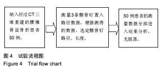
方法:选择2013年1月至2016年12月在山东省立第三医院接受骨盆病变、骨折治疗的患者50例。对患者进行骨盆CT扫描三维重建,将A点(S1骶后孔上缘连线与髂后上棘交点)位置作为髂骨钉置入点,在此点上分别测量3条路径:①髂前上棘路径标记为AD路径;②髂前下棘路径标记为AC路径;③坐骨大切迹上方至髋臼顶部路径标记为AB。在此3条路径平面上对髂骨做切面,分别在每个切面上设计髂骨钉所穿入的通道,测量出髂骨螺钉通道的直径、长度以及置入角度,根据测得的数据,选定合适的髂骨钉。
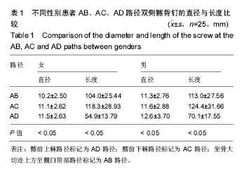
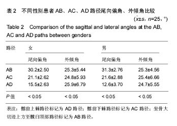
结果与结论:①同一置入路径的男性、女性髂骨钉通道长度比较,差异无显著性意义(P > 0.05)。经配对      t 检验,AB路径与AC路径,AC路径与AD路径的髂骨钉通道长度、直径比较,差异均有显著性意义(P < 0.05);②同一置入路径男性、女性的尾向偏角比较,差异无显著性意义(P > 0.05)。经配对t检验,男性、女性患者通过不同路径的尾向偏角比较,差异有显著性意义(P < 0.05);男性、女性患者通过不同路径的外倾角比较,差异亦有显著性意义(P < 0.05);③结果提示,骨盆CT三维重建可以测得可置入髂骨钉的长度、直径及角度,从而指导手术治疗。

中国组织工程研究杂志出版内容重点:人工关节;骨植入物;脊柱骨折;内固定;数字化骨科;组织工程
ORCID: 0000-0002-0418-1289(李春光)

关键词: 髂骨钉, 骨盆固定, 临床解剖, 骨科植入物

Abstract:

BACKGROUND: Iliac screw implantation has no unified standard. Because of the large individual differences, preoperative pelvic three-dimensional (3D) CT reconstruction can measure the diameter, depth and angle of the screws, which increases the placement accuracy.

OBJECTIVE: To measure the screw placement-related data by pelvic 3D CT reconstruction, so as to guide the screw placement and improve its accuracy.    
METHODS: Fifty patients with pelvic lesions or fractures admitted in Shandong Provincial Third Hospital from January 2013 to December 2016 were selected, and the pelvis was scanned with CT and then was reconstructed. The point A (the intersection of S1 posterior sacral posterior wall and posterior superior iliac spine) was as the screw placement point, and three paths were measured on the point: posterior superior iliac spine path (AD path); anterior inferior iliac spine path (AC path); top of greater sciatic notch to the acetabular roof (AB path). On the plane of these three pathes, the section was made on the iliac bone, respectively; in each section, the channel for iliac nail was designed.
RESULTS AND CONCLUSION: (1) In the same path, there was no significant difference in the path length of the screw between male and female patients (P > 0.05). Paired t test showed that the diameter and length depth and angle of the screws were significantly different between AD and AC paths, AC and AD (P < 0.05). (2) There was no significant difference in the inclination angle of the spiral channel between male and female patients (P > 0.05). The inclination angle of the spiral channel and lateral angle at different paths showed significant difference between male and female patients (P < 0.05). (3) To conclude, 3D reconstruction of pelvis CT can be used to measure the length, diameter and angle of the iliac screw, and to guide the surgical treatment. 

中国组织工程研究杂志出版内容重点:人工关节;骨植入物;脊柱骨折;内固定;数字化骨科;组织工程

Key words: Bone Nails, Pelvis, Internal Fixators, Tissue Engineering

中图分类号: