中国组织工程研究 ›› 2026, Vol. 30 ›› Issue (36): 9505-9518.doi: 10.12307/2026.903
• 口腔组织构建 oral tissue construction • 上一篇 下一篇
下颌阻生第三磨牙分冠治疗影响周围组织生物力学效应的三维有限元分析
李声昊1,张博锐1,马宇锋1,2
- 1山西医科大学口腔医学院,山西省太原市 030000;2山西医科大学第二医院口腔科,山西省太原市 030000
-
收稿日期:2025-10-15修回日期:2026-03-07出版日期:2026-12-28发布日期:2026-05-22 -
通讯作者:马宇锋,主任医师,山西医科大学口腔医学院,山西省太原市 030000;山西医科大学第二医院口腔科,山西省太原市 030000 -
作者简介:李声昊,男,1999年生,山西省运城市人,汉族,2025年山西医科大学毕业,硕士,医师,主要从事口腔颌面外科的研究。
Three-dimensional finite element analysis of the biomechanical effects on surrounding tissues following crown-by-crown treatment of impacted mandibular third molars
Li Shenghao1, Zhang Borui1, Ma Yufeng1, 2
- 1School of Stomatology, Shanxi Medical University, Taiyuan 030000, Shanxi Province, China; 2Department of Stomatology, The Second Hospital of Shanxi Medical University, Taiyuan 030000, Shanxi Province, China
-
Received:2025-10-15Revised:2026-03-07Online:2026-12-28Published:2026-05-22 -
Contact:Ma Yufeng, Chief physician, School of Stomatology, Shanxi Medical University, Taiyuan 030000, Shanxi Province, China; Department of Stomatology, The Second Hospital of Shanxi Medical University, Taiyuan 030000, Shanxi Province, China -
About author:Li Shenghao, MS, Physician, School of Stomatology, Shanxi Medical University, Taiyuan 030000, Shanxi Province, China

摘要:

文题释义:
三维有限元:三维有限元分析是计算力学和工程仿真中的一种强大的数值方法,其核心思想是将复杂的三维几何结构和物理场离散化(分割)成大量简单的、互相连接的小单元(如四面体、六面体、五面体),这些单元被称为有限元。然后通过求解一系列数学方程来模拟物理现象在这些单元上的行为,从而近似求解整个复杂系统的物理响应。
生物力学:是一门交叉学科,它应用力学(经典物理学中的分支,研究力、运动、变形和流动)的原理、理论和方法,来理解、分析、模拟和预测生物系统的结构、功能、运动、响应和调控,它关注的是生命体中的力学现象。
背景:阻生下颌第三磨牙拔除术是口腔颌面外科中常见的手术之一,其中传统劈冠去骨操作易引发邻牙损伤、下牙槽神经损伤及颌骨骨折等并发症。微创拔牙技术虽显著降低了术中创伤,但其分冠操作参数(如切削深度、宽度、角度)仍依赖经验性判断,缺乏生物力学量化标准。
目的:通过三维有限元构建下颌水平中位阻生第三磨牙及周围组织,分析不同切削深度、切削宽度和切削角度对牙冠断裂效应和周围组织生物力学的影响,为精准分冠操作提供量化标准。
方法:提取牙列完整的健康志愿者下颌第三磨牙区锥形束CT数据,通过三维重建软件Mimics 21.0、逆向工程软件Geomagic 21.0和三维计算机辅助设计软件Soildwork 2022,生成下颌第三磨牙区模型,包括下颌第三磨牙、下颌第二磨牙、颌骨(皮质骨和松质骨)、下牙槽神经管。采用Ansys 2021 R1有限元软件模拟临床分冠操作,通过施加35 N垂直载荷,探讨切削参数(深度:11 mm、10.5 mm、10 mm、9.5 mm、
9 mm;宽度:1.5 mm、1 mm;角度:0°,10°,20°,30°)对牙冠断裂效应、周围组织的应力分布特征和位移响应规律的影响。
结果与结论:成功构建了不同切削参数分冠的三维有限元模型,并通过三维有限元分析得出以下结论:①分冠时使用1.5 mm直径车针,在10-11 mm的切削深度和0°-10°的切削角度为最优参数组合,可在确保牙冠完全断裂(牙冠最大Von Mises应力值≥250 MPa)的同时,实现邻牙、下牙槽神经管和颌骨保护,为微创拔牙的分冠操作提供了精准的生物力学指导;②在水平中位阻生的下颌第三磨牙拔除操作中,切削深度和切削宽度的选择应建立在牙冠可以完全断裂的基础上,充分消除牙冠周围的阻力结构,为后续的操作提供理想空间,避免对周围组织的损伤;③在水平中位阻生的下颌第三磨牙拔除操作中,切削角度的选择应尽量与第二磨牙牙体长轴平行(0°-10°)以保证牙冠断裂效率以及对周围组织的保护。
https://orcid.org/0009-0003-7543-9165(李声昊)
中国组织工程研究杂志出版内容重点:干细胞;骨髓干细胞;造血干细胞;脂肪干细胞;肿瘤干细胞;胚胎干细胞;脐带脐血干细胞;干细胞诱导;干细胞分化;组织工程
中图分类号:
引用本文
李声昊, 张博锐, 马宇锋, . 下颌阻生第三磨牙分冠治疗影响周围组织生物力学效应的三维有限元分析[J]. 中国组织工程研究, 2026, 30(36): 9505-9518.
Li Shenghao, Zhang Borui, Ma Yufeng, . Three-dimensional finite element analysis of the biomechanical effects on surrounding tissues following crown-by-crown treatment of impacted mandibular third molars[J]. Chinese Journal of Tissue Engineering Research, 2026, 30(36): 9505-9518.
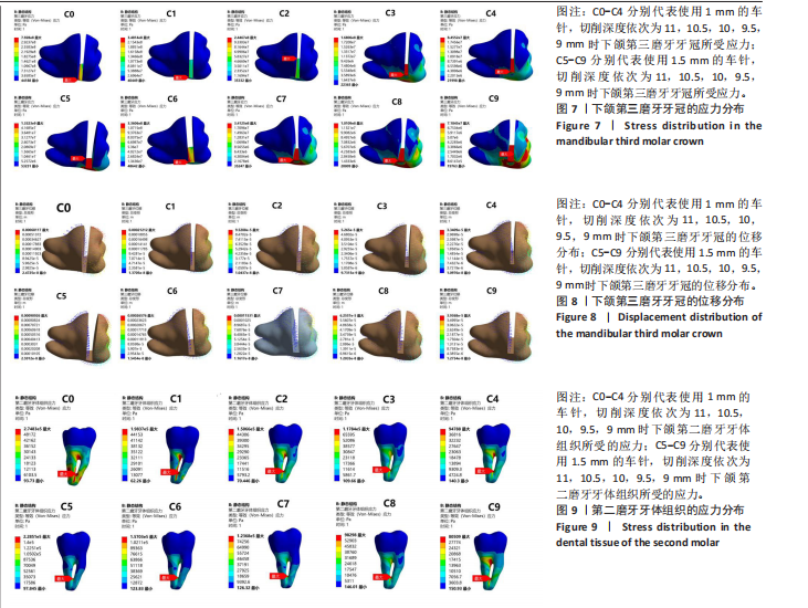
2.1.1 下颌第三磨牙的应力和位移分析 如图7所示,10组截冠模型的应力集中区主要分布在牙冠切削槽底部及附近、近中边缘嵴及附近、颊沟、舌沟、近中颊尖等区域。采用粗车针和细车针进行截冠后,二者的应力分布区域相似。
在10组模型牙冠断端的两侧,分别施加35 N的力,均垂直于表面,以模拟临床上使用牙挺断裂牙冠的操作后,C0-C9的最大应力集中区域均位于牙冠切削槽底部,C0(最大Von Mises应力值=792.81 MPa)、C1(最大Von Mises应力值=348.14 MPa)、C5(最大Von Mises应力值=1 332.34 MPa)、C6(最大Von Mises应力值=336.06 MPa)、C7(最大Von Mises应力值=341.25 MPa)均超过了牙体组织的最大屈服强度250 MPa[22-23],且最大应力集中区域均位于牙冠切削槽底部的远中,该应力传递路径完整,符合使牙冠断裂的生物力学特性,可以有效避免牙冠底部形成阶梯状残冠。细车针组的C2(最大Von Mises应力值=244.67 MPa)、C3(最大Von Mises应力值=168.06 MPa)、C4 (最大Von Mises应力值=84.55 MPa)牙冠切削槽底部最大应力值呈递减趋势,且其应力分布区域存在向牙冠切削槽底部的近中移动的趋势,说明该应力传递路径可能受阻,牙冠可能形成不完全断裂,有形成阶梯状残冠的风险,进而影响牙冠的取出。而粗车针组C9的最大应力分布区域位于牙冠切削槽底部的近中,同样存在牙冠不完全断裂的风险。
随着切削深度的减小,细车针组和粗车针粗的第三磨牙牙冠所受的最大Von Mises应力值呈非线性递减趋势,其中细车针组的C0-C1和粗车针组的C5-C6(即切削深度从11 mm减少至10.5 mm)的递减趋势最为显著,呈现骤降趋势,分别为56.09%和74.78%。
相同切削深度下,C5组对比C0组、C7组对比C2组产生的最大应力值增加了39.47%-68.01%,C6组对比C1组、C8组对比C3组、C9组对比C4组产生的最大应力值减少了3.47%-39.85%。
如图8所示,10组截冠模型的位移分布特点均为从牙冠切削槽顶部向底部及近远中逐级递减,符合生物力学能耗递减的分布模式。10种截冠模型的位移方向与牙冠断裂路径相一致,符合牙冠断裂的生物力学特性。随着切削深度的减小,两种车针的最大位移值呈非线性递减趋势,其中C0-C1和C5-C6,即切削深度从11 mm降低至10.5 mm的递减趋势最为明显,呈骤降趋势,分别降低了68.86%和70.77%。相同切削深度下,采用粗车针比采用细车针的牙冠最大位移值增加了17.84%-33.48%。
2.1.2 下颌第二磨牙牙体组织及牙周膜的应力和位移分析 如图9所示,在10组模型牙冠断端的两侧,分别施加35 N的力,均垂直于表面,以模拟临床上使用牙挺断裂牙冠的操作。10组模型的第二磨牙牙体组织的应力集中区主要分布在第二磨牙的根分叉处、近远中根融合部,该分布特点提示临床上拔除阻生牙时,应注意邻牙的保护和支点位置的选择,避免对第二磨牙牙根处产生损伤。同时,第二磨牙牙根的应力分布与牙周膜的生物学特性密切相关,根分叉区和近远中根融合部呈现最大Von Mises应力值,而牙颈部接近第三磨牙的部位因牙周膜的弹性作用,未出现应力集中。
粗车针组和细车针组的第二磨牙牙体组织最大Von Mises应力值出现区域相似,均位于第二磨牙近远中根融合部,且随着切削深度的减小,最大Von Mises应力值逐步减少,其中细车针组的C0-C1和粗车针组的C5-C6(即切削深度从11 mm减少至10.5 mm)的递减趋势最为显著,分别减少了27.82%和31.28%。相同切削深度下,粗车针组产生的最大Von Mises应力值均小于细车针组,粗车针组产生的最大Von Mises应力值相较细车针组减少了15.06%-20.84%。
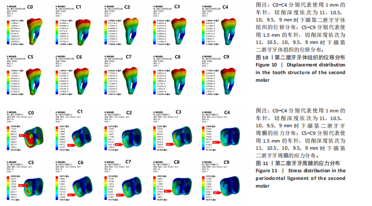
如图10所示,10组模型的第二磨牙主要位移中心相似,均位于第二磨牙牙冠远中边缘嵴和远中根尖处,且呈现向周围向近中逐步减少的趋势,在第二磨牙近中根的根中1/2达到最小位移。10种模型的最大位移处均位于第二磨牙牙冠的远中边缘嵴处。随着切削深度的减小,两种车针的最大位移呈非线性递减趋势,其中C0-C1组和C5-C6组的递减趋势较为显著,分别为27.82%和31.28%。相同切削深度下,粗车针组产生的最大位移较细车针组减少了3.21%-10.15%。
如图11所示,10组模型的主要应力分布区域相似,集中在第二磨牙牙颈部、远中根的远中面上1/3、远中根远中面的中1/3、根尖部等区域。C5组和C0组的最大Von Mises应力值主要集中在第二磨牙远中根远中面的中1/3处,分别为0.036 MPa和0.030 MPa,呈向周围递减趋势。而C1-C4组和C6-C9组的最大应力区域则位于第二磨牙远中颊颈部,呈向周围递减趋势。随着切削深度的减少,粗车针组和细车针组所产生的最大Von Mises应力值均呈非线性递减的趋势,其中细车针组的C0-C1和粗车针组的C5-C6(即切削深度从11 mm减少至10.5 mm)的递减趋势最为显著,分别为28.55%和17.22%。使用粗车针比使用细车针产生的最大Von Mises应力值减少了1.04%-16.19%。
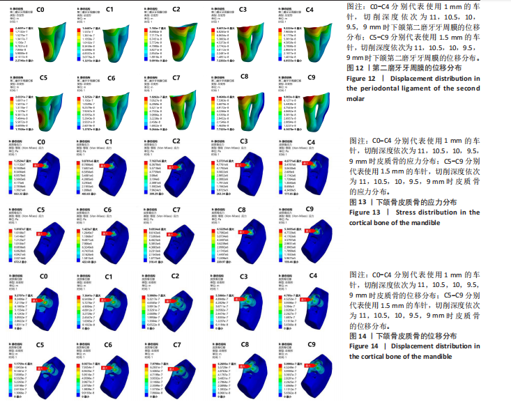
如图12所示,10组模型的主要位移分布区域相似,集中在第二磨牙远中颈部、远中根的下1/3处,并呈现向周围递减趋势。C0、C1、C5、C6的最大位移处在第二磨牙远中根的下1/3处,C2、C3、C4、C7、C8、C9的最大位移处在第二磨牙的远中颊颈部。随着切削深度的减小,粗车针组和细车针组的最大位移值均呈非线性递减趋势,其中细车针组的C0-C1和粗车针组的C5-C6(即切削深度从11 mm减少至10.5 mm)的递减趋势最为显著,分别为33.01%和34.82%。相同切削深度下,粗车针组比细车针组所产生的最大位移值减少了1.42%-19.23%。
2.1.3 下颌骨的应力和位移分析 如图13所示,10组模型皮质骨的应力集中区域相似,主要集中在下颌第三磨牙牙冠周围、颊舌侧骨板及根尖区域。10组模型皮质骨的最大Von Mises应力值区域相似,均位于下颌第三磨牙牙冠颊侧骨板。随着切削深度的减少,粗车针组和细车针组所产生的皮质骨最大Von Mises应力值呈非线性减小趋势,其中细车针组的C1(9.88 MPa)-C2(7.16 MPa)和粗车针组的C6(14.23 MPa)-C7(9.69 MPa),即切削深度从10.5 mm减小至10 mm的递减趋势最为显著,分别为27.48%和31.91%。相同切削深度下,粗车针组产生的皮质骨最大Von Mises应力值均大于细车针组。相同切削深度下,使用粗车针比使用细车针产生的皮质骨最大Von Mises应力值增加了14.80%-45.22%。
如图14所示,10组模型的皮质骨主要位移分布区域相似,主要分布在下颌第三磨牙牙冠下方骨板和颊侧骨板,并呈现向周围减小趋势。10组模型的最大位移区均位于下颌第三磨牙牙冠颊侧骨板,与图所示皮质骨最大Von Mises应力值区域相对应。随着切削深度的减小,10组模型的皮质骨位移呈非线性递减趋势,其中细车针组的C0(0.928 μm)-C1(0.736 μm)和粗车针组的C5(1.18 μm)-C6(0.893 μm),即切削深度从11 mm减少至10.5 mm的递减最为显著,分别为20.69%和24.32%。相同切削深度下,粗车针组产生的皮质骨位移均大于细车针组,增加了6.16%-26.73%。
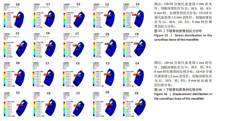
如图15所示,10组模型松质骨的主要应力集中区域相似,均位于下颌第三磨牙牙冠颊侧骨和牙冠下方的牙槽窝处。C0、C1、C2、C3、C5、C6的最大Von Mises应力值区域位于下颌第三磨牙牙冠下方的牙槽窝处,C4、C7、C8、C9的最大Von Mises应力值区域位于下颌第三磨牙牙冠颊侧骨处。随着切削深度的减小,粗车针和细车针所产生的松质骨最大Von Mises应力值呈非线性递减趋势,其中细车针组的C0(32.58 MPa)-C1(14.74 MPa)和粗车针组的C5(28.94 MPa)-C6(12.60 MPa),即切削深度从11 mm减少至10.5 mm的递减趋势最为显著,分别为54.76%和57.13%。相同切削深度下,粗车针组所产生的松质骨最大Von Mises应力值均小于细车针组。使用粗车针比使用细车针所产生的松质骨最大Von Mises应力值区域减小了1.50%-19.82%。
如图16所示,10组模型的松质骨最大位移处均位于下颌第三磨牙牙冠下方的牙槽窝处,并呈现向周围递减趋势。如图所示C0、C1、C2、C3、C5、C6的松质骨最大Von Mises应力值区域与最大位于区域相对应。随着切削深度的减小,松质骨的位移呈逐渐递减趋势,其中细车针组的C0(15.15 μm)-C1(7.42 μm)
和粗车针组的C5(19.04 μm)-C6(8.44 μm),即切削深度从11 mm减少至10.5 mm时的递减趋势最为显著,分别为51.02%和55.67%。相同切削深度下,粗车针组所产生的松质骨位移均大于细车针组。相同切削深度下,使用粗车针比使用细车针所产生的松质骨位移增加了1.70%-25.67%。
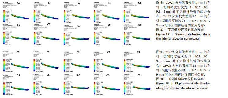
2.1.4 下牙槽神经管的应力和位移分析 如图17所示,下牙槽神经管与牙冠下方对应区域呈现出最大应力集中,该应力集中区域与上方的松质骨的位移(如图16所示)和第三磨牙牙冠的位移(如图8所示)相一致,说明在10组模型牙冠断端的两侧分别施加35 N的力(均垂直于断端表面)以模拟临床上使用牙挺断裂牙冠的操作后,神经管上部的松质骨和牙冠对其产生了挤压作用。随着切削深度的减小,下牙槽神经管所受的最大Von Mises应力值呈现出非线性递减趋势,其中C0(0.47 MPa)-C1(0.32 MPa)和C5(0.46 MPa)-C6(0.31 MPa),即切削深度从11 mm递减至10.5 mm的递减趋势最为显著,分别为31.91%和32.61%。相同切削深度下,粗车针组所产生下牙槽神经管的最大Von Mises应力值均小于细车针组。使用粗车针所产生的下牙槽神经管最大Von Mises应力值比使用细车针减少了1.11%-3.80%。
如图18所示10组模型的下牙槽神经管的最大位移区域与最大Von Mises应力值区域(如图17所示)相对应,呈现向周围递减趋势。下牙槽神经管的位移与下颌第三磨牙牙冠位移(如图8所示)和松质骨位移(如图16所示)相对于,说明了挤压作用的存在。随着切削深度的减小,下牙槽神经管的位移呈递减趋势,其中细车针组的C0(1.52 μm)-C1(1.10 μm)和粗车针组的C5(1.55 μm)-C6(1.09 μm),即在切削深度11 mm减小至10.5 mm时递减趋势最为明显,分别为27.63%和29.68%。在相同切削深度下,粗车针所产生的下牙槽神经管最大位移C5(1.55 μm)大于细车针组中的C0(1.52 μm),其余均小于细车针组。
2.2 不同切削角度所产生的应力与位移分析
2.2.1 下颌第三磨牙应力及位移分析 如图19所示,N0的应力集中区主要分布在牙冠切削槽底部及附近、近中边缘嵴及附近、颊沟、舌沟、近中颊尖等区域,N1、N2、N3的应力集中区主要分布在牙冠切削槽底部及附近。
N0、N1的最大应力集中区在牙冠切削槽底部颊侧偏远中处,而N2、N3的最大应力集中区在牙冠切削槽底部的近中处。N0(最大Von Mises应力值=341.25 MPa)、N1(最大Von Mises应力值=275.44 MPa)均超过了牙体组织的最大屈服强度250 MPa,且最大应力集中区域均位于牙冠切削槽底部的远中,该应力传递路径完整,符合使牙冠断裂的生物力学特性,可以有效避免牙冠底部形成阶梯状残冠。N2(最大Von Mises应力值=196.81 MPa)、N3(最大Von Mises应力值=137.73 MPa)牙冠切削槽底部最大应力值呈递减趋势,且其最大应力分布区域位于牙冠切削槽底部的近中,说明该应力传递路径可能受阻,牙冠可能形成不完全断裂,有形成阶梯状残冠的风险,进而影响牙冠的取出。同时,N3的牙冠切削槽底部近远中向出现应力中断带,说明在该负载情况下,牙冠可能形成不完全断裂。
随着切削角度的增大,下颌第三磨牙牙冠所受的最大Von Mises应力值呈非线性递减趋势,其中N2(196.81 MPa)-N3(137.73 MPa)的递减趋势最为显著,约为30.02%。
如图19所示,4组截冠模型的位移分布特点均为从牙冠切削槽顶部向底部及近远中逐级递减,符合生物力学能耗递减的分布模式。4组模型的最大位移处均位于牙冠远中边缘嵴处。4组截冠模型的位移方向与牙冠断裂路径相一致,符合牙冠断裂的生物力学特性。随着切削角度从0°增加到10°时,牙冠的最大位移值从115.31 μm增加至148.48 μm。但随着切削角度从10°增加到20°和30°时,牙冠的最大位移值呈现非线性递减趋势,裂纹扩展路径向根方偏移。其中N2(124.71 μm)-N3(49.16 μm)的递减趋势最为显著,呈骤降趋势,约为60.58%。
2.2.2 第二磨牙牙体及牙周膜的应力和位移分析 如图20所示,在4组模型牙冠断端的两侧,分别施加35 N的力,均垂直于表面,以模拟临床上使用牙挺断裂牙冠的操作。4组模型的第二磨牙牙体组织的应力集中区主要分布在第二磨牙的根分叉处、近远中根融合部,该分布特点提示临床上拔除阻生牙时,应注意邻牙的保护和支点位置的选择,避免对第二磨牙牙根处产生损伤。同时,第二磨牙牙根的应力分布与牙周膜的生物学特性密切相关,根分叉区和近远中根融合部呈现应力集中区,而牙颈部接近第三磨牙的部位因牙周膜的弹性作用,未出现应力集中。
4组模型的第二磨牙牙体组织最大Von Mises应力值出现区域相似,均位于第二磨牙近远中根融合部。且随着切削角度从0°增加到10°时,第二磨牙牙根的最大Von Mises应力值骤降,从0.12 MPa骤降到0.026 MPa,降低了约78.33%。随着切削角度从10°增加到20°时,第二磨牙牙根的最大Von Mises应力值持续骤降,降低了约65.58%。而随着切削角度从20°增加到30°时,第二磨牙牙根的最大Von Mises应力值骤增,增加了约63.9倍。
如图20所示,N0、N1、N2的第二磨牙最大位移中心相似,均位于第二磨牙牙冠远中边缘嵴,且呈现向周围向近中逐步减少的趋势。N3的第二磨牙最大位移中心第二磨牙根尖1/3的远中,并呈现向周围递减趋势。随着切削角度的增大,第二磨牙的牙根位移呈现先减小后增大的趋势:在切削角度从0°增加至10°时,第二磨牙牙体组织位移减少了约60.17%;在切削角度从10°增加至20°时,第二磨牙牙体组织位移减少了约48.60%,在切削角度从20°增加到30°时,第二磨牙牙根的最大位移增加了约2.48倍。
如图21所示,4组模型的最大应力集中区域相似,集中在第二磨牙牙颈部。在N0-N2组中,随着切削角度从0°(N0)依次递增至10°(N1)、20°(N2)时,最大Von Mises应力值呈非线性递减:N0(0.023 MPa)、N1(0.013 MPa)、N2(0.0017 MPa),最大Von Mises应力值的降低与切削角度呈负相关。而当切削角度从20°(N2)增加到30°(N3)时,最大Von Mises应力值骤增,增加了约14.21倍。

如图21所示,N0、N1、N2的最大位移中心均位于第二磨牙远中牙颈部,并呈现向周围递减趋势。N3的最大位移中心则位于远中根的根中1/3部,呈现向周围递减趋势。在N0-N2组中,随着切削角度从0°(N0)依次递增至10°(N1)、20°(N2)时,最大位移呈非线性递减:N0(0.11 μm)、N1(0.051 μm)、N2(0.027 μm)。
而当切削角度从20°(N2)增加到30°(N3)时,最大位移值骤增,增加了约3.79倍。
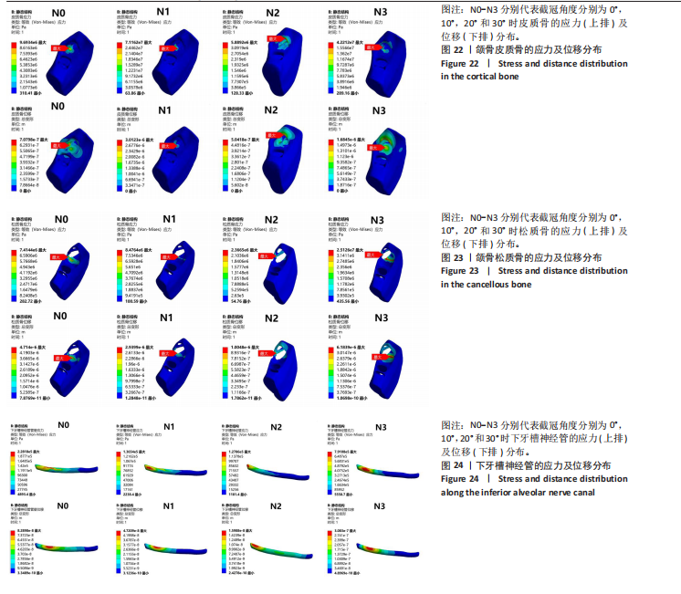
2.2.3 颌骨的应力和位移分析 如图22所示,N0、N1、N3的皮质骨最大应力集中区均位于下颌第三磨牙颊侧骨板,N2的最大应力集中区位于下颌第三磨牙远中骨板。随着切削角度从0°(N0)增大到10°(N1),皮质骨的最大Von Mises应力值从9.69 MPa增加到71.16 MPa;随着切削角度从10°(N1)增大到20°(N2),皮质骨的最大Von Mises应力值从71.16 MPa减少至5.89 MPa:随着切削角度从20°(N2)增大到30°(N3),皮质骨的最大Von Mises应力值从5.89 MPa增大至42.21 MPa。
如图22所示,N0的皮质骨最大位移中心位于下颌第三磨牙牙冠颊侧骨板,N1、N3的皮质骨最大位移中心均位于牙冠下方骨板、N2的皮质骨最大位移中心位于牙冠远中骨板。随着切削角度从0°(N0)增大到10°(N1),皮质骨的最大位移值从0.071 μm增加到3.01 μm;随着切削角度从10°(N1)增大到20°(N2),皮质骨的最大位移值从3.01 μm减少至0.50 μm:随着切削角度从20°(N2)增大到30°(N3),皮质骨的最大位移值从0.50 μm增大至1.68 μm。
如图23所示,N0、N2、N3松质骨的最大应力集中区均位于下颌第三磨牙牙冠颊侧骨,N1最大应力集中区位于牙冠下方骨。随着切削角度从0°(N0)增大到10°(N1),皮质骨的最大Von Mises应力值从7.41 MPa增加到8.48 MPa,增大了约14.44%;随着切削角度从10°(N1)增大到20°(N2),松质骨的最大Von Mises应力值从8.48 MPa减少至2.37 MPa,减少了约72.05%:随着切削角度从20°(N2)增大到30°(N3),松质骨的最大Von Mises应力值从2.37 MPa增大至25.13 MPa。
如图23所示,N0、N3的松质骨最大位移中心位于下颌第三磨牙牙冠下方骨,N1、N2的松质骨最大位移中心均位于下颌第三磨牙牙冠颊侧骨。随着切削角度从0°(N0)依次递增至10°(N1)、20°(N2)时,松质骨的最大位移值呈非线性递减:N0(4.71 μm)、N1(2.94 μm)、N2(1.00 μm),最大位移值的降低与切削角度呈负相关。而随着切削角度从20°(N2)增加到30°(N3),最大位移出现骤增趋势,从1.00 μm增长至6.18 μm。
2.2.4 下牙槽神经管的位移和应力分析 如图24所示,N0的下牙槽神经管与牙冠下方对应区域呈现出最大应力集中,该应力集中区域与上方松质骨的位移和第三磨牙牙冠的位移相一致,说明在牙冠断端的两侧分别施加35 N的力(均垂直于断端表面)以模拟临床上使用牙挺断裂牙冠的操作后,神经管上部的颌骨和牙冠对其产生了挤压作用。而N1、N2、N3的最大应力集中区出现向远中颊侧移动趋势。随着切削角度从0°(N0)依次递增至10°(N1)、20°(N2)时,最大Von Mises应力值呈非线性递减:N0(0.23 MPa)、N1(0.14 MPa)、N2(0.13 MPa),最大Von Mises应力值的降低与切削角度呈负相关。而随着切削角度从20°(N2)增加至30°(N3),下牙槽神经管的最大Von Mises应力值出现骤增趋势,增加了约5.23倍。
如图24所示,N0下牙槽神经管的最大位移区域与最大Von Mises应力值区域相对应,呈现向周围递减趋势。N0下牙槽神经管的位移与下颌第三磨牙牙冠位移和松质骨位移相对应,说明了挤压作用的存在。随着切削角度从0°(N0)依次递增至10°(N1)、20°(N2)时,最大位移值呈非线性递减:N0(0.083 μm)、N1(0.047 μm)、N2(0.016 μm),最大位移值的降低与切削角度呈负相关。而随着切削角度从20°(N2)增加至30°(N3),下牙槽神经管的最大位移值出现骤增趋势,从0.016 μm增加至0.31 μm。
| [1] STEEL BJ, SURENDRAN KSB, BRAITHWAITE C, et al. Current thinking in lower third molar surgery. Br J Oral Maxillofac Surg. 2022;60(3):257-265. [2] CANDOTTO V, OBERTI L, GABRIONE F, et al. Complication in third molar extractions. J Biol Regul Homeost Agents. 2019;33(3 Suppl. 1):169-172. [3] MARGHALANI AA, ALSAGGAF AU, HAZZAZI A, et al. Radiographic characteristics of impacted teeth: A retrospective study of 2199 radiograph. PLoS One. 2025; 20(2):e0316749. [4] WANG X. [Geostatics in impacted mandibular third molar surgury]. Zhonghua Kou Qiang Yi Xue Za Zhi. 2025;60(2):109-115. [5] 胡开进. 微创拔牙技术[J]. 现代口腔医学杂志,2010,24(4):241-243. [6] 胡开进. 现代标准拔牙术新理念[J]. 中国实用口腔科杂志,2010,3(10):577-581. [7] 张志霞, 周宏志, 刘瑶, 等. 微创拔牙术在下颌阻生智齿拔除中的应用[J]. 实用口腔医学杂志,2012,28(3):398-400. [8] 杨映阳, 杜胜男, 吕宗凯. 高速涡轮机头和微创拔牙刀在下颌阻生牙拔除术中的应用效果评价[J]. 上海口腔医学,2015,24(4):489-492. [9] 陈勇臻, 尹建超, 李森. 超声骨刀微创拔牙术与传统凿骨劈冠拔牙术治疗下颌阻生智齿患者的效果比较[J]. 现代临床医学,2025,51(1):16-19+37. [10] 柴娟,张昕,刘昌奎,等. 2023版《恒牙拔牙术临床操作规范》解读[J].华西口腔医学杂志,2025,43(2):158-162. [11] 鞠昊, 朱红华, 段涛, 等. CBCT的基本原理及在口腔各科的应用进展[J].医学影像学杂志,2015,25(5):907-909+942. [12] TAY WZ, SKLAVOS A, MIAN M, et al. Radiographic Predictors of Postoperative Inferior Alveolar Nerve Injury in Mandibular Third Molar Surgery. J Oral Maxillofac Surg. 2025;83(1):54-61. [13] POGORZELSKA A, MIELCZAREK A, REGULSKI PA, et al. Evaluation of the Inferior Alveolar Canal Course in Relation to Fully and Partially Impacted Lower Third Molar Roots via Cone-Beam Computed Tomography. Cureus. 2024;16(11):e74021. [14] SÁNCHEZ JORGE MI, OCAÑA RA, VALLE RODRÍGUEZ C, et al. Mandibular third molar extraction: perceived surgical difficulty in relation to professional training. BMC Oral Health. 2023;23(1):485. [15] 王岚. 种植修复义齿和天然邻牙生物力学的三维有限元分析[D]. 重庆:重庆医科大学,2022. [16] 蒋啸宇, 朱杰晶, 王栋才, 等. 上颌全牙列远移中牙周膜的应力分析[J]. 口腔医学研究,2023,39(4):341-344. [17] KUGUOGLU A, AKARSU-GUVEN B. Evaluation of the effects of the third molar on distalization and the effects of attachments on distalization and expansion with clear aligners: Three-dimensional finite element study. Korean J Orthod. 2025;55(1):69-81. [18] GAO H, LUO L, LIU J. Three-dimensional finite element analysis of maxillary molar distalization treated with clear aligners combined with different traction methods. Prog Orthod. 2024;25(1):47. [19] SU R, LI S, WANG W. Effect of high trimline aligners on distalizing mandibular molars: a three-dimensional finite element study. Eur J Med Res. 2024;29(1):623. [20] MEMON S, MEHTA S, MALIK S, et al. Three-dimensional finite element analysis of the stress distribution in the endodontically treated maxillary central incisor by glass fiber post and dentin post. J Indian Prosthodont Soc. 2016;16(1):70-74. [21] YALCIN S, AKTAS I, EMES Y, et al. A technique for atraumatic extraction of teeth before immediate implant placement using implant drills. Implant Dent. 2009; 18(6):464-472. [22] SAKAGUCHI RL, POWERS JM. Craig’s restorative dental materials. Mosby Elsevier, 2012. [23] CROTHERS JA. Fundamentals of operative dentistry. Br Dental J. 2006;201(3):180-181. [24] BAILEY E, KASHBOUR W, SHAH N, et al. Surgical techniques for the removal of mandibular wisdom teeth. Cochrane Database Syst Rev. 2020;7(7):Cd004345. [25] XU W, SHEN J, LI K, et al. A mechanical and three-dimensional finite element study of the optimum tooth sectioning depth during the extraction of low-level horizontally impacted mandibular third molar. Odontology. 2024;112(1):242-249. [26] YAN ZY, XU JY, ZHANG JY, et al. Optimal tooth sectioning using a surgical handpiece and elevator: a finite element study of horizontally deeply impacted mandibular third molars. Clin Oral Investig. 2024;28(8):439. [27] 刘平,胡开进,马洋,等.外科拔牙器械的选择及应用[J].中国实用口腔科杂志,2016,9(10):581-584. [28] SUEDBECK JR, ARMITANO-LAGO CN, LUDWIG EA. The Additive Effects of Cell Phone Use and Dental Hygiene Practice on Finger Muscle Strength: A Pilot Study. J Dent Hyg. 2020;94(2):45-53. [29] TRISI P, DE BENEDITTIS S, PERFETTI G, et al. Primary stability, insertion torque and bone density of cylindric implant ad modum Branemark: is there a relationship? An in vitro study. Clin Oral Implants Res. 2011;22(5):567-570. [30] BORRERO-LOPEZ O, RODRIGUEZ-ROJAS F, CONSTANTINO PJ, et al. Fundamental mechanics of tooth fracture and wear: implications for humans and other primates. Interface Focus. 2021;11(5):20200070. [31] ZENG J, JIANG Y, LIU Y, et al. Research on a novel digital tooth sectioning guide system for tooth sectioning during mandibular third molar extraction: An in vitro study. J Stomatol Oral Maxillofac Surg. 2023;124(1s):101383. [32] RESNICK CM, DANG RR, GLICK SJ, et al. Accuracy of three-dimensional soft tissue prediction for Le Fort I osteotomy using Dolphin 3D software: a pilot study. Int J Oral Maxillofac Surg. 2017;46(3):289-295. [33] ANAND M, PANWAR S. Role of Navigation in Oral and Maxillofacial Surgery: A Surgeon’s Perspectives. Clin Cosmet Investig Dent. 2021;13:127-139. [34] 陈新民,王劲茗,赵云凤,等.人体下颌骨强度研究[J]. 口腔颌面修复学杂志, 2002,3(2):113-115. [35] JIA B, WANG Q, CHEN J, et al. [Expert consensus on standardized clinical applications of minimally invasive tooth extraction techniques]. Nan Fang Yi Ke Da Xue Xue Bao. 2024;44(5):1004-1014. [36] LIU X, CHENG Y, QIN W, et al. Effects of upper-molar distalization using clear aligners in combination with Class II elastics: a three-dimensional finite element analysis. BMC Oral Health. 2022;22(1):546. [37] CHEN Q, LUO S, WANG Y, et al. Three-dimensional finite element analysis of occlusal stress on maxillary first molars with different marginal morphologies restored with occlusal veneers. BMC Oral Health. 2024;24(1):1349. [38] CHENG X, ZHANG XY, QIAN WH. Influence of different base materials and thicknesses on the fracture resistance of endocrown: A three-dimensional finite element analysis. BMC Oral Health. 2022;22(1):363. [39] VALERA-JIMÉNEZ JF, BURGUEÑO-BARRIS G, GÓMEZ-GONZÁLEZ S, et al. Finite element analysis of narrow dental implants. Dent Mater. 2020;36(7):927-935. [40] MAO B, TIAN Y, LI J, et al. Expansion rebound deformation of clear aligners and its biomechanical influence: a three-dimensional morphologic analysis and finite element analysis study. Angle Orthod. 2023;93(5):572-579. [41] GAO W, LU J, GAO X, et al. Biomechanical effects of joint disc perforation on the temporomandibular joint: a 3D finite element study. BMC Oral Health. 2023;23(1):943. [42] CHEN J, XU Y, LI C, et al. Displacement and stress distribution of the craniomaxillofacial complex under different surgical conditions: a three-dimensional finite element analysis of fracture mechanics. BMC Oral Health. 2021;21(1):596. [43] TRANG BNH, KANAZAWA M, MURAKAMI N, et al. Stress distribution of one-piece and two-piece mini-Implant overdentures with various attachment systems and diameters: A finite element analysis. J Prosthodont Res. 2023;67(3):430-436. |
| [1] | 樊佳兵, 付雪飞, 张军梅, 周索頔, 莫朝伦. 3D打印导板引导正畸微种植钉植入的精确性[J]. 中国组织工程研究, 2025, 29(24): 5102-5108. |
| [2] | 庄昕仪, 彭源浩, 喻 婷, 吕冬梅, 温秀杰, 程 钎. 不同颈椎骨龄青少年下颌骨外斜线区骨量的锥形束CT评价[J]. 中国组织工程研究, 2024, 28(8): 1253-1258. |
| [3] | 彭源浩, 吕冬梅, 庄昕仪, 喻 婷, 程 钎. 两种锥形束CT转化二维图像与传统头颅侧位片定量分析颈椎骨龄的一致性[J]. 中国组织工程研究, 2024, 28(18): 2881-2886. |
| [4] | 张 宁, 高思文, 郭 隽, 李泽奎, 张 娟. 青少年颞下颌关节骨关节病患者髁突在关节窝内间隙变化与骨质破坏位置的相关性[J]. 中国组织工程研究, 2022, 26(29): 4593-4597. |
| [5] | 周星宇, 贾 莹, 李仲伟, 丁 琪. 三维成像技术评估骨性Ⅱ类错颌不同垂直骨面型的牙轴倾斜特征[J]. 中国组织工程研究, 2022, 26(17): 2625-2630. |
| [6] | 顾月光, 沈剑欢, 倪洁丽, 郭舒瑜, 闫忠义, 张 阳. 应用体积分析牙槽嵴裂植骨治疗后的成骨效果[J]. 中国组织工程研究, 2022, 26(10): 1501-1504. |
| [7] | 樊佳兵, 张军梅. 成年女性不同垂直骨面型下颌骨形态的测量分析[J]. 中国组织工程研究, 2021, 25(8): 1177-1183. |
| [8] | 聂 晶, 石晓宇. 锥形束CT测量种植体支抗植入位点上颌前牙区牙槽骨厚度的性别差异[J]. 中国组织工程研究, 2021, 25(14): 2133-2136. |
| [9] | 张钰晶, 彭于治, 刘宝珍, 井 芳. 锥形束CT评价仙灵骨葆胶囊治疗绝经后女性牙周炎患者牙槽骨骨量的变化[J]. 中国组织工程研究, 2020, 24(17): 2694-2699. |
| [10] | 谢 慧,秦 波,王薮馨,莫朝伦,张军梅 . 多平面重建颧骨上颌骨复合体部软硬组织结构的测量分析[J]. 中国组织工程研究, 2019, 23(31): 4945-4949. |
| [11] | 王超然,李志勤,史培良,冯春月,殷 野. 锥形束CT测量分析安氏Ⅲ类错牙合畸形治疗前后颞下颌关节骨性结构的改变[J]. 中国组织工程研究, 2019, 23(31): 4950-4955. |
| [12] | 姜委杰,刘 珺,刘昌翠,张 晨 . 锥形束CT评价成人安氏Ⅱ1类拔牙后上前牙内收前后牙槽骨变化[J]. 中国组织工程研究, 2019, 23(19): 2986-2990. |
| [13] | 罗 艺,王恩群,许雅婷,任 伟. 改良型富血小板纤维蛋白修复下颌智齿拔除后邻牙远中骨缺损[J]. 中国组织工程研究, 2019, 23(15): 2314-2319. |
| [14] | 陈妍曲,唐 敏,黄旋平,周奉城,王家烯. 高精度三维整合牙颌模型个体化微种植体手术导板的计算机辅助设计与制作[J]. 中国组织工程研究, 2018, 22(10): 1529-1533. |
| [15] | 单健良,张 琴,胡 杨,姚 源,张 玮,何惠宇. 骨质疏松对种植义齿修复及预后的影响[J]. 中国组织工程研究, 2017, 21(4): 586-590. |
目前临床上对于下颌第三磨牙的治疗主要以牙拔除术为主。由于传统拔牙法中的锤、敲、劈冠、去骨等操作对周围软硬组织的损伤破坏较大,所以微创拔牙法现已逐渐运用于临床操作中[5]。微创拔牙是指通过精细化的操作技术和特殊器械,最大程度减少拔牙过程中硬组织的破坏和软组织损伤的临床技术。针对下颌第三磨牙这一拔除难度较高的解剖结构,其临床应用已逐渐取代传统骨凿敲击法。从操作原理分析,该技术通过精确的牙周膜切断和可控性分牙,显著降低了术中对下颌第三磨牙周围骨组织、软组织及邻牙的机械性冲击。在器械配置方面,现代微创拔牙系统主要包括:①高频超声骨刀,通过55-60 kHz的微振动实现选择性硬组织切割,可精确控制切割深度并避免热损伤;②微创牙挺系统,采用杠杆、楔力、轮轴的原理设计,能产生30-50 N的持续脱位力;③45°仰角设计的微创拔牙手机,配备直径1.0-1.5 mm的车针,可实现三维分牙[6-8]。临床数据显示,与传统方法相比,使用上述器械可使平均手术时间缩短40%,术中失血量更少,拔牙创口的肿胀程度更轻(95%CI)[9]。但该技术仍存在特定的并发症风险[10]:①下牙槽神经损伤,主要因超声骨刀工作尖或分牙操作时车针过度深入下颌管区域所致;②邻牙(第二磨牙)牙根微裂,多由分牙角度或深度偏差造成;③术后干槽症发生率,与微创操作中过度追求保留骨壁导致血凝块稳定性下降相关。
下颌第三磨牙拔除术的术前影像学评估是保障手术安全的核心环节,目前临床主要采用数字化曲面体层片和锥形束CT进行多维度分析。曲面体层片作为基础筛查手段,可明确阻生齿的倾斜角度、萌出深度以及与下颌管的空间关系,但其二维成像特性可能会导致多维度解剖信息的丢失,如牙根影像的重叠、与神经管颊舌向位置的模糊等,可能会低估术中造成神经管损伤、对邻牙损伤以及断根的风险。针对上述问题,锥形束CT的引入显著降低了术中风险的发生。锥形束CT通过三维重建技术,可在矢状位、冠状位及轴位多平面清晰显示下颌第三磨牙的牙根形态以及与下牙槽神经管的位置关系[11-12]。对于复杂病例(如牙根与下颌管接触、颊舌向异位),锥形束CT可提供0.2 mm3体素分辨率的三维重建影像,通过多平面重组技术精确测量牙根与下牙槽神经的接触面积,从而降低下牙槽神经损伤的风险[2,13]。
为了更好地减轻下颌第三磨牙拔除术中对软硬组织的破坏与损伤,口腔颌面外科医生往往会在术前和术中对阻生齿拔除过程的生物力学进行分析,以更好地应对牙拔除过程中可能出现的风险。低年资住院医师及规培学员在复杂阻生智齿拔除术中面临显著的技术挑战[14],其临床经验相对欠缺导致对生物力学响应机制的认知存在局限性,尤其未能充分掌握不同解剖条件牙-骨复合体的力学传导规律,致使理论认知与临床实践间存在转化障碍。在处理具有骨埋伏特征的病例时,此类医师在关键操作参数(如截冠角度、深度、挺出力度等)的量化控制方面表现出明显不确定性。目前国内外对于下颌第三磨牙分冠过程中生物力学的分析较为缺乏。此次研究基于三维有限元法,通过创建下颌第三磨牙及周围组织模型,分析比较不同宽度车针(切削宽度)和切削深度下进行分冠操作对下颌第三磨牙断裂效率及周围组织的影响,并分析比较相同切削宽度及切削深度在不同切削角度下截冠对下颌第三磨牙及周围组织的影响,为临床下颌第三磨牙拔除术前生物力学的分析及分冠策略的选择提供一定的理论依据和参考,进而减轻和预防下颌第三磨牙拔除过程中对周围软硬组织的损伤与破坏。
中国组织工程研究杂志出版内容重点:干细胞;骨髓干细胞;造血干细胞;脂肪干细胞;肿瘤干细胞;胚胎干细胞;脐带脐血干细胞;干细胞诱导;干细胞分化;组织工程
1.1 设计 三维模型的建立及三维有限元分析。
1.2 时间及地点 实验于2025年3-5月在山西医科大学第二医院口腔科内完成。
1.3 对象 选择山西医科大学第二医院口腔科1名牙列完整的健康成年人作为受试者,男性,24岁。
纳入标准:①下颌第三磨牙的锥形束CT影像学表现为中位水平阻生;②下颌第三磨牙牙冠完整无缺损,牙根发育完整,无明显牙根或牙槽骨吸收;③下颌第三磨牙邻近下颌第二磨牙存在,且牙根发育完整,无明显牙根或牙槽骨吸收;④无创伤史、正畸治疗史、正颌手术及其他颌面部手术史;⑤下牙槽神经管完整,无破坏或变异分支存在。
排除标准:①有影响牙槽骨吸收的系统疾病史或服药史;②颌骨创伤或手术治疗史;③有下颌第三磨牙或邻近下颌第二磨牙的根管治疗史或根尖周疾病史。
经受试者知情同意后,获取其锥形束CT影像资料,成像区域(FOV)18 cm×18 cm,扫描时间20 s,体素分辨率75 μm,共计扫描504层图像,使用Planmeca Romexis软件导出为DICMO格式数据。该受试者对此次实验知情,并签署知情同意书。此次研究经山西医科大学第二医院医学伦理委员会批准(批号:[2025]YX第327号)。
1.4 实验设备及软件
1.4.1 实验设备 Planmeca ProMax 3D Max 锥形束CT机(Planmeca
公司,芬兰);宏碁暗影骑士擎电脑一台,处理器:12th Gen Intel (R) Core(TM) i5-12500H 3.10 GHz,显示适配器:NVIDIA GeForce RTX3060 Laptop GPU,内存:16G,硬盘:512G SSD。
1.4.2 软件 Mimics 21.0(比利时,Materialise 公司);ANSYS 2021 R1(美国,Ansys 公司);Geomagic 21.0 (美国,Geomagic 公司) ;Soildwork 2022(法国 Dassault 公司)。
1.5 方法
1.5.1 下颌第三磨牙及其周围组织的三维有限元模型建立 在Mimics 21.0软件中新建项目后,导入锥形束CT的DICOM格式数据,系统自动重建冠状面、矢状面及水平面三维影像序列。通过分割模块(Segment)中的阈值分割功能创建初始蒙版,基于灰度值调整优化牙齿及颌骨结构边界,有效抑制背景噪点干扰(图1)。进一步采用交互式区域分割工具对连续断层图像进行多通道标记,通过逐层勾画建立右侧下颌骨、下颌第二磨牙和第三磨牙及下牙槽神经管等多结构蒙版(图2),最终整合生成包含目标解剖结构的数字化三维模型,并以STL格式导出供后续有限元分析。

在Ansys 2021 R1平台中导入X_T文件后,基于生物组织解剖特征实施网格优化策略:对下颌骨、牙体及下牙槽神经管采用混合网格划分技术,通过三角形/四边形单元实现复杂解剖形态的拓扑匹配,并执行网格敏感性分析以验证网格质量与数量对计算精度的收敛性影响。材料属性赋值依据口腔生物力学研究共识[17-20]:将骨组织、牙体组织等假设为各向同性线弹性体,其弹性模量及泊松比参数参照表1设置,其中下牙槽神经管壁材料属性映射至骨皮质参数。边界条件设定遵循临床力学加载机制:采用多点约束模拟下颌骨固定装置,对近远中截面及基底面施加全自由度约束(UX=UY=UZ=0);牙体-骨界面接触定义为绑定约束,消除界面滑移误差,确保拔牙阻力矩的精准传递。

此次研究基于Mimics 21.0-Geomagic 21.0-ANSYS 2021 R1跨平台技术集成框架,构建了具有解剖学特征保真度的下颌骨-牙列三维有限元模型(图3)。通过多模态影像配准算法与自适应曲面拟合技术,系统实现了下颌中位水平阻生第三磨牙、邻接第二磨牙、双层骨结构(皮质骨厚度2 mm/松质骨)及下牙槽神经管等复合解剖单元的三维可视化重构,有效克服了传统建模方法中因空间配准失真和组织边界模糊导致的阻生齿-神经管空间关系建模偏差。该模型通过亚体素级影像解析与多层级接触定义技术,显著提升生物力学仿真的临床相关性,为定量评估阻生牙-邻接第二磨牙-下牙槽神经管-周围骨组织生物力学风险提供了高精度数字化研究平台。

(1)10组截冠模型的建立:基于临床截冠术式标准化需求[10,21],此次研究建立多参数耦合的牙体切削有限元仿真框架:对高速涡轮仰角手机车针进行建模,其中细车针组(直径1.0 mm)与粗车针组(直径1.5 mm)分别对应临床常用车针规格。在下颌第三磨牙三维模型中,设置切削路径沿牙冠远中边缘嵴至最大颊舌径方向延伸,且与邻牙(第二磨牙)长轴保持平行关系(夹角偏差< 2°)。通过参数化建模技术生成10组实验模型:C0组(细车针/11 mm深度基准组)与C1-C4组(细径车针梯度递减组)、C5-C9组(粗径全深度组)(图4),各组模型通过布尔差运算精确构建牙釉质-牙本质-牙髓复合体与切削槽的几何拓扑关系。
为提升应力集中区域的数值精度,采用局部网格细化策略:在切削槽底部区域(Von Mises应力梯度> 15 MPa/mm2)设置0.5 mm高密度六面体单元,其他区域采用1.0 mm混合单元。

该建模方法通过网格收敛性分析验证了计算结果的稳定性,有效克服了传统建模中因牙髓腔形态简化导致的生物力学失真问题。
(2)材料属性与边界条件:模型的网格划分遵循表2,边界条件设定遵循临床力学加载机制:采用多点约束模拟下颌骨固定装置,对近远中截面及基底面施加全自由度约束;牙体-骨界面接触定义为绑定约束,消除界面滑移误差,确保拔牙阻力矩的精准传递。

使用Ansys 2021 R1软件在10组模型牙冠断端的两侧,采用双平面对称垂直加载模式,分别施加35 N的力,均垂直于表面,以模拟临床上使用牙挺断裂牙冠的操作(图5)。使用Ansys 2021 R1软件进行应力分析,观察下颌第三磨牙牙冠裂缝、周围皮质骨和松质骨、第二磨牙牙体及牙周膜、下牙槽神经管管壁的应力与变形情况。
1.5.3 不同切削角度对牙冠断裂效应及周围组织的影响
(1)4种截冠模型:由第二部分实验数据分析,C0、C1、C5、C6、C7所产生的第三磨牙最大Von Mises应力值均超过了牙体组织的最大屈服强度250 MPa,且最大应力集中区域均位于牙冠切削槽底部的远中,该应力传递路径完整,符合使牙冠

断裂的生物力学特性,可以有效避免牙冠底部形成阶梯状残冠。粗车针组和细车针组所产生的皮质骨最大Von Mises应力值呈非线性减小趋势,其中C6-C7递减最为显著,约为31.91%。且C7组相对于C0、C1、C2、C5、C6所产生的松质骨、下牙槽神经管、第二磨牙牙体组织和牙周膜最大Von Mises应力值均呈现最小数值,故此部分研究选择C7组为基础,即切削深度为10 mm,使用直径为1.5 mm的粗车针。
此章节在章节1.4建立的下颌第三磨牙及其周边组织模型上,确定从下颌第三磨牙的远中边缘嵴处,采用直径为1.5 mm的粗车针,切削深度为10 mm,考虑到高速涡轮仰角手机在口内的操作受限,分析不同的切削角度对下颌第三磨牙及周围组织的影响。如图6所示,在下颌第三磨牙三维模型中,设置切削路径从牙冠远中边缘嵴,沿着与下颌第二磨牙牙体长轴的不同角度,通过Parametric Design和生成4组实验模型:N0(与第二磨牙牙体长轴平行)、N1(与第二磨牙牙体长轴夹角为10°)、N2(与第二磨牙牙体长轴夹角为20°)、N3(与第二磨牙牙体长轴夹角为30°)。各组模型通过布尔差运算精确构建牙釉质-牙本质-牙髓复合体与切削槽的几何拓扑关系。

为提升应力集中区域的数值精度,采用局部网格细化策略:在切削槽底部区域(Von Mises应力梯度> 15 MPa/mm²)设置0.5 mm高密度六面体单元,其他区域采用1.0 mm混合单元。该建模方法通过网格收敛性分析验证了计算结果的稳定性,有效克服了传统建模中因牙髓腔形态简化导致的生物力学失真问题。
(2)材料属性与边界条件:模型的网格划分遵循表3,边界条件设定遵循临床力学加载机制:采用多点约束模拟下颌骨固定装置,对近远中截面及基底面施加全自由度约束;牙体-骨界面接触定义为绑定约束,消除界面滑移误差,确保拔牙阻力矩的精准传递。

(3)负荷加载及观察:使用Ansys 2021 R1软件在10组模型牙冠断端的两侧,采用双平面对称垂直加载模式,分别施加35 N的力,均垂直于表面,以模拟临床上使用牙挺断裂牙冠的操作。使用Ansys 2021 R1软件进行应力分析,观察下颌第三磨牙牙冠、周围皮质骨和松质骨、第二磨牙牙体及牙周膜、下牙槽神经管的应力与变形情况。
1.6 主要观察指标 使用Ansys 2021 R1软件进行应力分析,分析下颌第三磨牙牙冠断裂的效应及对下颌第二磨牙、下牙槽神经管、皮质骨和松质骨的应力和位移。
此次研究力学模型的构建需以临床实际操作载荷的精准量化为前提。此次研究所使用的1.0 mm和1.5 mm车针模型,取自临床使用的外科专用切割钻[27]。现有研究表明,牙科医师在高速手机操作中产生的肌力具有显著动态波动性[28-29]:手指肌肉施力在中等强度下可达(48.88±19.44) N,而在拔牙/去骨等高强度操作中,峰值握力范围集中于15-40 N,这与种植体植入时临床可感知的35 N/cm扭矩阈值存在力学等效性。值得注意的是,上述生物力学参数存在显著个体化差异,其波动源于三方面关键因素:①下颌第三磨牙解剖特征的特异性(与第二磨牙的关系、牙根的特异性、阻生方向等);②器械操作的杠杆放大效应(力学臂长与支点位置);③术者体位的生物力学传递效率差异。为建立具有普适性的力学评价体系,此次研究创新性地采用双平面对称垂直加载模式(近远中平面各35 N),该设计通过以下机制实现变量控制:①消除单侧施力导致的空间矢量偏移;②将复合杠杆效应量化为标准力学放大系数(×1.2);③与临床感知阈值形成生物力学等效映射。此参数设定有效模拟真实手术中的生物力学传递特征。
此次研究的第二部分通过三维有限元分析揭示了车针直径与切削深度对牙冠断裂效率的协同作用机制。实验表明,在11 mm的相同切削深度下,粗车针(1.5 mm)较细车针(1.0 mm)产生更高的最大Von Mises应力值(C5组较C0组的最大Von Mises应力值增加了约68.01%),这与其切削槽几何特征直接相关:粗车针切削截面宽度增加,导致应力集中效应增强,从而更易突破牙体屈服强度[30]。然而,切削深度的梯度递减显著改变了应力分布模式,在细车针组,当深度从11 mm降至10.5 mm时(C0-C1组)应力骤降56.09%,提示临界深度阈值的存在,即足够的切削深度才能确保35 N载荷下完全断裂,否则将形成阶梯状残冠(如C3、C4、C8、C9组),从而影响牙冠的取出,延长拔牙操作时间。临床操作中,建议结合锥形束CT三维测量技术精准控制切削深度至10-11 mm范围,既避免过度去骨损伤下颌管,又可利用杠杆效应提升断裂效率。
此次研究的第三部分通过三维有限元分析揭示了切削路径与邻牙牙体长轴的夹角对牙冠断裂效率的作用机制。三维有限元数据分析表明,在切削角度逐渐增大时,最大Von Mises应力值逐渐减小,应力中心逐渐向近中移动,这与牙冠剩余组织的几何特征直接相关:随着切削角度的增大,切削槽近中剩余牙冠组织更多,导致应力集中效应降低,从而增大牙冠完全断裂的难度。临床操作中,受限于患者口腔三维空间定位的解剖学屏障(如邻牙阻挡、张口度受限及骨性倒凹等),高速涡轮仰角手机往往难以实现理想工作角度,这可能导致截冠效率降低或意外骨损伤。临床中建议结合术前锥形束CT导航路径规划技术,开发具有动态角度补偿功能的智能截冠系统(如集成惯性测量单元与压力反馈装置),以提升复杂阻生病例的操作精准性[31-33]。
3.2 牙冠切削参数对周围组织的影响机制 此次实验的第二部分研究结果显示,第二磨牙牙根应力集中区(近远中根融合部)和牙周膜的应力集中区的最大Von Mises应力值随切削深度增加呈非线性递增(C0组较C4组的第二磨牙牙根应力高1.90倍,C0组较C4组的第二磨牙牙周膜应力增加了50.37%),表明深部切削会增强应力通过牙槽骨-牙周膜复合体的传导效率。值得注意的是,粗车针组在相同深度下较细车针组,降低第二磨牙牙体组织应力的15.06%-20.84%,降低第二磨牙牙周膜应力的1.04%-16.19,这可能与粗车针形成的宽截面切削槽分散了载荷传递路径有关。下牙槽神经管的位移也同样遵循上述规律:随着切削深度的减小,下牙槽神经管最大位移值逐渐降低;随着切削宽度的增加,下牙槽神经管的最大位移值逐渐降低。随着切削深度的增大,皮质骨和松质骨所受的最大Von Mises应力值逐渐增大,皮质骨C0比C4增加了1.68倍,松质骨C0比C4增加了5.41倍,这揭示了骨组织应力敏感性的显著差异,表明可能由于切削深度和宽度的增加,降低了剩余牙本质的支撑能力,导致施加于牙冠的载荷(35 N)更多通过牙根-牙周膜复合体传递至周围骨组织。临床工作中应注意切削深度及宽度的选择,可以在保证牙冠可以完全断裂的基础上,尽量减小切削的深度,增加剩余牙冠的厚度,减小对邻牙及下牙槽神经管的应力[25]。此次实验确定切削深度在10-11 mm之间为最佳切割范围。这一切削深度不仅能够有效维持较高的分冠效率,还显著降低了周围组织损伤的风险。
此次实验的第三部分研究结果显示,随着切削角度的增大(0°-10°、10°-20°),第二磨牙牙体组织和牙周膜所受的最大Von Mises应力值逐渐降低,表明随着切削角度的增大,增厚的牙本质通过弹性形变吸收机械能,影响了牙-牙周膜和牙-皮质骨-松质骨的传导,可以有效减少对周围组织的损伤。但随着切削角度增加到30°,第二磨牙牙体组织和牙周膜所受的最大Von Mises应力值骤增,裂纹扩展路径向根方偏移,破坏了牙本质和牙周膜的弹性缓冲作用。
应当注意,此次实验中的N1组皮质骨的最大Von Mises应力值达到了71.16 MPa,接近下颌骨的综合屈服强度阈值 (80 MPa)[34],临床操作时可能增加和骨折的风险。随着切削角度从20°(N2)增大到30°(N3),皮质骨的最大Von Mises应力值增加了约6.17倍,松质骨的最大Von Mises应力值增加了约9.60倍,下牙槽神经管的最大Von Mises应力值增加了约5.23倍。临床操作中应将切削角度控制在0°-10°的安全窗口,既可通过适度增厚的近中牙体实现应力衰减维持较高的分冠效率,又能避免过度切削造成周围组织损伤的风险[35]。
3.3 使用三维有限元的意义 近年来,三维有限元分析作为基于数值模拟的生物力学研究手段,在口腔医学领域展现出显著的学术价值与临床转化潜力[36]。该技术依托计算机建模技术的迭代更新,通过构建具有生物拟真度的三维数字化模型,实现了对复杂口腔组织结构应力场分布的非破坏性量化评估,有效克服了传统实验手段在活体测量及动态力学表征方面的技术瓶颈[37]。现阶段,三维有限元分析已形成覆盖牙体修复材料力学响应[38]、种植体系统生物力学适配性[39]、正畸矫治力系优化[40]、颞下颌关节动态载荷分析及颌骨骨折内固定稳定性评估的全领域研究体系[41-42]。值得关注的是,在个性化诊疗需求的持续增长驱动下,该技术通过与锥形束CT、逆向工程及生物材料本构建模技术的多模态融合,已构建从宏观解剖形态至微观骨小梁力学特性的多尺度建模体系,为临床诊疗方案的生物力学优化提供了科学量化的决策支持[43]。
针对下颌第三磨牙病例中普遍存在的解剖变异(与第二磨牙的关系、牙根的特异性、阻生方向等)及其与下牙槽神经管等关键解剖结构的空间交互特征,基于三维有限元分析的生物力学机制解析具有重要临床价值。通过建立包含颌骨各向异性骨组织、牙周膜黏弹性材料及神经束膜结构的病理-生理多组织集成有限元模型,可对比分析不同截冠术式及挺出轨迹下应力波在牙槽骨-牙周膜-神经束复合体中的传导特征,进而量化评估术区应力集中风险阈值,并建立基于力学响应的手术器械操作决策树,为临床操作路径的力学优化提供理论依据[26]。
此次研究通过三维有限元分析构建了下颌第三磨牙的生物力学模型,揭示了不同车针直接、切削深度和切削角度对牙体及周围组织的应力分布特征,体现了该技术在口腔颌面外科领域的独特价值。相较于传统二维影像评估和体外实验,三维有限元分析通过精准的几何重建和材料属性赋值(如表1所示),成功模拟了牙体切削过程中复杂的生物力学传导路径。
此次研究基于临床采集的锥形束CT影像数据,通过高保真度逆向重建技术建立了包含阻生第三磨牙、同向骨皮质和骨松质、黏弹性牙周膜及下牙槽神经管的多组织耦合有限元模型,实现了解剖标志点空间拓扑关系的定量化表征。在此基础上,构建了基于应力云图与位移矢量场的多维度力学可视化系统,通过动态载荷谱分析揭示了截冠施力过程中各个组织的应力集中区与位移集中区,为术前损伤概率预测提供了一定的数据参考。研究创新性地搭建了切削深度梯度(9-11 mm)、切削角度梯度(0°-30°)和切削宽度(1 mm,1.5 mm)的生物力学比较平台,为手术方案的选择提供了一定的数据参考。
3.4 实验的不足与改进 此次实验锥形束CT选取的是成年人下颌水平中位阻生的病例,距离下牙槽神经管的位置较为安全,神经损伤风险小。同时,该患者的下颌第二磨牙为双根但在根尖处融合,与常见的近远中双根下颌第二磨牙在生物力学特性上存在一定的差异。
此次研究基于Ansys 2021 R1有限元分析平台构建了第三磨牙牙冠-骨组织动态解耦仿真系统,采用基于节点自由度渐进式约束消除算法实现了临床去骨术式数字化重构。当前建模体系存在显著局限性:尚未构建基于术式偏好的生物力学响应谱系数据库(包含去骨部位、去骨范围和高速涡轮手机频率等离散化参数),难以实现个性化操作参数与组织应力响应的多维度关联映射。后续研究拟组织临床专家团队进行术式参数解构,通过建立包含去骨范围、高速涡轮手机频率等数据库,最终开发具有临床决策支持功能的智能预测模块,为精准去骨数值模型提供基于三维有限元分析的生物力学优化路径。
值得注意的是,此次研究仍存在模型简化带来的局限性,如将骨组织假设为各向同性材料,未能完全反映骨小梁结构的力学各向异性特征且以250 MPa作为牙体组织屈服强度,未区分釉质、本质及髓腔差异。未来研究可通过结合锥形束CT灰度值与弹性模量的映射关系,建立更为精确的骨密度-力学特性关联模型。同时,引入动态加载模拟和疲劳损伤分析,将有助于完善对反复操作应力累积效应的评估,为复杂阻生牙拔除术提供更全面的生物力学指导。
3.5 结论 此次研究采用有限元建模技术,构建了包含下颌水平中位阻生下颌第三磨牙及其周围组织(邻接第二磨牙、牙周膜、下牙槽神经管、皮质骨与松质骨)的生物力学模型。通过参数化建模系统实现了切削深度宽度和切削角度的两组实验,得出以下结论:
(1)分冠时使用1.5 mm直径车针,在10-11 mm的切削深度和0°-10°的切削角度为最优参数组合,可在确保牙冠完全断裂(牙冠最大Von Mises应力值≥250 MPa[22-23])的同时,实现邻牙、下牙槽神经管和颌骨保护,为微创拔牙的分冠操作提供了精准的生物力学指导。
(2)在水平中位阻生的下颌第三磨牙拔除操作中,切削深度和切削宽度的选择应建立在牙冠可以完全断裂的基础上,充分消除牙冠周围的阻力结构,为后续的操作提供理想空间,避免对周围组织的损伤。
(3)在水平中位阻生的下颌第三磨牙拔除操作中,切削角度的选择应尽量与第二磨牙牙体长轴平行(0°-10°),以保证牙冠断裂效率以及对周围组织的保护。
中国组织工程研究杂志出版内容重点:干细胞;骨髓干细胞;造血干细胞;脂肪干细胞;肿瘤干细胞;胚胎干细胞;脐带脐血干细胞;干细胞诱导;干细胞分化;组织工程
此文是一篇聚焦于口腔颌面外科微创拔牙技术的创新性研究,其核心在于通过高精度计算模型为临床操作提供量化依据。以下从特点、意义和创新点三方面展开分析:#br# 研究特点:本研究采用先进的三维有限元分析技术,基于真实患者的锥形束CT数据,构建了包含下颌第三磨牙、下颌第二磨牙、下颌骨及下牙槽神经管的解剖模型。通过参数化设计,系统模拟了不同切削参数(切削深度9–11 mm;切削宽度1.0 mm/1.5 mm;切削角度0°–30°)下的分冠操作,并施加35 N垂直载荷以模拟临床使用牙挺的过程;方法学上,研究整合了Mimics 21.0、Geomagic 21.0、SolidWorks 2022和ANSYS 2021 R1等多平台软件,实现了从影像重建到力学仿真的全流程闭环,模型网格敏感性分析和材料属性赋值均严格遵循生物力学共识,确保了结果的可靠性。#br# 科学意义:本研究的核心意义在于填补了微创拔牙领域生物力学量化指导的空白。传统阻生第三磨牙拔除术依赖经验性操作,易引发下牙槽神经损伤或邻牙损失等并发症。本文首次通过应力云图(如Von Mises应力≥250 MPa为牙冠完全断裂阈值)和位移响应规律,明确了“1.5 mm车针+10–11 mm深度+0°–10°角度”的最优参数组合。该组合在确保牙冠高效断裂的同时,显著降低周围组织应力,为临床操作提供可复现的量化标准。这不仅优化了手术安全性,还缩短了低年资医师的学习曲线,推动微创拔牙技术标准化。#br# 创新亮点创新点体现在三方面:1.参数化建模突破:首次构建切削深度、宽度与角度的梯度耦合模型,突破传统单一变量研究的局限。例如,揭示切削深度从11 mm降至10.5 mm时应力骤降56.09%的临界阈值,以及角度>20°时牙冠断裂效率显著降低的机制。2.生物力学机制新发现:通过位移矢量分析(如裂纹路径向根方偏移),阐明宽车针(1.5 mm)通过分散载荷路径降低邻牙损伤的机理,而平行于第二磨牙长轴的切削角度(0°–10°)则通过弹性缓冲减少骨组织应力集中(如皮质骨应力降幅达31.91%)。3.临床转化导向:结合三维有限元与临床需求,提出“分冠优先”策略,强调在消除冠部阻力基础上最小化组织损伤,为智能截冠导航系统(如角度动态补偿)的开发提供一定的理论基础。#br# 总结:文通过严谨的生物力学仿真,将微创拔牙从经验驱动提升至数据驱动阶段。其成果不仅为口腔颌面外科提供精准操作框架,还拓展了三维有限元在复杂解剖结构分析中的应用边界,对推动个性化诊疗和减少手术并发症具有深远影响。
中国组织工程研究杂志出版内容重点:干细胞;骨髓干细胞;造血干细胞;脂肪干细胞;肿瘤干细胞;胚胎干细胞;脐带脐血干细胞;干细胞诱导;干细胞分化;组织工程
| 阅读次数 | ||||||
|
全文 |
|
|||||
|
摘要 |
|
|||||