中国组织工程研究 ›› 2022, Vol. 26 ›› Issue (10): 1501-1504.doi: 10.12307/2022.194

• 组织工程口腔材料 tissue-engineered oral materials • 上一篇    下一篇

应用体积分析牙槽嵴裂植骨治疗后的成骨效果

顾月光1,沈剑欢1,倪洁丽2,郭舒瑜2,闫忠义1,张  阳2   

  1. 1徐州医科大学附属连云港市第一人民医院口腔科,江苏省连云港市  222000 ;2南京医科大学附属口腔医院正畸科,江苏省口腔疾病研究重点实验室,江苏省口腔转化医学工程研究中心,江苏省南京市  210029
  • 收稿日期:2021-05-11 修回日期:2021-05-12 接受日期:2021-06-17 出版日期:2022-04-08 发布日期:2021-10-25
  • 通讯作者: 张阳,博士,副主任医师,南京医科大学附属口腔医院正畸科,江苏省口腔疾病研究重点实验室,江苏省口腔转化医学工程研究中心,江苏省南京市 210029 闫忠义,博士,主治医师,徐州医科大学附属连云港市第一人民医院口腔科,江苏省连云港市 222000
  • 作者简介:顾月光,女,1985年生,汉族,硕士,主要从事正畸研究。
  • 基金资助:
    国家自然科学基金(81700942),项目负责人:张阳;江苏省高校优势学科建设工程资助项目(2018-87)

Effect of osteogenesis in patients with alveolar cleft after bone grafting investigated by volume analysis

Gu Yueguang1, Shen Jianhuan1, Ni Jieli2, Guo Shuyu2, Yan Zhongyi1, Zhang Yang2    

  1. 1Department of Stomatology, Lianyungang Hospital, Xuzhou Medical University, Lianyungang 222000, Jiangsu Province, China; 2Department of Orthodontics, Stomatological Hospital, Nanjing Medical University, Key Laboratory of Stomatological Disease of Jiangsu Province, Engineering Research Center for Translational Stomatology of Jiangsu Province, Nanjing 210029, Jiangsu Province, China 
  • Received:2021-05-11 Revised:2021-05-12 Accepted:2021-06-17 Online:2022-04-08 Published:2021-10-25
  • Contact: Zhang Yang, MD, Associate chief physician, Department of Orthodontics, Stomatological Hospital, Nanjing Medical University, Key Laboratory of Stomatological Disease of Jiangsu Province, Engineering Research Center for Translational Stomatology of Jiangsu Province, Nanjing 210029, Jiangsu Province, China Yan Zhongyi, MD, Attending physician, Department of Stomatology, Lianyungang Hospital, Xuzhou Medical University, Lianyungang 222000, Jiangsu Province, China
  • About author:Gu Yueguang, Master, Department of Stomatology, Lianyungang Hospital, Xuzhou Medical University, Lianyungang 222000, Jiangsu Province, China
  • Supported by:
    National Natural Science Foundation of China, No. 81700942 (to ZY); Jiangsu Province University Advantage Discipline Construction Project Foundation, No. 2018-87

摘要: 文题释义:
牙槽嵴裂:在胚胎的六七周,由于球状突与上颌突融合障碍所致,一般伴随着唇、腭裂形成。牙槽嵴裂的临床表现:首先,两侧的上颌骨、牙槽弓无法连接,可影响其稳定性;其次,裂隙处的牙齿往往缺如,如牙胚外萌时,尤其是侧切牙或尖牙正好萌到裂隙内,牙根包埋程度不深,裂隙的牙齿则不牢靠。此外,牙槽嵴裂容易引起牙列不整齐或出现其他咬合错乱的问题,影响咬合、美观和进食功能。
锥形束CT:是20世纪末发展起来的口腔三维影像诊断设备,锥形束CT的应用为口腔领域的诊断和治疗带来革命性的变化,它能在十几秒时间内完成患者口腔扫描,进而生成高精度的三维影像;与传统CT相比具有放射剂量低、价格低廉、成像清楚等优势。

背景:牙槽嵴裂患者植骨后骨存量的保留十分关键,目前对骨存量的研究多为二维测量,即使部分研究应用三维测量,也仅仅是应用锥形束CT进行单一的测量,测量的成骨量差异较大。
目的:通过锥形束CT结合Mimics软件探讨牙槽嵴裂患者植骨1年后哪个部位的成骨效果较好,并分析尖牙萌出及术前缺损区大小对骨存量的影响。
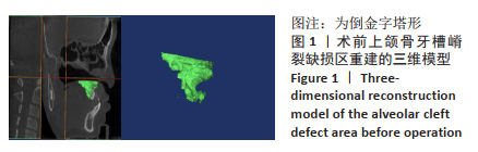
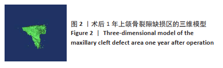
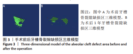
方法:纳入21例单侧完全性牙槽嵴裂髂骨移植1年后患者,采用锥形束CT结合Mimics软件分区域测量每例患者上颌骨牙槽嵴裂缺损区体积(整个牙槽嵴裂缺损区)及牙槽骨牙槽嵴裂缺损区体积(靠近口腔部缺损区),分析这两个区域的成骨效果,并分析尖牙萌出是否对骨存量产生影响。
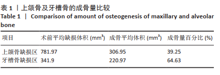
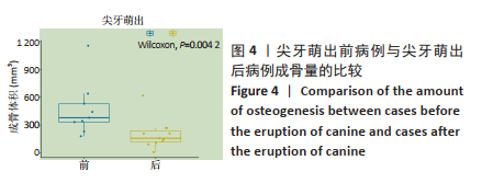
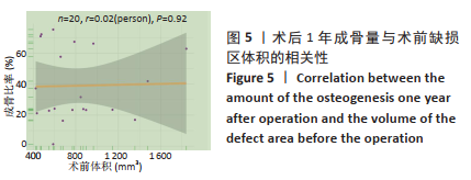
结果与结论:①牙槽嵴裂患者骨移植后,上颌骨牙槽嵴裂缺损区成骨量相对较低,牙槽骨牙槽嵴裂缺损区成骨量相对较高;尖牙萌出前缺损区的成骨量高于尖牙萌出后缺损区(P < 0.05);牙槽嵴裂缺损区术后成骨效果与术前缺损区体积大小无相关性。②结果表明,牙槽骨部分牙槽嵴缺损区成骨效果高于整个缺损区,临床应以牙槽骨缺损区成骨效果为基准指导后续的正畸及修复治疗;另外,植骨应在尖牙萌出前进行。

关键词: 牙槽嵴裂, 锥形束CT, 植骨术, 体积, 正畸, 口腔, 上颌骨, Mimics软件

Abstract: BACKGROUND: Preserving bone after bone grafting in patients with alveolar cleft is crucial. Currently, most studies on alveolar cleft used two-dimensional measurement; only a single measurement evaluation method was used even if some studies used three-dimensional measurement using cone beam CT. The amount of bone formation measured varies greatly.
OBJECTIVE: To investigate the osteogenic effect in patients with alveolar cleft 1 year after bone grafting and to analyze the influencing factors such as canine eruption and preoperative defect size using cone beam CT combined with Mimics software. 
METHODS: Twenty-one patients with unilateral complete alveolar cleft were included. One year after the iliac bone grafting, the volume of the alveolar cleft defect in maxillary bone (the entire alveolar cleft defect area) and alveolar bone (near the defect area of the oral cavity) were measured by cone beam CT combined with Mimics software. The osteogenic effect of these two areas and whether the eruption of the canine affected the bone stock were analyzed. 
RESULTS AND CONCLUSION: (1) After bone transplantation, the amount of osteogenesis in the maxillary cleft defect was ralatively low, while the amount of osteogenesis in the alveolar ridge cleft defect was relatively high. The amount of osteogenesis was higher before the eruption of canine than that after the eruption of canine at the defect area (P < 0.05). There was no correlation between the postoperative osteogenic effect of the alveolar cleft defect area and the preoperative volume of the defect area. (2) The results showed that the osteogenic effect of the alveolar ridge defect area was higher than that of the whole defect area, and the alveolar ridge defect area should be taken as the basis to guide the subsequent orthodontic and prosthodontics treatment. Moreover, bone grafting should be performed before the eruption of canine.  

Key words: alveolar cleft, cone beam CT, bone grating, volume, orthodontics, stomatology, maxillary bone, Mimics software

中图分类号: