[1] AHUJA CS, NORI S, TETREAULT L, et al. Traumatic Spinal Cord Injury-Repair and Regeneration. Neurosurgery. 2017; 80(3S):S9-S22.
[2] LU Y, SHANG Z, ZHANG W, et al. Global incidence and characteristics of spinal cord injury since 2000-2021: a systematic review and meta-analysis. BMC Med. 2024;22(1):285.
[3] HU S, WANG P, DONG Y, et al. Incidence, prevalence and disability of spinal cord injury in China from 1990 to 2019: a systematic analysis of the Global Burden of Disease Study 2019. Eur Spine J. 2023; 32(2):590-600.
[4] HE X, LI Y, DENG B, et al. The PI3K/AKT signalling pathway in inflammation, cell death and glial scar formation after traumatic spinal cord injury: Mechanisms and therapeutic opportunities. Cell Prolif. 2022;55(9):e13275.
[5] MA W, LI X. Spinal cord injury repair based on drug and cell delivery: From remodeling microenvironment to relay connection formation. Mater Today Bio. 2025;31:101556.
[6] LI D, LU X, XU G, et al. Dihydroorotate dehydrogenase regulates ferroptosis in neurons after spinal cord injury via the P53-ALOX15 signaling pathway. CNS Neurosci Ther. 2023;29(7):1923-1939.
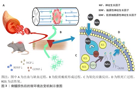
[7] FAN B, WEI Z, YAO X, et al. Microenvironment Imbalance of Spinal Cord Injury. Cell Transplant. 2018;27(6):853-866.
[8] FENG C, DENG L, YONG YY, et al. The Application of Biomaterials in Spinal Cord Injury. Int J Mol Sci. 2023;24(1):816.
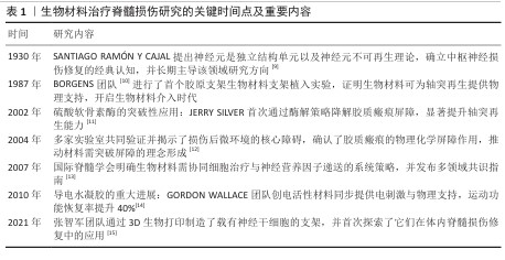
[9] D. B., D. Degeneration and Regeneration of the Nervous System. Nature. 1930;125: 230-231.
[10] BORGENS RB, BLIGHT AR, MCGINNIS ME. Behavioral recovery induced by applied electric fields after spinal cord hemisection in guinea pig. Science. 1987;238(4825): 366-369.
[11] BRADBURY EJ, MOON LD, POPAT RJ, et al. Chondroitinase ABC promotes functional recovery after spinal cord injury. Nature. 2002;416(6881):636-640.
[12] SILVER J, MILLER JH. Regeneration beyond the glial scar. Nat Rev Neurosci. 2004;5(2):146-156.
[13] FAWCETT JW, CURT A, STEEVES JD, et al. Guidelines for the conduct of clinical trials for spinal cord injury as developed by the ICCP panel: spontaneous recovery after spinal cord injury and statistical power needed for therapeutic clinical trials. Spinal Cord. 2007;45(3):190-205.
[14] THOMPSON BC, RICHARDSON RT, MOULTON SE, et al. Conducting polymers, dual neurotrophins and pulsed electrical stimulation--dramatic effects on neurite outgrowth. J Control Release. 2010;141(2):161-167.
[15] LIU X, HAO M, CHEN Z, et al. 3D bioprinted neural tissue constructs for spinal cord injury repair. Biomaterials. 2021;272:120771.
[16] LU Y, LIANG Z, WU Z, et al. Studying on the in vivo pathological evolution of spinal cord injury with the rat model by the method of integrated multispectral imaging and Raman spectroscopy. Talanta. 2024;279:126672.
[17] HUANG Y, WANG J, YUE C, et al. An In Situ Assembled Trapping Gel Repairs Spinal Cord Injury by Capturing Glutamate and Free Calcium Ions. Small. 2023;19(16):e2206229.
[18] LIU D, LU G, SHI B, et al. ROS-Scavenging Hydrogels Synergize with Neural Stem Cells to Enhance Spinal Cord Injury Repair via Regulating Microenvironment and Facilitating Nerve Regeneration. Adv Healthc Mater. 2023;12(18):e2300123.
[19] XIAO CL, LAI HT, ZHOU JJ, et al. Nrf2 Signaling Pathway: Focus on Oxidative Stress in Spinal Cord Injury. Mol Neurobiol. 2025;62(2):2230-2249.
[20] YAN L, HAN X, ZHANG M, et al. Melatonin exerts neuroprotective effects in mice with spinal cord injury by activating the Nrf2/Keap1 signaling pathway via the MT2 receptor. Exp Ther Med. 2023;27(1):37.
[21] HUA R, ZHAO C, XU Z, et al. ROS-responsive nanoparticle delivery of ferroptosis inhibitor prodrug to facilitate mesenchymal stem cell-mediated spinal cord injury repair. Bioact Mater. 2024;38:438-454.
[22] GONG L, GU Y, HAN X, et al. Spatiotemporal Dynamics of the Molecular Expression Pattern and Intercellular Interactions in the Glial Scar Response to Spinal Cord Injury. Neurosci Bull. 2023;39(2):213-244.
[23] HUSSEIN RK, MENCIO CP, KATAGIRI Y, et al.
Role of Chondroitin Sulfation Following Spinal Cord Injury. Front Cell Neurosci. 2020;14:208.
[24] AKRAM R, ANWAR H, JAVED MS, et al. Axonal Regeneration: Underlying Molecular Mechanisms and Potential Therapeutic Targets. Biomedicines. 2022;10(12):3186.
[25] ZHAO L, SHEN J, JIA K, et al. MicroRNA-24-3p Inhibits Microglia Inflammation by Regulating MK2 Following Spinal Cord Injury. Neurochem Res. 2021;46(4): 843-852.
[26] FANG B, WANG L, LIU S, et al. Sarsasapogenin regulates the immune microenvironment through MAPK/NF-kB signaling pathway and promotes functional recovery after spinal cord injury. Heliyon. 2024;10(3):e25145.
[27] MA J, LI J, WANG X, et al. GDNF-Loaded Polydopamine Nanoparticles-Based Anisotropic Scaffolds Promote Spinal Cord Repair by Modulating Inhibitory Microenvironment. Adv Healthc Mater. 2023;12(8):e2202377.
[28] SOSNOVTSEVA AO, STEPANOVA OV, STEPANENKO AA, et al. Recombinant Adenoviruses for Delivery of Therapeutics Following Spinal Cord Injury. Front Pharmacol. 2022;12:777628.
[29] SHA Q, WANG Y, ZHU Z, et al. A hyaluronic acid/silk fibroin/poly-dopamine-coated biomimetic hydrogel scaffold with incorporated neurotrophin-3 for spinal cord injury repair. Acta Biomater. 2023;167: 219-233.
[30] LI J, LUO W, XIAO C, et al. Recent advances in endogenous neural stem/progenitor cell manipulation for spinal cord injury repair. Theranostics. 2023;13(12):3966-3987.
[31] WANG YT, YUAN H. Research progress of endogenous neural stem cells in spinal cord injury. Ibrain. 2022;8(2):199-209.
[32] HELLENBRAND DJ, QUINN CM, PIPER ZJ, et al. Inflammation after spinal cord injury: a review of the critical timeline of signaling cues and cellular infiltration. J Neuroinflammation. 2021;18(1):284.
[33] 史旭,李瑞语,张兵,等.小胶质细胞极化介导炎症反应在脊髓损伤中的作用[J].中国组织工程研究,2023,27(1):121-129.
[34] SHEN J, GONG L, SUN Y, et al. Semaphorin3C identified as mediator of neuroinflammation and microglia polarization after spinal cord injury. iScience. 2024;27(5):109649.
[35] TANG H, GU Y, JIANG L, et al. The role of immune cells and associated immunological factors in the immune response to spinal cord injury. Front Immunol. 2023;13: 1070540.
[36] SHU H, ZHANG X, PU Y, et al. Fucoidan improving spinal cord injury recovery: Modulating microenvironment and promoting remyelination. CNS Neurosci Ther. 2024;30(8):e14903.
[37] GAO X, CHENG W, ZHANG X, et al. Nerve Growth Factor-Laden Anisotropic Silk Nanofiber Hydrogels to Regulate Neuronal/Astroglial Differentiation for Scarless Spinal Cord Repair. ACS Appl Mater Interfaces. 2022;14(3):3701-3715.
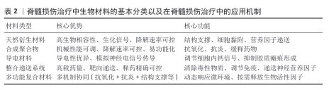
[38] SENSHARMA P, MADHUMATHI G, JAYANT RD, et al. Biomaterials and cells for neural tissue engineering: Current choices. Mater Sci Eng C Mater Biol Appl. 2017;77: 1302-1315.
[39] KAPLAN B, LEVENBERG S. The Role of Biomaterials in Peripheral Nerve and Spinal Cord Injury: A Review. Int J Mol Sci. 2022;23(3):1244.
[40] 薛学鑫,刘哲鹏.静电纺纤维神经组织工程支架:材料、功能及结构设计策略[J].中国组织工程研究,2022,26(28): 4575-4580.
[41] YU L, JIN H, XIA H, et al. Polylactic acid/chitosan-IKVAV Janus film serving as a dual functional platform for spinal cord injury repair. Nanoscale. 2024;16(47):21991-22000.
[42] ZENG X, WEI QS, YE JC, et al. A biocompatible gelatin sponge scaffold confers robust tissue remodeling after spinal cord injury in a non-human primate model. Biomaterials. 2023;299:122161.
[43] WALSH CM, WYCHOWANIEC JK, COSTELLO L, et al. An In Vitro and Ex Vivo Analysis of the Potential of GelMA Hydrogels as a Therapeutic Platform for Preclinical Spinal Cord Injury. Adv Healthc Mater. 2023; 12(26):e2300951.
[44] MUNGENAST L, NIEMINEN R, GAISER C,
et al. Electrospun decellularized extracellular matrix scaffolds promote the regeneration of injured neurons. Biomater Biosyst. 2023;11:100081.
[45] JIANG D, DING Y, HU S, et al. Broad-spectrum downregulation of inflammatory cytokines by polydopamine nanoparticles to protect the injured spinal cord. Acta Biomater. 2025;193:559-570.
[46] LIU Y, LIN F, WU C, et al. In Situ Reaction-Generated Aldehyde-Scavenging Polypeptides-Curcumin Conjugate Nanoassemblies for Combined Treatment of Spinal Cord Injury. ACS Nano. 2024; 18(10):7346-7362.
[47] MENG J, SUN J, KANG J, et al. Multifunctional hydrogels loaded with tellurium nanozyme for spinal cord injury repair. Mater Today Bio. 2024;29:101339.
[48] ZHANG Q, ZHENG J, LI L, et al. Bioinspired conductive oriented nanofiber felt with efficient ROS clearance and anti-inflammation for inducing M2 macrophage polarization and accelerating spinal cord injury repair. Bioact Mater. 2024;46:173-194.
[49] WANG G, LI Q, LIU S, et al. An injectable decellularized extracellular matrix hydrogel with cortical neuron-derived exosomes enhances tissue repair following traumatic spinal cord injury. Mater Today Bio. 2024; 28:101250.
[50] WU Z, ZHOU Y, HOU X, et al. Construction of functional neural network tissue combining CBD-NT3-modified linear-ordered collagen scaffold and TrkC-modified iPSC-derived neural stem cells for spinal cord injury repair. Bioact Mater. 2024;35:242-258.
[51] LIU Z, WAN X, WANG ZL, et al. Electroactive Biomaterials and Systems for Cell Fate Determination and Tissue Regeneration: Design and Applications. Adv Mater. 2021; 33(32):e2007429.
[52] GHANE N, BEIGI MH, LABBAF S, et al. Design of hydrogel-based scaffolds for the treatment of spinal cord injuries. J Mater Chem B. 2020, 21;8(47):10712-10738.
[53] SHAHEMI NH, MAHAT MM, ASRI NAN, et al.
Application of Conductive Hydrogels on Spinal Cord Injury Repair: A Review. ACS Biomater Sci Eng. 2023;9(7):4045-4085.
[54] YANG B, LIANG C, CHEN D, et al. A conductive supramolecular hydrogel creates ideal endogenous niches to promote spinal cord injury repair. Bioact Mater. 2021;15: 103-119.
[55] PENTLAVALLI S, COULTER S, LAVERTY G. Peptide Nanomaterials for Drug Delivery Applications. Curr Protein Pept Sci. 2020; 21(4):401-412.
[56] ZHOU L, QIU T, LV F, et al. Self-Assembled Nanomedicines for Anticancer and Antibacterial Applications. Adv Healthc Mater. 2018;7(20):e1800670.
[57] WU J, TANG J, ZHANG L, et al. Biomimetic “Trojan Horse” Fibers Modulate Innate Immunity Cascades for Nerve Regeneration. ACS Nano. 2025;19(1):781-802.
[58] SHA Q, WANG Y, ZHU Z, et al. A hyaluronic acid/silk fibroin/poly-dopamine-coated biomimetic hydrogel scaffold with incorporated neurotrophin-3 for spinal cord injury repair. Acta Biomater. 2023;167: 219-233.
[59] VERSTAPPEN K, KLYMOV A, CICUÉNDEZ M, et al. Biocompatible adipose extracellular matrix and reduced graphene oxide nanocomposite for tissue engineering applications. Mater Today Bio. 2024;26: 101059.
[60] TANG H, LI J, WANG H, et al. Human umbilical cord mesenchymal stem cell-derived exosomes loaded into a composite conduit promote functional recovery after peripheral nerve injury in rats. Neural Regen Res. 2024;19(4):900-907.
[61] LI Y, CHENG S, WEN H, et al. Coaxial 3D printing of hierarchical structured hydrogel scaffolds for on-demand repair of spinal cord injury. Acta Biomater. 2023;168: 400-415.
[62] 文峰,周磊,李扬,等.通腑逐瘀法指导下抵当汤加减可抑制大鼠急性脊髓损伤后胶质瘢痕的形成[J].中国组织工程研究,2023,27(20):3180-3187.
[63] RAO S, LIN Y, LIN R, et al. Traditional Chinese medicine active ingredients-based selenium nanoparticles regulate antioxidant selenoproteins for spinal cord injury treatment. J Nanobiotechnology. 2022;20(1):278.
[64] MA W, LI X. Spinal cord injury repair based on drug and cell delivery: From remodeling microenvironment to relay connection formation. Mater Today Bio. 2025;31:101556.
[65] SHEN H, FAN C, YOU Z, et al. Advances in Biomaterial - Based Spinal Cord Injury Repair. Advanced Functional Materials. 2022;32:2110628.
|