[1] LV T, LI Z, WANG D, et al. Role of exosomes in prostate cancer bone metastasis. Arch Biochem Biophys. 2023;748:109784.
[2] XU S, LI X, DU Y, et al. Decoding the past and future of breast cancer bone metastasis: A bibliometric analysis from 2003 to 2022. Asian J Surg. 2023:S1015-9584(23)01400-8. doi: 10.1016/j.asjsur.2023.08.231.
[3] TSENG YD. Radiation Therapy for Painful Bone Metastases: Fractionation, Recalcification, and Symptom Control. Semin Radiat Oncol. 2023;33(2):139-147.
[4] CHOY E, COTE GM, MICHAELSON MD, et al. Phase II Study of Cabozantinib in Patients With Bone Metastasis. Oncologist. 2022;27(7):600-606.
[5] NISHIMURA K. Management of bone metastasis in prostate cancer. J Bone Miner Metab. 2023;41(3):317-326.
[6] DAI X, LIU B, HOU Q, et al. Global and local fat effects on bone mass and quality in obesity. Bone Joint Res. 2023;12(9):580-589.
[7] LI J, LU L, LIU L, et al. The unique role of bone marrow adipose tissue in ovariectomy-induced bone loss in mice. Endocrine. 2023 Sep 8. doi: 10.1007/s12020-023-03504-6.
[8] ROSEN CJ, HOROWITZ MC. Nutrient regulation of bone marrow adipose tissue: skeletal implications of weight loss. Nat Rev Endocrinol. 2023; 19(11):626-638.
[9] TODOSENKO N, KHAZIAKHMATOVA O, MALASHCHENKO V, et al. Adipocyte- and Monocyte-Mediated Vicious Circle of Inflammation and Obesity (Review of Cellular and Molecular Mechanisms). Int J Mol Sci. 2023;24(15):12259.
[10] CHENG F, HE J, YANG J. Bone marrow microenvironment: roles and therapeutic implications in obesity-associated cancer. Trends Cancer. 2023; 9(7):566-577.
[11] LUO G, HE Y, YU X. Bone Marrow Adipocyte: An Intimate Partner With Tumor Cells in Bone Metastasis. Front Endocrinol (Lausanne). 2018;9:339.
[12] SALAMANNA F, CONTARTESE D, ERRANI C, et al. Role of bone marrow adipocytes in bone metastasis development and progression: a systematic review. Front Endocrinol (Lausanne). 2023;14:1207416.
[13] WITKOWSKI MT, KOUSTENI S, AIFANTIS I. Mapping and targeting of the leukemic microenvironment. J Exp Med. 2020;217(2):e20190589.
[14] GALÁN-DÍEZ M, CUESTA-DOMÍNGUEZ Á, KOUSTENI S. The Bone Marrow Microenvironment in Health and Myeloid Malignancy. Cold Spring Harb Perspect Med. 2018;8(7):a031328.
[15] SABBAH R, SAADI S, SHAHAR-GABAY T, et al. Abnormal adipogenic signaling in the bone marrow mesenchymal stem cells contributes to supportive microenvironment for leukemia development. Cell Commun Signal. 2023; 21(1):277.
[16] LWIN ST, OLECHNOWICZ SW, FOWLER JA, et al. Diet-induced obesity promotes a myeloma-like condition in vivo. Leukemia. 2015;29(2):507-510.
[17] TABE Y, YAMAMOTO S, SAITOH K, et al. Bone Marrow Adipocytes Facilitate Fatty Acid Oxidation Activating AMPK and a Transcriptional Network Supporting Survival of Acute Monocytic Leukemia Cells. Cancer Res. 2017; 77(6):1453-1464.
[18] DIRAT B, BOCHET L, DABEK M, et al. Cancer-associated adipocytes exhibit an activated phenotype and contribute to breast cancer invasion. Cancer Res. 2011;71(7):2455-2465.
[19] FAIRFIELD H, FALANK C, FARRELL M, et al. Development of a 3D bone marrow adipose tissue model. Bone. 2019;118:77-88.
[20] SCHELLER EL, DOUCETTE CR, LEARMAN BS, et al. Region-specific variation in the properties of skeletal adipocytes reveals regulated and constitutive marrow adipose tissues. Nat Commun. 2015;6:7808.
[21] 张纯希,李想,周钰翔,等.骨骼微环境中谁决定间充质干细胞的分化命运[J].中国组织工程研究,2021,25(25):4045-4052.
[22] YUE R, ZHOU BO, SHIMADA IS, et al. Leptin Receptor Promotes Adipogenesis and Reduces Osteogenesis by Regulating Mesenchymal Stromal Cells in Adult Bone Marrow. Cell Stem Cell. 2016;18(6):782-796.
[23] CHI M, CHEN J, YE Y, et al. Adipocytes contribute to resistance of human melanoma cells to chemotherapy and targeted therapy. Curr Med Chem. 2014; 21(10):1255-1267.
[24] WALTER M, LIANG S, GHOSH S, et al. Interleukin 6 secreted from adipose stromal cells promotes migration and invasion of breast cancer cells. Oncogene. 2009;28(30):2745-2755.
[25] HOLT V, CAPLAN AI, HAYNESWORTH SE. Identification of a subpopulation of marrow MSC-derived medullary adipocytes that express osteoclast-regulating molecules: marrow adipocytes express osteoclast mediators. PLoS One. 2014; 9(10):e108920.
[26] YANG HL, LIU T, WANG XM, et al. Diagnosis of bone metastases: a meta-analysis comparing ¹⁸FDG PET, CT, MRI and bone scintigraphy. Eur Radiol. 2011;21(12): 2604-2617.
[27] HOTTAT NA, BADR DA, BEN GHANEM M, et al. Assessment of whole-body MRI including diffusion-weighted sequences in the initial staging of breast cancer patients at high risk of metastases in comparison with PET-CT: a prospective cohort study. Eur Radiol. 2023 Aug 9. doi: 10.1007/s00330-023-10060-0.
[28] MONTOYA-BORDÓN J, ELVIRA-RUIZ P, CARRIAZO-JIMÉNEZ B, et al. Imaging diagnosis of vertebral metastasis. Rev Esp Cir Ortop Traumatol. 2023;67(6): 511-522.
[29] MORAWITZ J, BRUCKMANN NM, JANNUSCH K, et al. Conventional Imaging, MRI and 18F-FDG PET/MRI for N and M Staging in Patients with Newly Diagnosed Breast Cancer. Cancers (Basel). 2023;15(14):3646.
[30] RUAN D, SUN L. Diagnostic Performance of PET/MRI in Breast Cancer: A Systematic Review and Bayesian Bivariate Meta-analysis. Clin Breast Cancer. 2023;23(2):108-124.
[31] ZHAN Y, ZHANG G, LI M, et al. Whole-Body MRI vs. PET/CT for the Detection of Bone Metastases in Patients With Prostate Cancer: A Systematic Review and Meta-Analysis. Front Oncol. 2021;11:633833.
[32] BRUCKMANN NM, KIRCHNER J, UMUTLU L, et al. Prospective comparison of the diagnostic accuracy of 18F-FDG PET/MRI, MRI, CT, and bone scintigraphy for the detection of bone metastases in the initial staging of primary breast cancer patients. Eur Radiol. 2021;31(11):8714-8724.
[33] BHALUDIN BN, TUNARIU N, KOH DM, et al. A review on the added value of whole-body MRI in metastatic lobular breast cancer. Eur Radiol. 2022;32(9):6514-6525.
[34] NAKANISHI K, TANAKA J, NAKAYA Y, et al. Whole-body MRI: detecting bone metastases from prostate cancer. Jpn J Radiol. 2022;40(3):229-244.
[35] CHEN R, YANG Q, CHEN W, et al. Whole-body MRI-based multivariate prediction model in the assessment of bone metastasis in prostate cancer. World J Urol. 2021;39(8):2937-2943.
[36] GONG XQ, TAO YY, WANG R, et al. Application of Diffusion Weighted Imaging in Prostate Cancer Bone Metastasis: Detection and Therapy Evaluation. Anticancer Agents Med Chem. 2021;21(15):1950-1956.
[37] VAN NIEUWENHOVE S, VAN DAMME J, PADHANI AR, et al. Whole-body magnetic resonance imaging for prostate cancer assessment: Current status and future directions. J Magn Reson Imaging. 2022;55(3):653-680.
[38] CERANKA J, WUTS J, CHIABAI O, et al. Computer-aided diagnosis of skeletal metastases in multi-parametric whole-body MRI. Comput Methods Programs Biomed. 2023;242:107811.
[39] RAYA JG, DUARTE A, WANG N, et al. Applications of Diffusion-Weighted MRI to the Musculoskeletal System. J Magn Reson Imaging. 2023 Jul 21. doi: 10.1002/jmri.28870.
[40] 朱柳红,刘豪,周建军.磁共振T2 mapping技术在体部恶性肿瘤中的研究进展[J].磁共振成像,2020,11(5):398-400.
[41] HAMILTON G, MIDDLETON MS, BYDDER M, et al. Effect of PRESS and STEAM sequences on magnetic resonance spectroscopic liver fat quantification. J Magn Reson Imaging. 2009;30(1):145-152.
[42] BYDDER M, GIRARD O, HAMILTON G. Mapping the double bonds in triglycerides. Magn Reson Imaging. 2011;29(8):1041-1046.
[43] REEDER SB, HU HH, SIRLIN CB. Proton density fat-fraction: a standardized MR-based biomarker of tissue fat concentration. J Magn Reson Imaging. 2012;36(5): 1011-1014.
[44] 赵秀芹,狄玉进,徐金法,等.单体素氢质子磁共振波谱在脊柱转移瘤中的初步应用研究[J].医学影像学杂志,2012,22(4):624-626.
[45] 樊秋菊,谭辉,于楠,等.IVIM-DWI联合MRS鉴别诊断骨质疏松与转移性椎体压缩性骨折[J].中国医学影像技术,2018,34(2):297-301.
[46] TAN H, XU H, LUO F, et al. Combined intravoxel incoherent motion diffusion-weighted MR imaging and magnetic resonance spectroscopy in differentiation between osteoporotic and metastatic vertebral compression fractures. J Orthop Surg Res. 2019;14(1):299.
[47] 刘斌,董国礼,郭静,等.椎体~1H-MRS的临床应用研究进展[J].国际医学放射学杂志,2012,35(6):553-556.
[48] MA J. Dixon techniques for water and fat imaging. J Magn Reson Imaging. 2008; 28(3):543-558.
[49] VAN VUCHT N, SANTIAGO R, LOTTMANN B, et al. The Dixon technique for MRI of the bone marrow. Skeletal Radiol. 2019;48(12):1861-1874.
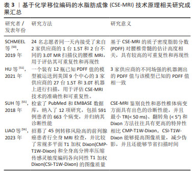
[50] SCHMEEL FC, VOMWEG T, TRÄBER F, et al. Proton density fat fraction MRI of vertebral bone marrow: Accuracy, repeatability, and reproducibility among readers, field strengths, and imaging platforms. J Magn Reson Imaging. 2019; 50(6):1762-1772.
[51] HU HH, YOKOO T, BASHIR MR, et al. Linearity and Bias of Proton Density Fat Fraction as a Quantitative Imaging Biomarker: A Multicenter, Multiplatform, Multivendor Phantom Study. Radiology. 2021;298(3):640-651.
[52] BAUM T, YAP SP, DIECKMEYER M, et al. Assessment of whole spine vertebral bone marrow fat using chemical shift-encoding based water-fat MRI. J Magn Reson Imaging. 2015;42(4):1018-1023.
[53] KARAMPINOS DC, RUSCHKE S, DIECKMEYER M, et al. Modeling of T2* decay in vertebral bone marrow fat quantification. NMR Biomed. 2015; 28(11): 1535-1542.
[54] REEDER SB, ROBSON PM, YU H, et al. Quantification of hepatic steatosis with MRI: the effects of accurate fat spectral modeling. J Magn Reson Imaging. 2009; 29(6):1332-1339.
[55] SUH CH, YUN SJ, JIN W, et al. Diagnostic Performance of In-Phase and Opposed-Phase Chemical-Shift Imaging for Differentiating Benign and Malignant Vertebral Marrow Lesions: A Meta-Analysis. AJR Am J Roentgenol. 2018;211(4): W188-W197.
[56] LIAO Z, LIU G, MING B, et al. Evaluating prostate cancer bone metastasis using accelerated whole-body isotropic 3D T1-weighted Dixon MRI with compressed SENSE: a feasibility study. Eur Radiol. 2023;33(3):1719-1728.
[57] SCHMEEL FC, LUETKENS JA, ENKIRCH SJ, et al. Proton density fat fraction (PDFF) MR imaging for differentiation of acute benign and neoplastic compression fractures of the spine. Eur Radiol. 2018;28(12):5001-5009.
[58] JUNG Y, JEON SW, KWACK KS, et al. Differentiation of Vertebral Metastases From Focal Hematopoietic Marrow Depositions on MRI: Added Value of Proton Density Fat Fraction. AJR Am J Roentgenol. 2021;216(3):734-741.
[59] PARK S, DO HUH J. Differentiation of bone metastases from benign red marrow depositions of the spine: the role of fat-suppressed T2-weighted imaging compared to fat fraction map. Eur Radiol. 2022;32(10):6730-6738.
[60] SCHMEEL FC, ENKIRCH SJ, LUETKENS JA, et al. Diagnostic Accuracy of Quantitative Imaging Biomarkers in the Differentiation of Benign and Malignant Vertebral Lesions : Combination of Diffusion-Weighted and Proton Density Fat Fraction Spine MRI. Clin Neuroradiol. 2021;31(4):1059-1070.
[61] DONNERS R, FIGUEIREDO I, TUNARIU N, et al. Multiparametric bone MRI can improve CT-guided bone biopsy target selection in cancer patients and increase diagnostic yield and feasibility of next-generation tumour sequencing. Eur Radiol. 2022;32(7):4647-4656.
[62] LECOUVET FE, PASOGLOU V, VAN NIEUWENHOVE S, et al. Shortening the acquisition time of whole-body MRI: 3D T1 gradient echo Dixon vs fast spin echo for metastatic screening in prostate cancer. Eur Radiol. 2020; 30(6):3083-3093.
[63] 罗云,高新.2021版欧洲泌尿外科学会前列腺癌诊疗指南更新要点解读[J].中华腔镜泌尿外科杂志(电子版),2022,16(2):97-100.
[64] COOK GJR. Imaging of Bone Metastases in Breast Cancer. Semin Nucl Med. 2022;52(5):531-541.
[65] COOK GJR, GOH V. Molecular Imaging of Bone Metastases and Their Response to Therapy. J Nucl Med. 2020;61(6):799-806.
[66] OPREA-LAGER DE, CYSOUW MCF, BOELLAARD R, et al. Bone Metastases Are Measurable: The Role of Whole-Body MRI and Positron Emission Tomography. Front Oncol. 2021;11:772530.
[67] 李利,谢莎,敬宗林,等.影像学检查技术在前列腺癌骨转移诊断中的应用进展[J].山东医药,2021,61(36):105-109. |
