中国组织工程研究 ›› 2022, Vol. 26 ›› Issue (23): 3733-3737.doi: 10.12307/2022.675

• 组织构建实验造模 experimental modeling in tissue construction • 上一篇    下一篇

心肌梗死大鼠模型的建立及疾病进程评价

杨  钤,张轶欧,贾力莉,谢  君,冯玛莉,李庭凯   

  1. 山西省中医药研究院,山西省太原市   030012
  • 收稿日期:2021-05-06 接受日期:2021-06-24 出版日期:2022-08-18 发布日期:2022-02-15
  • 通讯作者: 李庭凯,主任医师,山西省中医药研究院,山西省太原市 030012
  • 作者简介:杨钤,女,1990年生,山西省河津市人,汉族,2013年山西大学毕业,硕士,主管技师,主要从事中药药性理论分子机制研究。
  • 基金资助:
    山西省自然科学基金面上项目(201801D121299),项目负责人:李庭凯;山西省自然科学基金青年项目(201901D211525),项目负责人:杨钤;山西中医药大学科技创新能力培育计划项目(2019PY-021),项目负责人:杨钤

Establishment and disease progression in a rat myocardial infarction model

Yang Qian, Zhang Yiou, Jia Lili, Xie Jun, Feng Mali, Li Tingkai   

  1. Shanxi Province Academy of Traditional Chinese Medicine, Taiyuan 030012, Shanxi Province, China
  • Received:2021-05-06 Accepted:2021-06-24 Online:2022-08-18 Published:2022-02-15
  • Contact: Li Tingkai, Chief physician, Shanxi Province Academy of Traditional Chinese Medicine, Taiyuan 030012, Shanxi Province, China
  • About author:Yang Qian, Master, Technologist-in-charge, Shanxi Province Academy of Traditional Chinese Medicine, Taiyuan 030012, Shanxi Province, China
  • Supported by:
    the Natural Science Foundation of Shanxi Province (General Project), No. 201801D121299 (to LTK); the Natural Science Foundation of Shanxi Province (Youth Project), No. 201901D211525 (to YQ); Science and Technology Innovation Capability Cultivation Project of Shanxi University of Chinese Medicine, No. 2019PY-021 (to YQ)

摘要:

文题释义:
心肌梗死:是由于血栓或栓子堵塞冠状动脉,心肌血液灌注减少、供氧降低,心肌氧耗增加,导致的一种贫血性坏死的病理状态。心肌梗死后,缺血短期内心肌细胞坏死,激发促炎因子释放、中性粒细胞浸润和单核细胞动员,继而胶原纤维沉积和瘢痕形成。数周至数月后,左心室腔扩张、瘢痕变薄、细胞外间质纤维化。
大鼠模型:大鼠易饲养、繁殖快,个体差异小,与人类有相似的血管解剖特点,有极强的抗感染能力等,是基础研究中的重要疾病模型动物。借助大鼠模型研究人类疾病,有助于快速、有效地探索疾病病因和发病机制、研究防治措施。

背景:心肌梗死是冠心病最严重的一种临床类型,抗心肌缺血类药物的研发已成为研究热点。建立合适的动物模型并探讨其病理生理机制,可为新药研发过程中药物的药效学评价提供有效的工具,促进心血管疾病有效治疗药物的开发。
目的:评价急性心肌梗死损伤后大鼠的心脏功能、组织形态和心肌细胞超微结构改变。
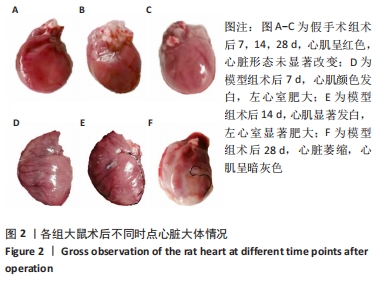
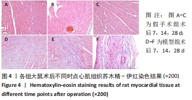
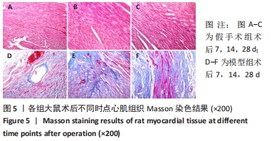
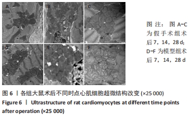
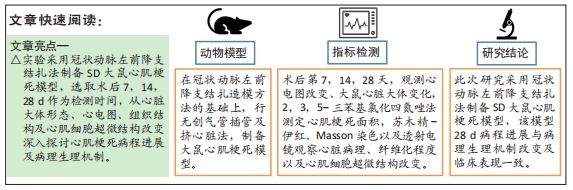
方法:30只雄性SD大鼠随机分为假手术组及模型组,每组15只。模型组采用冠状动脉左前降支结扎法制备心肌梗死模型,假手术组只穿线不结扎。分别在造模后第7,14,28天,心电图观测大鼠Ⅱ导联ST段改变情况;开胸观察心脏大体改变情况;2,3,5-三苯基氯化四氮唑染色法测定心肌梗死面积;苏木精-伊红染色法观察心脏组织病理改变;Masson染色法观察心肌组织纤维化程度;透射电镜观察心肌细胞超微结构改变。
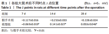
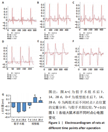
结果与结论:①与假手术组比较,模型组大鼠术后7 d心电图可见J点明显抬高,14 d可见Q波的波形增宽、振幅增大;②心脏大体观察可见,模型组大鼠术后7,14 d时左心室肥大,28 d时心脏萎缩;③2,3,5-三苯基氯化四氮唑染色显示,模型组大鼠心梗面积进展性增加;④苏木精-伊红和Masson染色显示,心肌细胞排列紊乱、心肌细胞坏死、炎细胞浸润及纤维增生逐渐加重;⑤透射电镜观察可见,心肌细胞内线粒体肿胀加剧,线粒体嵴数量显著减少;⑥提示SD大鼠心肌梗死模型心脏功能、组织形态以及心肌细胞超微结构改变符合心肌梗死发生、发展规律,可为心肌梗死治疗药物研发提供实验证据。
缩略语:2,3,5-三苯基氯化四氮唑:2,3,5-Triphenyltetrazolium chloride,TTC

https://orcid.org/0000-0003-4351-8267 (杨钤) 

中国组织工程研究杂志出版内容重点:组织构建;骨细胞;软骨细胞;细胞培养;成纤维细胞;血管内皮细胞;骨质疏松;组织工程

关键词: 心肌梗死, 冠状动脉, 左前降支, 心电图, 动物模型, 大鼠

Abstract: BACKGROUND: Myocardial infarction is the most serious clinical type of coronary heart disease. The development of anti-myocardial ischemia drugs has become a research hotspot. Establishing an appropriate animal model and investigating its pathophysiological mechanism can provide an effective tool for pharmacodynamics evaluation in new drug development and promote the development of anti-myocardial infarction drugs for myocardial infarction. 
OBJECTIVE: To evaluate the changes of cardiac function, tissue morphology and cardiomyocyte ultrastructure in rats after acute myocardial infarction. 
METHODS: Thirty male Sprague-Dawley rats were randomly divided into two groups (n=15 per group): a sham surgery group and a model group. Rats in the model group were subjected to ligation of the left anterior descending coronary artery, while in the sham surgery group, the left anterior descending coronary artery was only threaded without ligation. On the 7th, 14th, and 28th days after modeling, changes of ST segment in lead II were observed by electrocardiogram, and the general changes of the heart were observed by thoracotomy. Myocardial infarction area was determined by 2,3,5-triphenyltetrazolium chloride staining. Hematoxylin-eosin staining was used for pathological observation of the heart, and Masson staining was adopted for the determination of myocardial fibrosis degree. Moreover, the cardiomyocyte ultrastructure was observed by transmission electron microscope. 
RESULTS AND CONCLUSION: Compared with the sham surgery group, the results of electrocardiogram showed that the J point was obviously elevated on the 7th day, and the Q wave waveform was broadened and the amplitude was increased on the 14th day. The gross observation of the heart showed that rats in the model group had left ventricular hypertrophy on the 7th and 14th days and cardiac atrophy on the 28th day after operation. The results of 2,3,5-triphenyltetrazolium chloride staining showed myocardial infarction area in the model group was increased progressively. Hematoxylin-eosin and Masson staining results indicated that myocardial cells arranged disorderly, accompanied by myocyte necrosis, inflammatory cell infiltration, and fibrous proliferation that were gradually aggravated. Under the transmission electron microscope, the mitochondrial swelling was worsened in the cardiomyocytes, and the number of mitochondrial cristae was significantly reduced. All these findings reveal that the changes of cardiac function, tissue morphology, and ultrastructure of cardiomyocytes in the Sprague-Dawley rat model of myocardial infarction accord with the occurrence and development of myocardial infarction, which can provide experimental evidence for the development of therapeutic drugs for myocardial infarction.

Key words: myocardial infarction, coronary artery, left anterior descending artery, electrocardiogram, animal model, rat

中图分类号: