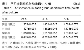
| [1] World Health Organization.Global Tuberculosis Report, 2014. [2] 庄伟,石仕元,庄汝杰.骨与关节结核患者病灶感染病原菌检测结果分析[J].中华医院感染学杂志, 2015,25(2): 401-403.[3] Ramírez-Lapausa M,Menéndez-Saldaña A, Noguerado-Asensio A.Extrapulmonary tuberculosis. Rev Esp Sanid Penit.2015;17(1):3-11.[4] Golden MP,Vikram HP.Extrapulmonary tuberculosis:an overview.Am Fam Physician.2005;72(9):1761-1768. [5] Grosskopf I,Ben David A,Charach G,et al.Bone and joint tuberculosis—a 10-year review.Isr J Med Sci. 1994;30:278-283.[6] Lifeso RM,Weaver P,Harder EH.Tuberculous spondylitis in adults.J Bone Joint Surg Am. 1985;67: 1405-1413.[7] Hoshino A,Hanada S,Yamada H,et al.Mycobacterium tuberculosis escapes from the phagosomes of infected human osteoclasts reprograms osteoclast development via dysregulation of cytokines and chemokines.Pathog Dis.2014;70(1):28-39. [8] Wu J, Zuo Y, Hu Y, et al. Development and in vitro characterization of drug delivery system of rifapentine for osteoarticular tuberculosis.Drug Des Devel Ther. 2015;9:1359-1366.[9] 2006-09-30.中华人民共和国科学技术部.关于善待实验动物的指导性意见.2006-09-30.[10] USP 32-NF27 biological reactivity tests,in vivo.[11] Hu J,Hou Y,Park H,et al.Beta-tricalcium phosphate particles as a controlled release carrier of osteogenic proteins for bone tissue engineering.J Biomed Mater Res A. 2012;100(7):1680-1686. [12] Chou J, Valenzuela SM, Santos J,et al.Strontium- and magnesium-enriched biomimetic β-TCP macrospheres with potential for bone tissue morphogenesis.J Tissue Eng Regen Med. 2014 ;8(10):771-778.[13] Lee M,Wu BM.Recent advances in 3D printing of tissue engineering scaffolds. Methods Mol Biol. 2012; 868:257-267. [14] Gross BC,Erkal JL,Lockwood SY,et al.Evaluation of 3D printing and its potential impact on biotechnology and the chemical sciences.Anal Chem.2014;86:3240-3253. [15] Fullerton JN,Frodsham GC,Day RM.3D printing for the many, not the few.Nat Biotechnol. 2014;32:1086-1087. [16] Hoy MB.3D printing: making things at the library.Med Ref Serv Q.2013;32:94-99. [17] Cox SC,Thornby JA,Gibbons GJ,et al.3D printing of porous hydroxyapatite scaffolds intended for use in bone tissue engineering applications.Mater Sci Eng C Mater Biol Appl.2015;47:237-247.[18] Kao CT,Lin CC,Chen YW,et al.Poly(dopamine) coating of 3D printed poly(lactic acid) scaffolds for bone tissue engineering.Mater Sci Eng C Mater Biol Appl. 2015;56: 165-173.[19] Murphy SV,Atala A.3D bioprinting of tissues and organs.Nat Biotechnol.2014;32(8):773-785.[20] Butscher A,Bohner M,Doebelin N,et al.New depowdering-friendly designs for three-dimensional printing of calcium phosphate bone substitutes.Acta Biomater.2013;9(11):9149-9158.[21] Gatto M,Memoli G,Shaw A,et al.Three-dimensional printing (3DP) of neonatal head phantom for ultrasound: thermocouple embedding and simulation of bone.Med Eng Phys. 2012;34(7):929-937. [22] Sherwood JK,Riley SL,Palazzolo R,et al.A three-dimensional osteochondral composite scaffold for articular cartilage repair.Biomaterials. 2002;23(24): 4739-4751.[23] Seitz H,Rieder W,Irsen S,et al.Three-dimensional printing of porous ceramic scaffolds for bone tissue engineering.J Biomed Mater Res B Appl Biomater. 2005;74(2):782-788.[24] Warnke PH,Seitz H,Warnke F,et al.Ceramic scaffolds produced by computer-assisted 3D printing and sintering:characterization and biocompatibility investigations.J Biomed Mater Res B Appl Biomater. 2010;93(1):212-217.[25] Carletti E,Motta A,Migliaresi C.Scaffolds for tissue engineering and 3D cell culture.Methods Mol Biol. 2011;695:17-39.[26] 袁景,甄平,赵红斌.高性能多孔β-磷酸三钙骨组织工程支架的3D打印[J].中国组织工程研究, 2014,18(43):6914-6921. [27] 付维力,李箭,王践云,等.壳聚糖聚磷酸钙复合支架的生物相容性研究[J].中国运动医学杂志,2015,34(2):117-122.[28] 杨晓芳.生物材料生物相容性评价研究进展[J].生物医学工程学杂志,2001,18(1):123-128.[29] 张丹,南黎,任玲,等.CCK-8法和MTT法评价医用抗菌不锈钢细胞毒性的比较研究[J].口腔医学,2012,31(10):5-8.[30] Su H,Chen YL,Chen SY,et al.Preparation and bioactivity evalution of streptavidin-tagged human interleukin-15 fusion protein. Nan fang Yi Ke Da Xue Xue Bao. 2009;29(3):397-401. |
.jpg)


.jpg)
.jpg)