[1] HORVATH A, SZUCS A, CSUKLY G, et al. EEG and ERP biomarkers of Alzheimer’s disease: a critical review. Front Biosci (Landmark Ed). 2018;23:183-220.
[2] CHEN YG. Research Progress in the Pathogenesis of Alzheimer’s Disease. Chin Med J (Engl). 2018;131(13):1618-1624.
[3] MARESOVA P, TOMSONE S, LAMESKI P, et al. Technological Solutions for Older People with Alzheimer’s Disease: Review. Curr Alzheimer Res. 2018;15(10):975-983.
[4] ROSS CA, AKIMOV SS. Human-induced pluripotent stem cells: potential for neurodegenerative diseases. Hum Mol Genet. 2014;23(R1):R17-26.
[5] JIANG Y, FAY JM, POON CD, et al. Nanoformulation of Brain-Derived Neurotrophic Factor with Target Receptor-Triggered-Release in the Central Nervous System. Adv Funct Mater. 2018;28(6):1703982.
[6] YÜCEL F, YAMAN SO, OZBABALIK D, et al. Morphometric measurements of MRI findings in patients with Alzheimer’s disease. Adv Clin Exp Med. 2014;23(1):91-96.
[7] IULITA MF, BISTUÉ MILLÓN MB, PENTZ R, et al. Differential deregulation of NGF and BDNF neurotrophins in a transgenic rat model of Alzheimer’s disease. Neurobiol Dis. 2017;108:307-323.
[8] LI LM, HUANG LL, JIANG XC, et al. Transplantation of BDNF Gene Recombinant Mesenchymal Stem Cells and Adhesive Peptide-modified Hydrogel Scaffold for Spinal Cord Repair. Curr Gene Ther. 2018;18(1): 29-39.
[9] HEI WH, ALMANSOORI AA, SUNG MA, et al. Adenovirus vector-mediated ex vivo gene transfer of brain-derived neurotrophic factor (BDNF) tohuman umbilical cord blood-derived mesenchymal stem cells (UCB-MSCs) promotescrush-injured rat sciatic nerve regeneration. Neurosci Lett. 2017;643:111-120.
[10] WANG Z, XU P, CHEN B, et al. Identifying circRNA-associated-ceRNA networks in the hippocampus of Aβ1-42-induced Alzheimer’s disease-like rats using microarray analysis. Aging (Albany NY). 2018;10(4): 775-788.
[11] WANG Y, HAN S, HAN R, et al. Propofol-induced downregulation of NR2B membrane translocation in hippocampus and spatial memory deficits of neonatal mice. Brain Behav. 2017;7(7):e00734.
[12] CHEN X, XIAO JW, CAO P, et al. Brain-derived neurotrophic factor protects against acrylamide-induced neuronal and synaptic injury via the TrkB-MAPK-Erk1/2 pathway. Neural Regen Res. 2021;16(1):150-157.
[13] AHMED S, REYNOLDS BA, WEISS S. BDNF enhances the differentiation but not the survival of CNS stem cell-derived neuronal precursors. J Neurosci. 1995;15(8):5765-5778.
[14] MA H, YU B, KONG L, et al. Neural stem cells over-expressing brain-derived neurotrophic factor (BDNF) stimulate synaptic protein expression and promote functional recovery following transplantation in rat model of traumatic brain injury. Neurochem Res. 2012;37(1):69-83.
[15] YANG JW, MA W, LUO T, et al. BDNF promotes human neural stem cell growth via GSK-3β-mediated crosstalk with the wnt/β-catenin signaling pathway. Growth Factors. 2016;34(1-2):19-32.
[15] ROSENBLUM S, SMITH TN, WANG N, et al. BDNF Pretreatment of Human Embryonic-Derived Neural Stem Cells Improves Cell Survival and Functional Recovery After Transplantation in Hypoxic-Ischemic Stroke. Cell Transplant. 2015;24(12):2449-2461.
[17] MARSH SE, BLURTON-JONES M. Neural stem cell therapy for neurodegenerative disorders: The role of neurotrophic support. Neurochem Int. 2017;106:94-100.
[18] WU CC, LIEN CC, HOU WH, et al. Gain of BDNF Function in Engrafted Neural Stem Cells Promotes the Therapeutic Potential for Alzheimer’s Disease. Sci Rep. 2016;6:27358.
[19] RUAN Q, YU Z, ZHANG W, et al. Cholinergic Hypofunction in Presbycusis-Related Tinnitus With Cognitive Function Impairment: Emerging Hypotheses. Front Aging Neurosci. 2018;10:98.
[20] JAMAL M, ITO A, TANAKA N, et al. The Role of Apolipoprotein E and Ethanol Exposure in Age-Related Changes in Choline Acetyltransferase and Brain-Derived Neurotrophic Factor Expression in the Mouse Hippocampus. J Mol Neurosci. 2018;65(1):84-92.
[21] RAFII MS, TUSZYNSKI MH, THOMAS RG, et al. Adeno-Associated Viral Vector (Serotype 2)-Nerve Growth Factor for Patients With Alzheimer Disease: A Randomized Clinical Trial. JAMA Neurol. 2018;75(7):834-841.
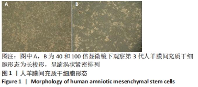
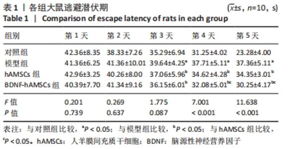
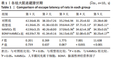
|