Chinese Journal of Tissue Engineering Research ›› 2018, Vol. 22 ›› Issue (30): 4864-4869.doi: 10.3969/j.issn.2095-4344.0986
Previous Articles Next Articles
In vitro bioactivity of silicon-incorporated titanium dioxide nanotubes
Zhang Xian-jun, Zhao Xi-jiang
- Department of Orthopaedics, Affiliated Hospital of Jiangnan University, Wuxi 214062, Jiangsu Province, China
-
Received:2018-05-16Online:2018-10-28Published:2018-10-28 -
Contact:Zhao Xi-jiang, MD, Associate chief physician, Department of Orthopaedics, Affiliated Hospital of Jiangnan University. Wuxi 214062, Jiangsu Province, China -
About author:Zhang Xian-jun, Master, Physician, Department of Orthopaedics, Affiliated Hospital of Jiangnan University. Wuxi 214062, Jiangsu Province, China -
Supported by:the Scientific Research Project of Wuxi Municipal Health and Family Planning, No. MS201726
CLC Number:
Cite this article
Zhang Xian-jun, Zhao Xi-jiang. In vitro bioactivity of silicon-incorporated titanium dioxide nanotubes[J]. Chinese Journal of Tissue Engineering Research, 2018, 22(30): 4864-4869.
share this article

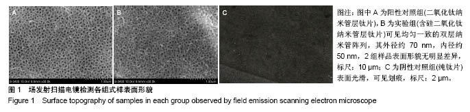
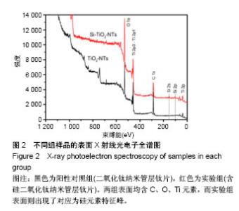
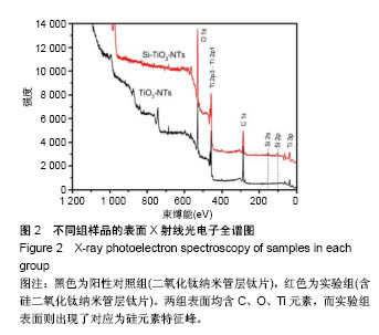
2.1 各组钛片表征 各组钛片表面形貌见图1。扫描电镜下显示阳性对照组(图1A)和实验组(图1B)表面形貌无明显差异,这表明硅离子注入不会破坏二氧化钛纳米管的管状形貌。各组钛片表面元素分析见图2。X射线光电子能谱仪显示实验组与阳性对照组式样表面含C、O、Ti元素,而实验组管层表面的全分辨图谱在100-150 eV附近出现了对应为硅元素特征峰,其含量约为0.51%。表示硅元素已成功注入氧化钛纳米管层中。"
"

2.2 各组钛片与MC3T3-E1细胞共培养后的细胞形态 见图3。培养1 d后,阴性对照组的MC3T3-E1细胞呈扁平形态,同时细胞在阳性对照组和实验组表面显示圆形和不规则的形状,此外,在实验组可以观察到细胞上有丝状伪足扩展。第3天,阴性对照组细胞显示拉长和扁平形态,在阳性对照组上开始观察到丝状伪足延伸,而在实验组上丝状伪足活性增加并开始合并。 第5天见细胞覆盖3组材料的表面。与阴性对照组表面相比,更多的网状丝状伪足出现在阳性对照组表面,而实验组表面的丝状网络结构较其他2组明显增多。"

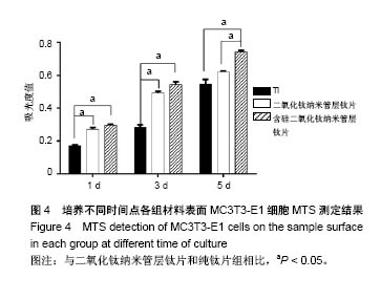
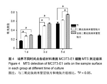
2.3 各组细胞的黏附 各组随时间延长细胞黏附增殖活性有增加趋势。培养第1及3天,MTS检测示黏附于实验组表面及阳性对照组表面的细胞数量比阴性对照组显著增多(P < 0.05);培养第5天时实验组表面的细胞增殖显著高于其他2组(P < 0.05),见图4。"
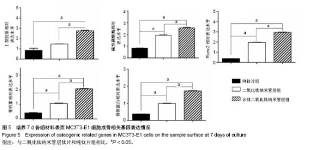
2.4 细胞分化相关基因的检测 培养7 d时,实验组成骨分化相关基因Ⅰ型胶原、碱性磷酸酶、Runx2、骨钙素和骨桥蛋白的表达水平显着高于其他2组(P < 0.05)。提示在含硅二氧化钛纳米管层钛片表面培养的成骨活性得到改善,见图5。"

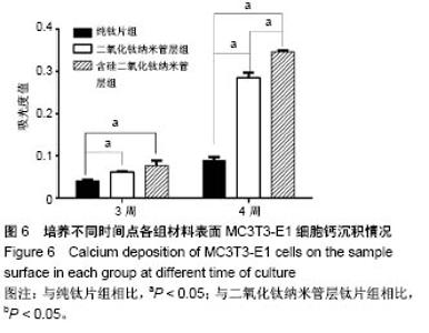
2.5 细胞茜素红染色定性检测 与阴性对照组相比,阳性对照组和实验组材料上的细胞在第3及4周的钙矿物结节形成明显增多(P < 0.05)。 此外,在第4周时,实验组材料上沉积的钙矿物显著多于阳性对照组(P < 0.05),见图6。"

| [1] Cipriano AF, Miller C, Liu H. Anodic growth and biomedical applications of Ti02 nanotubes. J Biomed Nanotechnol. 2014;10(10):2977-3003.[2] Munirathinam B, Neelakantan L. Titania nanotubes from weak organic acid electrolyte: fabrication, characterization and oxide film properties.Mater Sci Eng C Mater Biol Appl. 2015;49:567-578.[3] Khan AF, Saleem M, Afzal A, et al. Bioactive behavior of silicon substituted calcium phosphate based bioceramics for bone regeneration. Mater Sci Eng C Mater Biol Appl. 2014;35:245-252.[4] Shi X, Nakagawa M, Kawachi G, et al. Surface modification of titanium by hydrothermal treatment in Mg-containing solution and early osteoblast responses. Mater Sci Mater Med. 2012;23(5):1281-1290.[5] Bauer S, Park J, Faltenbacher J, et al. Size selective behavior of mesenchymal stem cells on ZrO(2) and TiO(2) nanotube arrays. Integr Biol (Camb).2009;1(8-9):525-532.[6] Lou W, Dong Y, Zhang H, et al. Preparation and Characterization of Lanthanum-Incorporated Hydroxyapatite Coatings on Titanium Substrates. Int J Mol Sci. 2015;16(9):21070-21086.[7] Rao PJ, Pelletier MH, Walsh WR, Mobbs RJ. Spine interbody implants: material selection and modification, functionalization and bioactivation of surfaces to improve osseointegration. Orthop Surg.2014;6(2):81-89.[8] Matena J, Petersen S, Gieseke M, et al. SLM produced porous titanium implant improvements for enhanced vascularization and osteoblast seeding. Int J Mol Sci. 2015;16(4):7478-7492.[9] Chen WC, Chen YS, Ko CL, et al. Interaction of progenitor bone cells with different surface modifications of titanium implant. Mater Sci Eng C Mater Biol Appl.2014;37:305-313.[10] Kulkarui M, Mazare A, Gongadze E, et al. Titanium nanostruetnres for biomedical applications. Nanoteehnology. 2015;26(6):062002.[11] 杜庆华,曹均凯,董溪溪,等.镁黄长石浸提液诱导多潜能干细胞的成骨分化[J].中国组织工程研究,2015,19(14):2236-2242.[12] Zhang W, Jin Y, Qian S, et al. Vacuum extraction enhances rhPDGF-BB immobilization on nanotubes to improve implant osseointegration in ovariectomized rats. Nanomedicine. 2014;10(8):1809-1818.[13] Buttke TM, McCubrey JA, Owen TC. Use of an aqueous soluble tetrazolium/formazan assay to measure viability and proliferation of lymphokine-dependent cell lines. J Immunol Methods. 1993;157(1-2): 233-240.[14] Yamamoto O,Alvarez K,Kashiwaya Y,et al. Surface characterization and biological response of carbon-coated oxygen-diffused titanium having different topographical surfaces. J Mater Sci Mater Med. 2011;22(4):977-987.[15] Gong T, Xie J, Liao J, et al. Nanomaterials and bone regeneration. Bone Res.2015;3:15029.[16] Qiao Y, Zhang W, Tian P, et al. Stimulation of bone growth following zinc incorporation into biomaterials. Biomaterials. 2014;35(25):6882-6897.[17] Wang G, Li J, Zhang W, et al. Magnesium ion implantation on a micro/nanostructured titanium surface promotes its bioactivity and osteogenic differentiation function. Int J Nanomedicine. 2014;9:2387-2398.[18] Macdonald HM, Hardcastle AC, Jugdaohsingh R, et al. Dietary silicon interacts with oestrogen to influence bone health: evidence from the Aberdeen Prospective Osteoporosis Screening Study. Bone. 2012;50(3): 681-687.[19] Honda M, Kikushima K, Kawanobe Y, et al. Enhanced early osteogenic differentiation by silicon-substituted hydroxyapatite ceramics fabricated via ultrasonic spray pyrolysis route. J Mater Sci Mater Med. 2012;23(12): 2923-2932.[20] Rosemary FL, Suswillo, Behzad Javaheri, et al. Strain uses gap junctions to reverse stimulation of osteoblast proliferation by osteocytes. Cell Biochem Funct. 2017;35(1):56-65.[21] Minagar S, Li Y, Berndt CC, Wen C. The influence of titania-zirconia- zirconium titanate nanotube characteristics on osteoblast cell adhesion. Acta Biomater. 2015;12:281-289.[22] Sawada R, Kono K, Isama K, et al. Calcium-incorporated titanium surfaces influence the osteogenic differentiation of human mesenchymal stem cells. J Biomed Mater Res A. 2013;101(9):2573-2585.[23] Liu W, Arnaud Chaix, Magali Gary-Bobo, et al. Stealth Biocompatible Si-Based Nanoparticles for Biomedical Applications. Nanomaterials (Basel).2017;7(10):288.[24] Li J, Wei L, Sun J, et al. Effect of ionic products of dicalcium silicate coating on osteoblast differentiation and collagen production via TGF-beta1 pathway. J Biomater Appl. 2013;27(5):595-604.[25] Juliana AD, Mariana M, Celso AB, et al. Addition of Wollastonite Fibers to Calcium Phosphate Cement Increases Cell Viability and Stimulates Differentiation of Osteoblast-Like Cells. ScientificWorldJournal. 2017; 2017: 5260106.[26] Shahdadfar A, Fronsdal K, Haug T, et al. In vitro expansion of human mesenchymal stem cells: choice of serum is a determinant of cell proliferation, differentiation, gene expression, and transcriptome stability. Stem Cells. 2005;23(9):1357-1366.[27] Premnath P, Tan B, Venkatakrishnan K. Programming cell fate on bio-functionalized silicon. Colloids and surfaces B. Biointerfaces. 2015; 128:100-105.[28] Ling M, Huang PX, Islam S, et al. Epigenetic regulation of Runx2 transcription and osteoblast differentiation by nicotinamide phosphoribosyltransferase. Cell Biosci. 2017;7:27.[29] Ercan B, Webster TJ. The effect of biphasic electrical stimulation on osteoblast function at anodized nanotubular titanium surfaces. Biomaterials. 2010;31(13):3684-3693.[30] Wang C, Duan Y, Markovic B, et al. Phenotypic expression of bone-related genes in osteoblasts grown on calcium phosphate ceramics with different phase compositions. Biomaterials.2004;25(13):2507-2514.[31] Ducy P, Zhang R, Geoffroy V, et al. Osf2/Cbfa1: a transcriptional activator of osteoblast differentiation. Cell. 1997;89(5):747-754.[32] Geoffroy V, Kneissel M, Fournier B, et al. High bone resorption in adult aging transgenic mice overexpressing cbfa1/runx2 in cells of the osteoblastic lineage. Mol Cell Biol.2002;22(17):6222-6233.[33] Franceschi RT, Xiao G, Jiang D, et al. Multiple signaling pathways converge on the Cbfa1/Runx2 transcription factor to regulate osteoblast differentiation. Connect Tissue Res. 2003;44(Suppl 1):109-116. |
| [1] | Zhang Tongtong, Wang Zhonghua, Wen Jie, Song Yuxin, Liu Lin. Application of three-dimensional printing model in surgical resection and reconstruction of cervical tumor [J]. Chinese Journal of Tissue Engineering Research, 2021, 25(9): 1335-1339. |
| [2] | Zeng Yanhua, Hao Yanlei. In vitro culture and purification of Schwann cells: a systematic review [J]. Chinese Journal of Tissue Engineering Research, 2021, 25(7): 1135-1141. |
| [3] | Xu Dongzi, Zhang Ting, Ouyang Zhaolian. The global competitive situation of cardiac tissue engineering based on patent analysis [J]. Chinese Journal of Tissue Engineering Research, 2021, 25(5): 807-812. |
| [4] | Li Xingping, Xiao Dongqin, Zhao Qiao, Chen Shuo, Bai Yiguang, Liu Kang, Feng Gang, Duan Ke. Preparation and properties of copper-loaded antibacterial functional film on titanium surface [J]. Chinese Journal of Tissue Engineering Research, 2021, 25(4): 553-557. |
| [5] | Ma Zhijie, Li Jingyu, Cao Fang, Liu Rong, Zhao Dewei. Influencing factors and biological property of novel biomedical materials: porous silicon carbide coated with bioactive tantalum [J]. Chinese Journal of Tissue Engineering Research, 2021, 25(4): 558-563. |
| [6] | Wu Zijian, Hu Zhaoduan, Xie Youqiong, Wang Feng, Li Jia, Li Bocun, Cai Guowei, Peng Rui. Three-dimensional printing technology and bone tissue engineering research: literature metrology and visual analysis of research hotspots [J]. Chinese Journal of Tissue Engineering Research, 2021, 25(4): 564-569. |
| [7] | Shi Xiaoxiu, Mao Shilong, Liu Yang, Ma Xingshuang, Luo Yanfeng. Comparison of tantalum and titanium (alloy) as orthopedic materials: physical and chemical indexes, antibacterial and osteogenic ability [J]. Chinese Journal of Tissue Engineering Research, 2021, 25(4): 593-599. |
| [8] | Chang Wenliao, Zhao Jie, Sun Xiaoliang, Wang Kun, Wu Guofeng, Zhou Jian, Li Shuxiang, Sun Han. Material selection, theoretical design and biomimetic function of artificial periosteum [J]. Chinese Journal of Tissue Engineering Research, 2021, 25(4): 600-606. |
| [9] | Liu Fei, Cui Yutao, Liu He. Advantages and problems of local antibiotic delivery system in the treatment of osteomyelitis [J]. Chinese Journal of Tissue Engineering Research, 2021, 25(4): 614-620. |
| [10] | Li Xiaozhuang, Duan Hao, Wang Weizhou, Tang Zhihong, Wang Yanghao, He Fei. Application of bone tissue engineering materials in the treatment of bone defect diseases in vivo [J]. Chinese Journal of Tissue Engineering Research, 2021, 25(4): 626-631. |
| [11] | Zhang Zhenkun, Li Zhe, Li Ya, Wang Yingying, Wang Yaping, Zhou Xinkui, Ma Shanshan, Guan Fangxia. Application of alginate based hydrogels/dressings in wound healing: sustained, dynamic and sequential release [J]. Chinese Journal of Tissue Engineering Research, 2021, 25(4): 638-643. |
| [12] | Chen Jiana, Qiu Yanling, Nie Minhai, Liu Xuqian. Tissue engineering scaffolds in repairing oral and maxillofacial soft tissue defects [J]. Chinese Journal of Tissue Engineering Research, 2021, 25(4): 644-650. |
| [13] | Xing Hao, Zhang Yonghong, Wang Dong. Advantages and disadvantages of repairing large-segment bone defect [J]. Chinese Journal of Tissue Engineering Research, 2021, 25(3): 426-430. |
| [14] | Chen Siqi, Xian Debin, Xu Rongsheng, Qin Zhongjie, Zhang Lei, Xia Delin. Effects of bone marrow mesenchymal stem cells and human umbilical vein endothelial cells combined with hydroxyapatite-tricalcium phosphate scaffolds on early angiogenesis in skull defect repair in rats [J]. Chinese Journal of Tissue Engineering Research, 2021, 25(22): 3458-3465. |
| [15] | Wang Hao, Chen Mingxue, Li Junkang, Luo Xujiang, Peng Liqing, Li Huo, Huang Bo, Tian Guangzhao, Liu Shuyun, Sui Xiang, Huang Jingxiang, Guo Quanyi, Lu Xiaobo. Decellularized porcine skin matrix for tissue-engineered meniscus scaffold [J]. Chinese Journal of Tissue Engineering Research, 2021, 25(22): 3473-3478. |
| Viewed | ||||||
|
Full text |
|
|||||
|
Abstract |
|
|||||