Chinese Journal of Tissue Engineering Research ›› 2024, Vol. 28 ›› Issue (22): 3517-3523.doi: 10.12307/2024.531
Previous Articles Next Articles
Preparation and characterization of methacryloylated hyaluronic acid/acellular Wharton’s jelly composite hydrogel scaffold
Yuan Xun1, 2, Ding Zhengang1, Fu Liwei2, Wu Jiang2, Zheng Yazhe1, 2, Zhang Zhichao2, Tian Guangzhao2, Sui Xiang2, Liu Shuyun2, Guo Quanyi1, 2
- 1Department of Orthopedics, Affiliated Hospital of Guizhou Medical University, Guiyang 550004, Guizhou Province, China; 2Institute of Orthopedics, First Medical Center, General Hospital of Chinese PLA, Beijing Key Laboratory of Orthopedic Regenerative Medicine, Military Key Laboratory of Orthopedic Warfare Trauma, Beijing 100853, China
-
Received:2023-09-14Accepted:2023-11-02Online:2024-08-08Published:2024-01-20 -
Contact:Guo Quanyi, MD, Chief physician, Professor, Department of Orthopedics, Affiliated Hospital of Guizhou Medical University, Guiyang 550004, Guizhou Province, China; Institute of Orthopedics, First Medical Center, General Hospital of Chinese PLA, Beijing Key Laboratory of Orthopedic Regenerative Medicine, Military Key Laboratory of Orthopedic Warfare Trauma, Beijing 100853, China -
About author:Yuan Xun, Master candidate, Department of Orthopedics, Affiliated Hospital of Guizhou Medical University, Guiyang 550004, Guizhou Province, China; Institute of Orthopedics, First Medical Center, General Hospital of Chinese PLA, Beijing Key Laboratory of Orthopedic Regenerative Medicine, Military Key Laboratory of Orthopedic Warfare Trauma, Beijing 100853, China -
Supported by:National Key Research and Development Plan Project, No. 2019YFA0110600 (to GQY)
CLC Number:
Cite this article
Yuan Xun, Ding Zhengang, Fu Liwei, Wu Jiang, Zheng Yazhe, Zhang Zhichao, Tian Guangzhao, Sui Xiang, Liu Shuyun, Guo Quanyi. Preparation and characterization of methacryloylated hyaluronic acid/acellular Wharton’s jelly composite hydrogel scaffold[J]. Chinese Journal of Tissue Engineering Research, 2024, 28(22): 3517-3523.
share this article
Add to citation manager EndNote|Reference Manager|ProCite|BibTeX|RefWorks
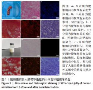
2.1 脱细胞前后华通胶的大体观和组织学染色 见图1。"
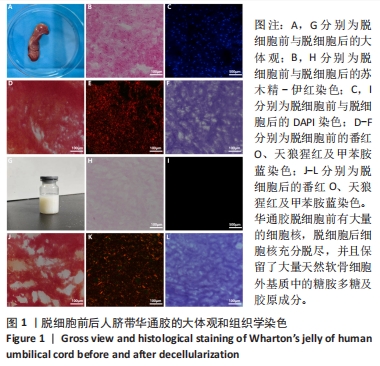

图1A为人脐带大体观,图1G为脱细胞并冻干后平衡盐液重悬的脱细胞华通胶溶液,呈白色偏透明胶状。 为了检测华通胶脱细胞情况,使用苏木精-伊红染色和DAPI染色检测脱细胞前后华通胶中的细胞核存在情况,脱细胞前华通胶中存在大量的细胞核结构(图1B,C),经脱细胞处理后无明显的细胞核结构(图1H,I),表明脱细胞成功。 为了评估脱细胞后华通胶中成分的保留情况分别进行了番红O、甲苯胺蓝、天狼猩红染色,实验结果表明,脱细胞前华通胶中富含大量的糖胺多糖及胶原成分(图1D-F),经脱细胞处理后仍保留了大量的糖胺多糖及胶原成分(图1J-L)。 2.2 两组水凝胶支架的大体观及微观结构 见图2。"
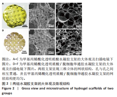
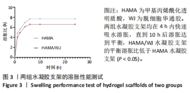
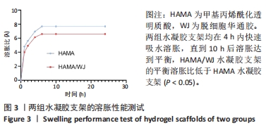
实验成功打印构建了两种生物墨水支架,支架大体为圆形,经纬交叉呈网络状结构,网状结构整齐,加入华通胶后的支架呈黄白色。扫描电镜下可见两组支架呈现三维立体的网状结构,孔与孔之间相互贯通,大孔径基本相似,为500-600 μm大小,加入华通胶后纤维间连接更加均匀,有利于细胞的活动和伸展。 2.3 两组水凝胶支架的溶胀性能 图3为两组水凝胶支架的动态溶胀曲线,在前4 h内两组水凝胶支架均能快速吸收水分溶胀增重,6-8 h达到巅峰,10 h后基本达到溶胀平衡,HAMA/WJ水凝胶支架的平衡溶胀比低于HAMA水凝胶支架(P < 0.05)。"

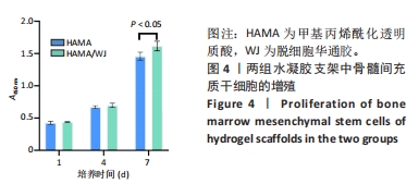
2.4 两组含细胞水凝胶支架中的细胞增殖情况 如图4所示,随着骨髓间充质干细胞在水凝胶支架中培养时间的延长,细胞的增殖能力逐渐增强,两组培养第1,4天的细胞增殖吸光度值比较差异无显著性意义(P > 0.05),HAMA/WJ水凝胶支架组培养第7天的细胞增殖吸光度值高于HAMA水凝胶支架组(P < 0.05)。说明两组水凝胶支架的生物相容性良好,同时华通胶的加入使得水凝胶支架具有更强的促进骨髓间充质干细胞生长和增殖作用。"

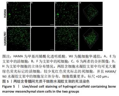
2.5 两组含细胞水凝胶支架的死活染色 死活染色显示,两组含细胞水凝胶支架中均可见大量绿色荧光标记的活细胞,较少见红色荧光标志的死细胞,并且HAMA/WJ水凝胶支架中的细胞呈立体分布、细胞数量更多,见图5,说明骨髓间充质干细胞在两种支架上均能良好存活,并且在HAMA/WJ水凝胶支架中细胞增殖更多。"
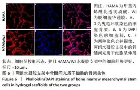
2.6 两组含细胞水凝胶支架中的细胞伸展及形态观察 鬼笔环肽染色显示两组水凝胶支架中的骨髓间充质干细胞呈伸展状态,细胞呈梭形形态,并且HAMA/WJ水凝胶支架中的细胞舒展更好,见图6。"

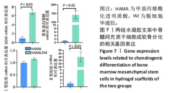
2.7 两组含细胞水凝胶支架的体外成软骨分化基因表达检测 qRT-PCR检测结果显示,HAMA/WJ水凝胶支架组聚集蛋白聚糖、SOX9、Ⅱ型胶原mRNA表达量均高于HAMA水凝胶支架组(P < 0.05,P < 0.01),两组Ⅰ型胶原mRNA表达量比较差异无显著性意义(P > 0.05),见图7。"

| [1] JIANG S, TIAN G, YANG Z, et al. Enhancement of acellular cartilage matrix scaffold by Wharton’s jelly mesenchymal stem cell-derived exosomes to promote osteochondral regeneration. Bioact Mater. 2021;6(9):2711-2728. [2] MADRY H, GRÜN UW, KNUTSEN G. Cartilage repair and joint preservation: medical and surgical treatment options. Dtsch Arztebl Int. 2011;108(40):669-677. [3] YANG Z, CAO F, LI H, et al. Microenvironmentally optimized 3D-printed TGFβ-functionalized scaffolds facilitate endogenous cartilage regeneration in sheep. Acta Biomater. 2022;150:181-198. [4] DALY AC, FREEMAN FE, GONZALEZ-FERNANDEZ T, et al. 3D Bioprinting for Cartilage and Osteochondral Tissue Engineering. Adv Healthc Mater, 2017;6(22).doi: 10.1002/adhm.201700298. [5] LEE A, HUDSON AR, SHIWARSKI DJ, et al. 3D bioprinting of collagen to rebuild components of the human heart. Science. 2019;365(6452): 482-487. [6] WANG S, ZHAO S, YU J, et al. Advances in Translational 3D Printing for Cartilage, Bone, and Osteochondral Tissue Engineering. Small. 2022; 18(36):e2201869. [7] LIU Y, PENG L, LI L, et al. 3D-bioprinted BMSC-laden biomimetic multiphasic scaffolds for efficient repair of osteochondral defects in an osteoarthritic rat model. Biomaterials. 2021;279:121216. [8] SCHUURMANS C, MIHAJLOVIC M, HIEMSTRA C, et al. Hyaluronic acid and chondroitin sulfate (meth)acrylate-based hydrogels for tissue engineering: Synthesis, characteristics and pre-clinical evaluation. Biomaterials. 2021;268:120602. [9] LEE JM, YEONG WY. Design and Printing Strategies in 3D Bioprinting of Cell-Hydrogels: A Review. Adv Healthc Mater. 2016;5(22):2856-2865. [10] JONIDI SHARIATZADEH F, SOLOUK A, BAGHERI KHOULENJANI S, et al. Injectable and reversible preformed cryogels based on chemically crosslinked gelatin methacrylate (GelMA) and physically crosslinked hyaluronic acid (HA) for soft tissue engineering. Colloids Surf B Biointerfaces. 2021;203:111725. [11] YANG C, WANG X, YAO X, et al. Hyaluronic acid nanogels with enzyme-sensitive cross-linking group for drug delivery. J Control Release. 2015; 205:206-217. [12] SCOTT JM, WILKINSON R. Further studies on the umbilical cord and its water content. J Clin Pathol. 1978;31(10):944-948. [13] CHAN RW, RODRIGUEZ ML, MCFETRIDGE PS. The human umbilical vein with Wharton’s jelly as an allogeneic, acellular construct for vocal fold restoration. Tissue Eng Part A. 2009;15(11):3537-3546. [14] VALIYAVEETTIL M, ACHUR RN, MUTHUSAMY A, et al. Characterization of chondroitin sulfate and dermatan sulfate proteoglycans of extracellular matrices of human umbilical cord blood vessels and Wharton’s jelly. Glycoconj J. 2004;21(6):361-375. [15] WU J, FU L, YAN Z, et al. Hierarchical porous ECM scaffolds incorporating GDF-5 fabricated by cryogenic 3D printing to promote articular cartilage regeneration. Biomater Res. 2023;27(1):7. [16] GOGIEL T, BAŃKOWSKI E, JAWORSKI S. Proteoglycans of Wharton’s jelly. Int J Biochem Cell Biol. 2003;35(10):1461-1469. [17] XU HQ, LIU JC, ZHANG ZY, et al. A review on cell damage, viability, and functionality during 3D bioprinting. Mil Med Res. 2022;9(1):70. [18] SHEN Y, TANG H, HUANG X, et al. DLP printing photocurable chitosan to build bio-constructs for tissue engineering. Carbohydr Polym. 2020; 235:115970. [19] 殷瀚,吴江,严子能,等.基于低温沉积打印技术构建分级多孔脱细胞华通胶支架[J].中国医药生物技术,2022,17(6):488-496. [20] 杨振,李浩,付力伟,等. 3D生物打印负载转化生长因子β3的软骨复合支架[J].中国组织工程研究,2021,25(34):5445-5452. [21] LEE S, SANI ES, SPENCER AR, et al. Human-Recombinant-Elastin-Based Bioinks for 3D Bioprinting of Vascularized Soft Tissues. Adv Mater. 2020;32(45):e2003915. [22] 吴江,殷瀚,严子能,等.低温沉积3D打印负载基质细胞衍生因子1的分级多孔软骨复合支架[J].中国组织工程研究,2023,27(30): 4776-4782. [23] SHI W, SUN M, HU X, et al. Structurally and Functionally Optimized Silk-Fibroin-Gelatin Scaffold Using 3D Printing to Repair Cartilage Injury In Vitro and In Vivo. Adv Mater. 2017;29(29).doi: 10.1002/adma. 201701089. [24] RAIO L, CROMI A, GHEZZI F, et al. Hyaluronan content of Wharton’s jelly in healthy and Down syndrome fetuses. Matrix Biol. 2005;24(2):166-174. [25] LIU J, LI L, SUO H, et al. 3D printing of biomimetic multi-layered GelMA/nHA scaffold for osteochondral defect repair. ScienceDirect. 2019;171:107708-107708. [26] QI J, CHEN A, YOU H, et al. Proliferation and chondrogenic differentiation of CD105-positive enriched rat synovium-derived mesenchymal stem cells in three-dimensional porous scaffolds. Biomed Mater. 2011;6(1):015006. [27] LIU L, XIONG Z, YAN YN, et al. Porous morphology, porosity, mechanical properties of poly(α-hydroxy acid)–tricalcium phosphate composite scaffolds fabricated by low-temperature deposition. J Biomed Mater Res A. 2007;82A(3):618-629. [28] MIAO X, TAN DM, LI J, et al. Mechanical and biological properties of hydroxyapatite/tricalcium phosphate scaffolds coated with poly(lactic-co-glycolic acid). Acta Biomater. 2008;4(3):638-645. [29] CHEN M, LI Y, LIU S, et al. Hierarchical macro-microporous WPU-ECM scaffolds combined with Microfracture Promote in Situ Articular Cartilage Regeneration in Rabbits. Bioact Mater. 2021;6(7):1932-1944. [30] TOLABI H, DAVARI N, KHAJEHMOHAMMADI M, et al. Progress of Microfluidic Hydrogel-Based Scaffolds and Organ-on-Chips for the Cartilage Tissue Engineering. Adv Mater. 2023;35(26):e2208852. [31] GRIFFIN DR, ARCHANG MM, KUAN CH, et al. Activating an adaptive immune response from a hydrogel scaffold imparts regenerative wound healing. Nat Mater. 2021;20(4):560-569. [32] JIANG G, LI S, YU K, et al. A 3D-printed PRP-GelMA hydrogel promotes osteochondral regeneration through M2 macrophage polarization in a rabbit model. Acta Biomater. 2021;128:150-162. [33] SHI W, FANG F, KONG Y, et al. Dynamic hyaluronic acid hydrogel with covalent linked gelatin as an anti-oxidative bioink for cartilage tissue engineering. Biofabrication. 2021;14(1). doi: 10.1088/1758-5090/ac42de. [34] FENG Y, XIAO K, CHEN J, et al. Immune-microenvironment modulatory polyurethane-hyaluronic acid hybrid hydrogel scaffolds for diabetic wound treatment. Carbohydr Polym. 2023;320:121238. [35] GUO C, CAO Z, PENG Y, et al. Subchondral bone-inspired hydrogel scaffold for cartilage regeneration. Colloids Surf B Biointerfaces. 2022;218:112721. [36] LI M, YIN H, YAN Z, et al. The immune microenvironment in cartilage injury and repair. Acta Biomater. 2022;140:23-42. [37] YANG Z, ZHAO T, GAO C, et al. 3D-Bioprinted Difunctional Scaffold for In Situ Cartilage Regeneration Based on Aptamer-Directed Cell Recruitment and Growth Factor-Enhanced Cell Chondrogenesis. ACS Appl Mater Interfaces. 2021;13(20):23369-23383. [38] HULL SM, BRUNEL LG, HEILSHORN SC. 3D Bioprinting of Cell-Laden Hydrogels for Improved Biological Functionality. Adv Mater. 2022;34(2):e2103691. [39] KRUJATZ F, DANI S, WINDISCH J, et al. Think outside the box: 3D bioprinting concepts for biotechnological applications -recent developments and future perspectives. Biotechnol Adv. 2022;58: 107930. [40] XU J, ZHANG M, DU W, et al. Chitosan-based high-strength supramolecular hydrogels for 3D bioprinting. Int J Biol Macromol. 2022;219:545-557. [41] LI M, SUN D, ZHANG J, et al. Application and development of 3D bioprinting in cartilage tissue engineering. Biomater Sci. 2022;10(19): 5430-5458. |
| [1] | Zhou Shijie, Li Muzhe, Yun Li, Zhang Tianchi, Niu Yuanyuan, Zhu Yihua, Zhou Qinfeng, Guo Yang, Ma Yong, Wang Lining. Effect of Wenshen Tongluo Zhitong formula on mouse H-type bone microvascular endothelial cell/bone marrow mesenchymal stem cell co-culture system [J]. Chinese Journal of Tissue Engineering Research, 2025, 29(1): 8-15. |
| [2] | Wang Chenglong , Yang Zhilie , Chang Junli , Zhao Yongjian , Zhao Dongfeng , Dai Weiwei , Wu Hongjin , Zhang Jie , Wang Libo , Xie Ying , Tang Dezhi , Wang Yongjun , Yang Yanping. Restoration of osteogenic differentiation of bone marrow mesenchymal stem cells in mice inhibited by cyclophosphamide with psoralen [J]. Chinese Journal of Tissue Engineering Research, 2025, 29(1): 16-23. |
| [3] | Wang Zhikun, Bai Shaoxuan, Zhao Wei, Wang Chenyu. Exercise preconditioning combined with bone marrow mesenchymal stem cell transplantation for myocardial infarction in rats [J]. Chinese Journal of Tissue Engineering Research, 2025, 29(1): 65-73. |
| [4] | Yang Yufang, Yang Zhishan, Duan Mianmian, Liu Yiheng, Tang Zhenglong, Wang Yu. Application and prospects of erythropoietin in bone tissue engineering [J]. Chinese Journal of Tissue Engineering Research, 2024, 28(9): 1443-1449. |
| [5] | Chen Kaijia, Liu Jingyun, Cao Ning, Sun Jianbo, Zhou Yan, Mei Jianguo, Ren Qiang. Application and prospect of tissue engineering in treatment of osteonecrosis of the femoral head [J]. Chinese Journal of Tissue Engineering Research, 2024, 28(9): 1450-1456. |
| [6] | Wang Shanshan, Shu Qing, Tian Jun. Physical factors promote osteogenic differentiation of stem cells [J]. Chinese Journal of Tissue Engineering Research, 2024, 28(7): 1083-1090. |
| [7] | Mei Jingyi, Liu Jiang, Xiao Cong, Liu Peng, Zhou Haohao, Lin Zhanyi. Proliferation and metabolic patterns of smooth muscle cells during construction of tissue-engineered blood vessels [J]. Chinese Journal of Tissue Engineering Research, 2024, 28(7): 1043-1049. |
| [8] | Wang Wu, Fan Xiaolei, Xie Jie, Hu Yihe, Zeng Min. Hydroxyapatite-polyvinyl alcohol/collagen-chitosan-gelatin composite hydrogel for repairing rabbit osteochondral defect [J]. Chinese Journal of Tissue Engineering Research, 2024, 28(5): 682-689. |
| [9] | Zhang Ya, Mu Qiuju, Wang Zilin, Liu Hongjie, Zhu Lili. Hydrogel loaded with platelet-rich plasma promotes wound healing in diabetic rats [J]. Chinese Journal of Tissue Engineering Research, 2024, 28(5): 690-696. |
| [10] | Shen Ziqing, Xia Tian, Shan Yibo, Zhu Ruijun, Wan Haoxin, Ding Hao, Pan Shu, Zhao Jun. Vascularized tracheal substitutes constructed by exosome-load hydrogel-modified 3D printed scaffolds [J]. Chinese Journal of Tissue Engineering Research, 2024, 28(5): 697-705. |
| [11] | Wang Jianchun, Yang Shuqing, Su Xin, Wang Hongyuan. Different contents of B2O3 affect mechanical properties and bioactivity of bioactive glass scaffolds [J]. Chinese Journal of Tissue Engineering Research, 2024, 28(5): 712-716. |
| [12] | Zhang Yihai, Shang Peng, Ma Benyuan, Hou Guanghui, Cui Lunxu, Song Wanzhen, Qi Dexuan, Liu Yancheng. Structural design and mechanical property analysis of trabecular scaffold of triply periodic minimal surface with a radial gradient [J]. Chinese Journal of Tissue Engineering Research, 2024, 28(5): 741-746. |
| [13] | Zhu Liwei, Wang Jiangyue, Bai Ding. Application value of nanocomposite gelatin methacryloyl hydrogels in different bone defect environments [J]. Chinese Journal of Tissue Engineering Research, 2024, 28(5): 753-758. |
| [14] | Chen Xiaofang, Zheng Guoshuang, Li Maoyuan, Yu Weiting. Preparation and application of injectable sodium alginate hydrogels [J]. Chinese Journal of Tissue Engineering Research, 2024, 28(5): 789-794. |
| [15] | Wang Jiani, Chen Junyu. Angiogenesis mechanism of metal ions and their application in bone tissue engineering [J]. Chinese Journal of Tissue Engineering Research, 2024, 28(5): 804-812. |
| Viewed | ||||||
|
Full text |
|
|||||
|
Abstract |
|
|||||