Chinese Journal of Tissue Engineering Research ›› 2024, Vol. 28 ›› Issue (5): 697-705.doi: 10.12307/2023.885
Previous Articles Next Articles
Vascularized tracheal substitutes constructed by exosome-load hydrogel-modified 3D printed scaffolds
Shen Ziqing1, Xia Tian1, Shan Yibo2, Zhu Ruijun1, Wan Haoxin1, Ding Hao1, Pan Shu1, Zhao Jun1
- 1Department of Thoracic Surgery, The First Affiliated Hospital of Soochow University, Suzhou 215006, Jiangsu Province, China; 2Institute of Translational Research, College of Medicine, Yangzhou University, Yangzhou 225000, Jiangsu Province, China
-
Received:2022-11-24Accepted:2023-01-05Online:2024-02-18Published:2023-08-16 -
Contact:Zhao Jun, PhD, Professor, Chief physician, Department of Thoracic Surgery, The First Affiliated Hospital of Soochow University, Suzhou 215006, Jiangsu Province, China -
About author:Shen Ziqing, Master candidate, Department of Thoracic Surgery, The First Affiliated Hospital of Soochow University, Suzhou 215006, Jiangsu Province, China -
Supported by:Natural Science Foundation for the Youth of Jiangsu Province, No. BK20200196 (to PS); Enterprise Commissioned Project of Soochow University, No. H221026 (to ZJ)
CLC Number:
Cite this article
Shen Ziqing, Xia Tian, Shan Yibo, Zhu Ruijun, Wan Haoxin, Ding Hao, Pan Shu, Zhao Jun. Vascularized tracheal substitutes constructed by exosome-load hydrogel-modified 3D printed scaffolds[J]. Chinese Journal of Tissue Engineering Research, 2024, 28(5): 697-705.
share this article
Add to citation manager EndNote|Reference Manager|ProCite|BibTeX|RefWorks
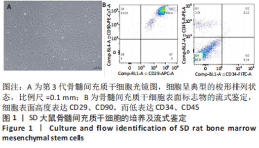
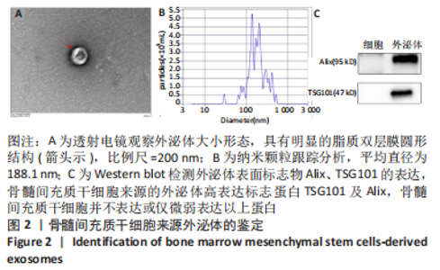
2.1 骨髓间充质干细胞的培养与鉴定结果 成功从SD大鼠双下肢骨髓中分离出骨髓间充质干细胞,培养至P3代细胞呈典型的梭形排列状态,见图1A。流式分析结果可以看到,细胞表面高度表达CD29、CD90,而低表达CD34、CD45,见图1B,符合骨髓间充质干细胞表面标志物的表达,该结果表明所提取培养的细胞为骨髓间充质干细胞。"
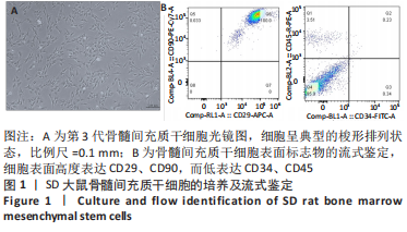
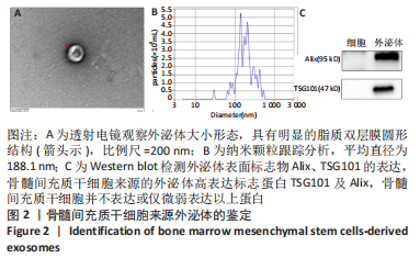
2.2 外泌体的鉴定结果 采用BCA法检测外泌体中的蛋白质量浓度为0.503 65 μg/μL。透射电镜下可见,骨髓间充质干细胞来源的外泌体具有明显的脂质双层膜圆形结构,直径约为150 nm,见图2A,即为外泌体。结果表示该法提取的外泌体符合广泛接受的标准特征。粒径检测结果表明,所提取及纯化外泌体悬液中的颗粒平均直径为188.1 nm,所提取外泌体的浓度约为1.0×10¹² L-1,见图2B。Western blot检测结果显示,骨髓间充质干细胞来源的外泌体高表达标志蛋白TSG101及Alix,骨髓间充质干细胞并不表达或仅微弱表达以上蛋白,见图2C。"

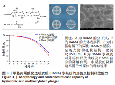
2.3 HAMA水凝胶的形貌及控释能力检测结果 图3A为HAMA的化学式,其大体形态如图3B所示。扫描电镜下可见固化HAMA水凝胶呈现光滑的孔状结构,见图3C所示。图3D显示了HAMA水凝胶对外泌体的控释能力及HAMA本身的降解特性。可以看出,随着时间的延长,外泌体被逐渐释放,当浸泡到30 d时,经分析计算,仍有约20%的外泌体保存于固化HAMA水凝胶中。同样,随着时间的延长,HAMA水凝胶自身也在降解,至30 d时剩约30%的HAMA,说明水凝胶的降解速率慢于外泌体的释放速率。该结果表明,通过固化HAMA水凝胶来实现对外泌体的控制释放的方法是可行的。"
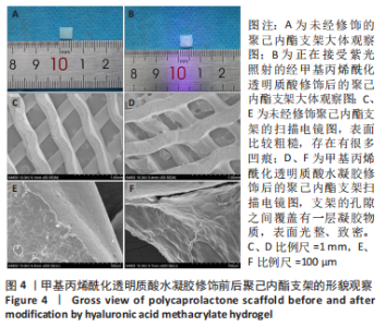
2.4 3D打印多孔聚己内酯支架的形貌观察结果 实验打印出直径约200 μm、大小约0.5 cm×0.5 cm的聚己内酯支架,见图4A;在纯聚己内酯支架上滴加少量HAMA水凝胶溶液,经紫灯照射5 min后等待其固化,见图4B。扫描电镜下可以观察到,原先的聚己内酯材料表面比较粗糙,存在有很多凹痕,见图4C、E。经HAMA修饰处理后,支架的孔隙之间覆盖有一层凝胶物质,见图4D;高倍镜下可见材料之前变得光整、致密,原先的凹痕基本消失,见图4F。"

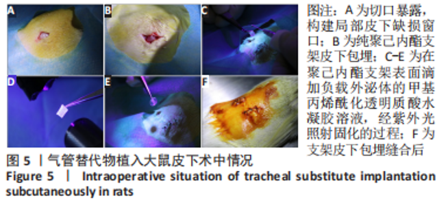
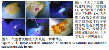
2.5 气管替代物大鼠皮下包埋实验结果 2.5.1 实验动物数量分析 30只大鼠全部进入结果分析。 2.5.2 大鼠术中情况、术后生存情况及标本收获 构建大鼠皮囊模型后,将补片植入皮下,术中出血少,采取间断缝合法缝合伤口。单纯支架组切开皮肤后立即将单纯聚己内酯支架植入皮下。将提前制备好的HAMA及负载外泌体的HAM水凝胶溶液抽吸于2 mL避光针筒中,紫光短暂照射针尖,使其达到滴出便可固化状态,将其滴加于聚己内酯支架表面,分别获得水凝胶修饰的支架与外泌体修饰的支架,植入皮下,见图5。"
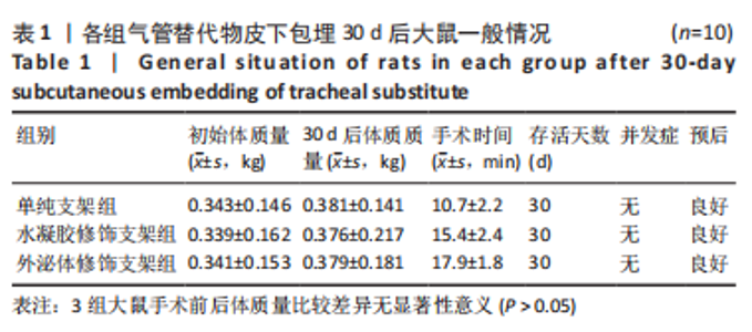
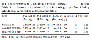
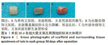
各组大鼠术后均未出现免疫排斥反应或其他并发症,术后一般情况均良好,如表1所示。于术后30 d时收获大体标本,见图6,单纯支架组表面粗糙,未见明显组织新生;水凝胶修饰支架组可见表面覆盖凝胶仍未完全降解,形态良好;外泌体修饰支架组支架表面有明显的新生血管形成。"

"
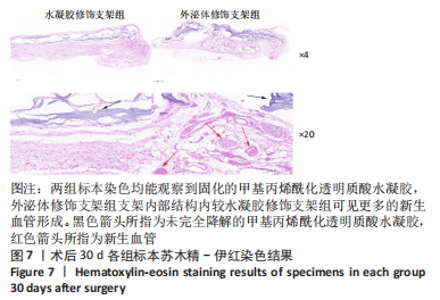
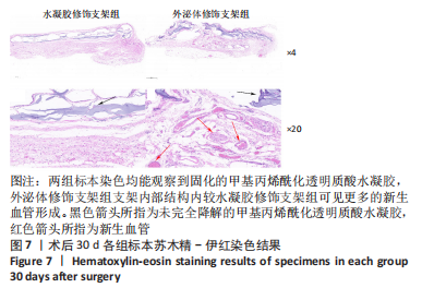
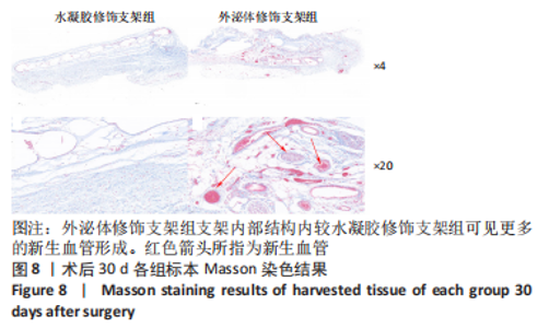
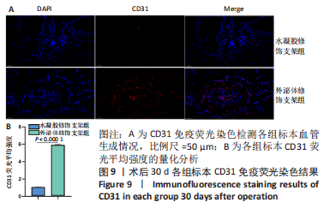
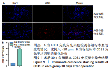
2.5.3 各组标本组织学分析结果 由于聚己内酯材料是酯溶性的,因此在制蜡过程中会发生溶解,因而后续实验仅进行了水凝胶修饰支架及外泌体修饰支架组的切片组织学分析。 术后30 d的苏木精-伊红及Masson染色可以观察到,外泌体修饰支架组支架内部结构内较水凝胶修饰支架组可见更多的新生血管形成,两组标本染色均能观察到固化的HAMA水凝胶,支架表面的HAMA于30 d时仍未完全降解,见图7,8,不仅提高了气管替代物支架的生物相容性,还实现了对外泌体的控制释放,使其更精准和更长时间的作用于靶点。免疫荧光染色显示,外泌体修饰支架组CD31表达高于水凝胶修饰支架组(P < 0.000 1),见图9。"
"

"

2.6 支架的生物相容性 聚己内酯是一种非免疫原性、非致癌性、可生物降解、降解产物无毒、生物相容性良好的疏水材料[47]。透明质酸是动物组织的细胞外基质的成分,具有良好的锁水保湿性能,在细胞增殖、分化、形态发生、炎症和伤口愈合中发挥重要作用[48]。外泌体是细胞分泌的纳米级膜囊泡物质,拥有良好的稳定性和极低的免疫原性[49]。该研究将经负载外泌体HAMA水凝胶修饰的聚己内酯支架种植到大鼠皮下,均未出现皮肤溃疡、腹泻、体质量下降等表现,未见明显的移植物抗宿主反应,具有良好的生物相容性。"

| [1] LEE H, MARIN-ARAUJO AE, AOKI FG, et al. Computational fluid dynamics for enhanced tracheal bioreactor design and long-segment graft recellularization. Sci Rep. 2021;11(1):1187. [2] CHOI JS, LEE MS, KIM J, et al. Hyaluronic Acid Coating on Hydrophobic Tracheal Scaffold Enhances Mesenchymal Stem Cell Adhesion and Tracheal Regeneration. Tissue Eng Regen Med. 2021;18:225-233. [3] XU C, MA Y, HUANG H, et al. A Review of Woven Tracheal Stents: Materials, Structures, and Application. J Funct Biomater. 2022;13(3):96. [4] YU YS, AHN CB, SON KH, et al. Motility Improvement of Biomimetic Trachea Scaffold via Hybrid 3D-Bioprinting Technology. Polymers (Basel) 2021;13(6):971. [5] SORIANO L, KHALID T, WHELAN D, et al. Development and clinical translation of tubular constructs for tracheal tissue engineering: a review. Eur Respir Rev. 2021;30:(162):210154. [6] VRANCKX JJ, DELAERE P. The current status and outlook of trachea transplantation. Curr Opin Organ Transplant. 2020;25:601-608. [7] GAO B, JING H, GAO M, et al. Long-segmental tracheal reconstruction in rabbits with pedicled Tissue-engineered trachea based on a 3D-printed scaffold. Acta Biomater. 2019;97:177-186. [8] Kim IG, Park SA, Lee SH, et al. Transplantation of a 3D-printed tracheal graft combined with iPS cell-derived MSCs and chondrocytes. Sci Rep. 2020;10(1):4326. [9] XIA D, JIN D, WANG Q, et al. Tissue-engineered trachea from a 3D-printed scaffold enhances whole-segment tracheal repair in a goat model. J Tissue Eng Regen Med. 2019;13:694-703. [10] YAO ZY, FENG BW, LIU CS, et al. The Application of a Bone Marrow Mesenchymal Stem Cell Membrane in the Vascularization of a Decellularized Tracheal Scaffold. Stem Cells Int. 2021;2021:6624265. [11] XIANG Y, WANG W, GAO Y, et al. Production and Characterization of an Integrated Multi-Layer 3D Printed PLGA/GelMA Scaffold Aimed for Bile Duct Restoration and Detection. Front Bioeng Biotechnol. 2020;8:971. [12] GRIGORYAN B, PAULSEN SJ, CORBETT DC, et al. Multivascular networks and functional intravascular topologies within biocompatible hydrogels. Science. 2019;364:458-464. [13] UDELSMAN B, MATHISEN DJ, OTT HC. A reassessment of tracheal substitutes-a systematic review. Ann Cardiothorac Surg. 2018;7:175-182. [14] HUO Y, XU Y, WU X, et al. Functional Trachea Reconstruction Using 3D-Bioprinted Native-Like Tissue Architecture Based on Designable Tissue-Specific Bioinks. Adv Sci (Weinh). 2022;9:e2202181. [15] PARK JY, RYU H, LEE B, et al. Development of a functional airway-on-a-chip by 3D cell printing. Biofabrication. 2018;11:015002. [16] DHASMANA A, SINGH A, RAWAL S. Biomedical grafts for tracheal tissue repairing and regeneration “Tracheal tissue engineering: an overview”. J Tissue Eng Regen Med. 2020; 14:653-672. [17] MAUGHAN EF, BUTLER CR, CROWLEY C, et al. A comparison of tracheal scaffold strategies for pediatric transplantation in a rabbit model. Laryngoscope. 2017;127:E449-e457. [18] REHMANI SS, AL-AYOUBI AM, AYUB A, et al. Three-Dimensional-Printed Bioengineered Tracheal Grafts: Preclinical Results and Potential for Human Use. Ann Thorac Surg. 2017; 104:998-1004. [19] LEE JS, PARK J, SHIN DA, et al. Characterization of the biomechanical properties of canine trachea using a customized 3D-printed apparatus. Auris Nasus Larynx. 2019;46:407-416. [20] BAE SW, LEE KW, PARK JH, et al. 3D Bioprinted Artificial Trachea with Epithelial Cells and Chondrogenic-Differentiated Bone Marrow-Derived Mesenchymal Stem Cells. Int J Mol Sci. 2018;19(6):1624. [21] MACHINO R, MATSUMOTO K, TANIGUCHI D, et al. Replacement of Rat Tracheas by Layered, Trachea-Like, Scaffold-Free Structures of Human Cells Using a Bio-3D Printing System. Adv Healthc Mater. 2019;8:e1800983. [22] KATAGIRI W, TAKEUCHI R, SAITO N, et al. Migration and phenotype switching of macrophages at early-phase of bone-formation by secretomes from bone marrow derived mesenchymal stem cells using rat calvaria bone defect model. J Dent Sci. 2022;17:421-429. [23] AL-QADHI G, ABOUSHADY I, AL-SHARABI N. The Gingiva from the Tissue Surrounding the Bone to the Tissue Regenerating the Bone: A Systematic Review of the Osteogenic Capacity of Gingival Mesenchymal Stem Cells in Preclinical Studies. Stem Cells Int. 2021; 2021:6698100. [24] NASSERI MALEKI S, ABOUTALEB N, NAZARINIA D, et al. Conditioned medium obtained from human amniotic membrane-derived mesenchymal stem cell attenuates heart failure injury in rats. Iran J Basic Med Sci. 2019;22:1253-1258. [25] SISSUNG TM, FIGG WD. Stem cell clinics: risk of proliferation. Lancet Oncol. 2020;21:205-206. [26] BIAN X, MA K, ZHANG C, et al. Therapeutic angiogenesis using stem cell-derived extracellular vesicles: an emerging approach for treatment of ischemic diseases. Stem Cell Res Ther. 2019;10:158. [27] JESKE R, BEJOY J, MARZANO M, et al. Human Pluripotent Stem Cell-Derived Extracellular Vesicles: Characteristics and Applications. Tissue Eng Part B Rev. 2020;26:129-144. [28] CAO J, ZHANG M, XIE F, et al. Exosomes in head and neck cancer: Roles, mechanisms and applications. Cancer Lett. 2020;494:7-16. [29] ROCCARO AM, SACCO A, MAISO P, et al. BM mesenchymal stromal cell-derived exosomes facilitate multiple myeloma progression. J Clin Invest. 2013;123:1542-1555. [30] WU D, CHANG X, TIAN J, et al. Bone mesenchymal stem cells stimulation by magnetic nanoparticles and a static magnetic field: release of exosomal miR-1260a improves osteogenesis and angiogenesis. J Nanobiotechnology. 2021;19:209. [31] HUANG Y, HE B, WANG L, et al. Bone marrow mesenchymal stem cell-derived exosomes promote rotator cuff tendon-bone healing by promoting angiogenesis and regulating M1 macrophages in rats. Stem Cell Res Ther. 2020;11:496. [32] LIAO W, NING Y, XU HJ, et al. BMSC-derived exosomes carrying microRNA-122-5p promote proliferation of osteoblasts in osteonecrosis of the femoral head. Clin Sci (Lond). 2019;133:1955-1975. [33] LIANG B, LIANG JM, DING JN, et al. Dimethyloxaloylglycine-stimulated human bone marrow mesenchymal stem cell-derived exosomes enhance bone regeneration through angiogenesis by targeting the AKT/mTOR pathway. Stem Cell Res Ther. 2019;10:335. [34] XU H, WANG Z, LIU L, et al. Exosomes derived from adipose tissue, bone marrow, and umbilical cord blood for cardioprotection after myocardial infarction. J Cell Biochem. 2020;121:2089-2102. [35] SHABBIR A, COX A, RODRIGUEZ-MENOCAL L, et al. Mesenchymal Stem Cell Exosomes Induce Proliferation and Migration of Normal and Chronic Wound Fibroblasts, and Enhance Angiogenesis In Vitro. Stem Cells Dev. 2015;24:1635-1647. [36] ZENG T, YUAN P, LIANG L, et al. Cartilaginous Extracellular Matrix Enriched with Human Gingival Mesenchymal Stem Cells Derived “Matrix Bound Extracellular Vesicles” Enabled Functional Reconstruction of Tracheal Defect. Adv Sci (Weinh). 2022;9:e2102735. [37] HUA T, YANG M, SONG H, et al. Huc-MSCs-derived exosomes attenuate inflammatory pain by regulating microglia pyroptosis and autophagy via the miR-146a-5p/TRAF6 axis. J Nanobiotechnology. 2022;20:324. [38] GUO L, CHEN Y, FENG X, et al. Oxidative stress-induced endothelial cells-derived exosomes accelerate skin flap survival through Lnc NEAT1-mediated promotion of endothelial progenitor cell function. Stem Cell Res Ther. 2022;13:325. [39] WANG B, WANG ZM, JI JL, et al. Macrophage-Derived Exosomal Mir-155 Regulating Cardiomyocyte Pyroptosis and Hypertrophy in Uremic Cardiomyopathy. JACC Basic Transl Sci. 2020;5:148-166. [40] YERNENI SS, LATHWAL S, CUTHBERT J, et al. Controlled Release of Exosomes Using Atom Transfer Radical Polymerization-Based Hydrogels. Biomacromolecules. 2022;23:1713-1722. [41] YANG J, CHEN Z, PAN D, et al. Umbilical Cord-Derived Mesenchymal Stem Cell-Derived Exosomes Combined Pluronic F127 Hydrogel Promote Chronic Diabetic Wound Healing and Complete Skin Regeneration. Int J Nanomedicine. 2020;15:5911-5926. [42] KAWANO Y, PATRULEA V, SUBLET E, et al. Wound Healing Promotion by Hyaluronic Acid: Effect of Molecular Weight on Gene Expression and In Vivo Wound Closure. Pharmaceuticals (Basel) 2021;14(4):301. [43] XIA H, ZHAO D, ZHU H, et al. Lyophilized Scaffolds Fabricated from 3D-Printed Photocurable Natural Hydrogel for Cartilage Regeneration. ACS Appl Mater Interfaces. 2018;10: 31704-31715. [44] CHEN P, ZHENG L, WANG Y, et al. Desktop-stereolithography 3D printing of a radially oriented extracellular matrix/mesenchymal stem cell exosome bioink for osteochondral defect regeneration. Theranostics. 2019;9:2439-2459. [45] WANG C, DU H, HOU J, et al. Chaihulonggumulitang Shows Psycho-cardiology Therapeutic Effects on Acute Myocardial Infarction by Enhancing Bone Marrow Mesenchymal Stem Cells Mobilization. Sci Rep. 2018;8:3724. [46] PAN S, ZHONG Y, SHAN Y, et al. Selection of the optimum 3D-printed pore and the surface modification techniques for tissue engineering tracheal scaffold in vivo reconstruction. J Biomed Mater Res A. 2019;107:360-370. [47] HUANG HY, CHEN LQ, SUN W, et al. Collagenase IV and clusterin-modified polycaprolactone-polyethylene glycol nanoparticles for penetrating dense tumor tissues. Theranostics. 2021;11:906-924. [48] INUBUSHI T, NAKANISHI Y, ABE M, et al. The cell surface hyaluronidase TMEM2 plays an essential role in mouse neural crest cell development and survival. PLoS Genet. 2022; 18(7):e1009765. [49] WAN Z, GAN X, MEI R, et al. ROS triggered local delivery of stealth exosomes to tumors for enhanced chemo/photodynamic therapy. J Nanobiotechnology 2022;20(1):385. [50] BATIOGLU-KARAALTIN A, OVALI E, KARAALTIN MV, et al. Decellularization of Trachea With Combined Techniques for Tissue-Engineered Trachea Transplantation. Clin Exp Otorhinolaryngol. 2019;12(1):86-94. [51] KIM H, LEE JY, HAN H, et al. Improved chondrogenic performance with protective tracheal design of Chitosan membrane surrounding 3D-printed trachea. Sci Rep. 2021;11(1):9258. [52] LIU L, DHARMADHIKARI S, SPECTOR BM, et al. Tissue-engineered composite tracheal grafts create mechanically stable and biocompatible airway replacements. J Tissue Eng. 2022;13:20417314221108791. [53] MASSOUMI B, HATAMZADEH M, FIROUZI N, et al. Electrically conductive nanofibrous scaffold composed of poly(ethylene glycol)-modified polypyrrole and poly(ε-caprolactone) for tissue engineering applications. Mater Sci Eng C Mater Biol Appl. 2019;98:300-310. [54] LUO Y, GUI R. Circulating exosomal circFoxp1 confers cisplatin resistance in epithelial ovarian cancer cells. J Gynecol Oncol 2020;31(5):e75. [55] ZHOU Z, WANG R, WANG J, et al. Melatonin pretreatment on exosomes: Heterogeneity, therapeutic effects, and usage. Front Immunol 2022;13:933736. [56] WEI Q, WANG Y, MA K, et al. Extracellular Vesicles from Human Umbilical Cord Mesenchymal Stem Cells Facilitate Diabetic Wound Healing Through MiR-17-5p-mediated Enhancement of Angiogenesis. Stem Cell Rev Rep. 2022;18:1025-1040. [57] XIAO S, XIAO C, MIAO Y, et al. Human acellular amniotic membrane incorporating exosomes from adipose-derived mesenchymal stem cells promotes diabetic wound healing. Stem Cell Res Ther. 2021;12(1):255. [58] ZHANG X, JIANG Y, HUANG Q, et al. Exosomes derived from adipose-derived stem cells overexpressing glyoxalase-1 protect endothelial cells and enhance angiogenesis in type 2 diabetic mice with limb ischemia. Stem Cell Res Ther. 2021;12(1):403. [59] MAO Q, LIANG XL, ZHANG CL, et al. LncRNA KLF3-AS1 in human mesenchymal stem cell-derived exosomes ameliorates pyroptosis of cardiomyocytes and myocardial infarction through miR-138-5p/Sirt1 axis. Stem Cell Res Ther. 2019;10(1):393. [60] NI J, LIU X, YIN Y, et al. Exosomes Derived from TIMP2-Modified Human Umbilical Cord Mesenchymal Stem Cells Enhance the Repair Effect in Rat Model with Myocardial Infarction Possibly by the Akt/Sfrp2 Pathway. Oxid Med Cell Longev. 2019;2019:1958941. [61] ZHU D, WANG Y, THOMAS M, et al. Exosomes from adipose-derived stem cells alleviate myocardial infarction via microRNA-31/FIH1/HIF-1α pathway. J Mol Cell Cardiol. 2022; 162:10-19. [62] ABBASS MMS, EL-RASHIDY AA, SADEK KM, et al. Hydrogels and Dentin-Pulp Complex Regeneration: From the Benchtop to Clinical Translation. Polymers (Basel). 2020;12(12): 2935. [63] PAN S, SHEN Z, XIA T, et al. Hydrogel modification of 3D printing hybrid tracheal scaffold to construct an orthotopic transplantation. Am J Transl Res. 2022;14:2910-2925. [64] YAZDANI M, SHAHDADFAR A, JACKSON CJ, et al. Hyaluronan-Based Hydrogel Scaffolds for Limbal Stem Cell Transplantation: A Review. Cells. 2019;8(3):245. [65] WIKLANDER OPB, BRENNAN M, LÖTVALL J, et al. Advances in therapeutic applications of extracellular vesicles. Sci Transl Med. 2019;11(492):eaav8521. |
| [1] | Wang Menghan, Qi Han, Zhang Yuan, Chen Yanzhi. Three kinds of 3D printed models assisted in treatment of Robinson type II B2 clavicle fracture [J]. Chinese Journal of Tissue Engineering Research, 2024, 28(9): 1403-1408. |
| [2] | Yang Yufang, Yang Zhishan, Duan Mianmian, Liu Yiheng, Tang Zhenglong, Wang Yu. Application and prospects of erythropoietin in bone tissue engineering [J]. Chinese Journal of Tissue Engineering Research, 2024, 28(9): 1443-1449. |
| [3] | Chen Kaijia, Liu Jingyun, Cao Ning, Sun Jianbo, Zhou Yan, Mei Jianguo, Ren Qiang. Application and prospect of tissue engineering in treatment of osteonecrosis of the femoral head [J]. Chinese Journal of Tissue Engineering Research, 2024, 28(9): 1450-1456. |
| [4] | Mei Jingyi, Liu Jiang, Xiao Cong, Liu Peng, Zhou Haohao, Lin Zhanyi. Proliferation and metabolic patterns of smooth muscle cells during construction of tissue-engineered blood vessels [J]. Chinese Journal of Tissue Engineering Research, 2024, 28(7): 1043-1049. |
| [5] | Wang Shanshan, Shu Qing, Tian Jun. Physical factors promote osteogenic differentiation of stem cells [J]. Chinese Journal of Tissue Engineering Research, 2024, 28(7): 1083-1090. |
| [6] | Liu Hanfeng, Wang Jingjing, Yu Yunsheng. Artificial exosomes in treatment of myocardial infarction: current status and prospects [J]. Chinese Journal of Tissue Engineering Research, 2024, 28(7): 1118-1123. |
| [7] | Ma Shuwei, He Sheng, Han Bing, Zhang Liaoyun. Exosomes derived from mesenchymal stem cells in treatment of animals with acute liver failure: a meta-analysis [J]. Chinese Journal of Tissue Engineering Research, 2024, 28(7): 1137-1142. |
| [8] | Ning Tianliang, Wang Kun, Wang Lingbiao, Han Pengfei. Finite element analysis on correction effect of varus foot orthosis based on the three-point force principle [J]. Chinese Journal of Tissue Engineering Research, 2024, 28(6): 891-899. |
| [9] | Wang Wu, Fan Xiaolei, Xie Jie, Hu Yihe, Zeng Min. Hydroxyapatite-polyvinyl alcohol/collagen-chitosan-gelatin composite hydrogel for repairing rabbit osteochondral defect [J]. Chinese Journal of Tissue Engineering Research, 2024, 28(5): 682-689. |
| [10] | Zhang Ya, Mu Qiuju, Wang Zilin, Liu Hongjie, Zhu Lili. Hydrogel loaded with platelet-rich plasma promotes wound healing in diabetic rats [J]. Chinese Journal of Tissue Engineering Research, 2024, 28(5): 690-696. |
| [11] | Zhu Liwei, Wang Jiangyue, Bai Ding. Application value of nanocomposite gelatin methacryloyl hydrogels in different bone defect environments [J]. Chinese Journal of Tissue Engineering Research, 2024, 28(5): 753-758. |
| [12] | Chen Xiaofang, Zheng Guoshuang, Li Maoyuan, Yu Weiting. Preparation and application of injectable sodium alginate hydrogels [J]. Chinese Journal of Tissue Engineering Research, 2024, 28(5): 789-794. |
| [13] | Wang Jiani, Chen Junyu. Angiogenesis mechanism of metal ions and their application in bone tissue engineering [J]. Chinese Journal of Tissue Engineering Research, 2024, 28(5): 804-812. |
| [14] | Yang Yuqing, Chen Zhiyu. Role and application of early transient presence of M1 macrophages in bone tissue engineering [J]. Chinese Journal of Tissue Engineering Research, 2024, 28(4): 594-601. |
| [15] | Dai Jing, Liu Shasha, Shen Mingjing. Exosome-loaded injectable hydrogel for repairing bone defects around implants [J]. Chinese Journal of Tissue Engineering Research, 2024, 28(3): 347-354. |
| Viewed | ||||||
|
Full text |
|
|||||
|
Abstract |
|
|||||