Chinese Journal of Tissue Engineering Research ›› 2024, Vol. 28 ›› Issue (3): 347-354.doi: 10.12307/2023.975
Previous Articles Next Articles
Exosome-loaded injectable hydrogel for repairing bone defects around implants
Dai Jing, Liu Shasha, Shen Mingjing
- Second Affiliated Hospital of Soochow University, Suzhou 215000, Jiangsu Province, China
-
Received:2022-12-09Accepted:2023-01-10Online:2024-01-28Published:2023-07-08 -
Contact:Shen Mingjing, MD, Associate chief physician, Second Affiliated Hospital of Soochow University, Suzhou 215000, Jiangsu Province, China -
About author:Dai Jing, Attending physician, Second Affiliated Hospital of Soochow University, Suzhou 215000, Jiangsu Province, China -
Supported by:the Provincial and Municipal Joint Construction of the National Key Laboratory of Radiology and Protection Open Fund in 2019, No. GZK1201904 (to SMJ)
CLC Number:
Cite this article
Dai Jing, Liu Shasha, Shen Mingjing. Exosome-loaded injectable hydrogel for repairing bone defects around implants[J]. Chinese Journal of Tissue Engineering Research, 2024, 28(3): 347-354.
share this article
Add to citation manager EndNote|Reference Manager|ProCite|BibTeX|RefWorks
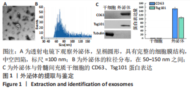
2.1 外泌体的提取与鉴定 透射电镜下观察可见提取的外泌体呈椭圆形,具有完整的细胞膜结构,中空凹陷(图1A);qNano纳米分析仪检测显示,外泌体的粒径在50-150 nm之间(图1B);Western blot检测显示,骨髓间充质干细胞中的CD63、Tsg101蛋白呈阴性表达,而外泌体高表达CD63、Tsg101蛋白(图1C)。"

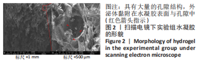
2.2 可注射水凝胶的形貌 扫描电镜下观察可见,实验组水凝胶具有大量的孔隙结构,外泌体黏附在水凝胶表面与孔隙中(图中红色箭头指示),见图2。"
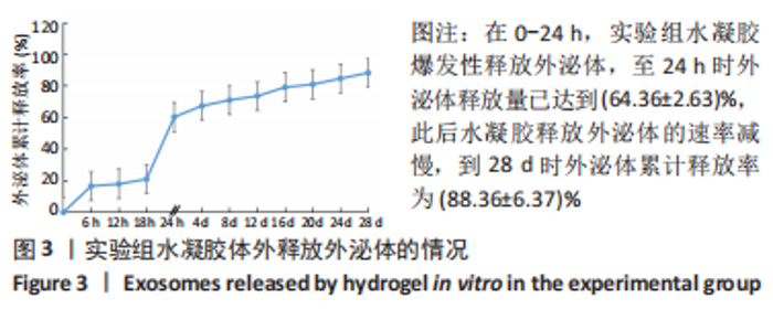
2.3 可注射水凝胶的缓释性能 在0-24 h,实验组水凝胶爆发性释放外泌体,至24 h时外泌体释放量已达到(64.36±2.63)%,此后水凝胶释放外泌体的速率减慢,到28 d时外泌体累计释放率为(88.36±6.37)%,见图3。"

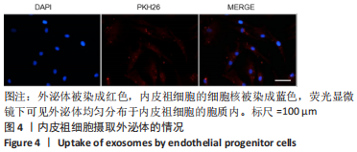
2.4 可注射水凝胶的体外实验 2.4.1 内皮祖细胞摄取实验结果 外泌体被染成红色,内皮祖细胞的细胞核被染成蓝色,荧光显微镜下可见外泌体均匀分布于内皮祖细胞的胞质内,说明内皮祖细胞可摄取外泌体见图4。"
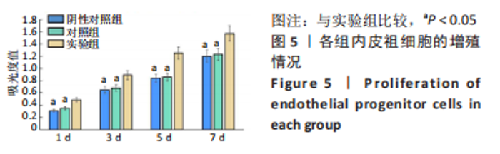
2.4.2 CCK-8实验检测结果 随着培养时间的延长,3组细胞呈现持续增殖的状态,与阴性对照组比较,实验组1,3,5,7 d的细胞增殖吸光度值增加(P < 0.05),对照组1,3,5,7 d的细胞增殖吸光度值未见明显增加(P > 0.05),见图5。"

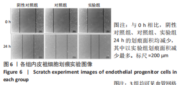
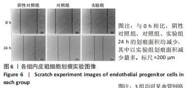
2.4.3 细胞划痕实验结果 与0 h相比,阴性对照组、对照组、实验组24 h的划痕面积均减少,其中以实验组划痕面积减少最多,见图6。通过统计分析得出,阴性对照组、对照组、实验组24 h细胞迁移面积依次为(25.05±3.16)%,(27.32±2.58)%,(54.72±8.53)%,实验组细胞迁移面积大于其他两组(P < 0.05),阴性对照组、对照组细胞迁移面积比较差异无显著性意义(P > 0.05)。"
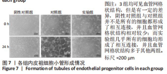
2.4.4 细胞成管实验结果 培养24 h后,3组均可见血管网格状结构,但是有一定的差异,阴性对照组与对照组并不是所有的细胞都形成了相互连接,并且血管网格状结构相对较少,而实验组几乎所有的细胞均形成了相互连接,并且血管网格状结构多于其他两组,见图7。"
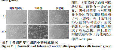
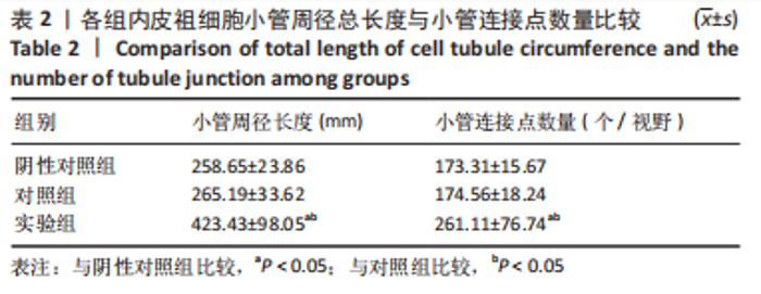
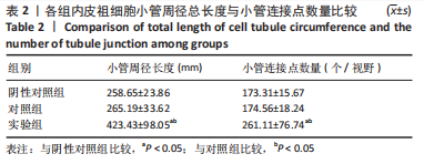
利用ImageJ软件统计3组的小管长度与小管连接点情况,具体结果见表2,显示实验组小管周径总长度大于其他两组(P < 0.05),小管连接点数量多于其他两组(P < 0.05)。"
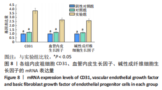
2.4.5 细胞成血管基因检测结果 RT-PCR检测结果显示,与阴性对照组比较,实验组CD31、血管内皮生长因子、碱性成纤维细胞生长因子的mRNA表达量显著增加(P < 0.05),对照组CD31、血管内皮生长因子、碱性成纤维细胞生长因子的mRNA表达量未见明显的变化(P > 0.05),见图8。"

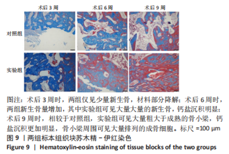
2.5 水凝胶的体内实验 2.5.1 实验动物数量分析 术后实验兔的精神状态与食欲较差,3 d后恢复正常;术后切口愈合良好。12只兔全部进入结果分析。 2.5.2 标本苏木精-伊红染色结果 两组标本苏木精-伊红染色结果见图9。对照组术后3周时仅见少量新生骨,稀疏的骨小梁周围可见线性排列的成骨细胞,材料部分降解,无炎性细胞浸润;术后6周时新生骨增加,成骨细胞排列于骨小梁周围,未见明显的钙盐沉积,无炎性细胞浸润;术后9周时,新生骨进一步增加,但是骨小梁仍然较稀疏。实验组术后3周时仅见少量新生骨,稀疏的骨小梁周围可见线性排列的成骨细胞,材料部分降解,无炎性细胞浸润;术后6周时可见大量的新生骨,钙盐沉积明显,无炎性细胞浸润;术后9周时可见大量粗大于成熟的骨小梁,钙盐沉积更加明显,骨小梁周围可见大量排列的成骨细胞。"

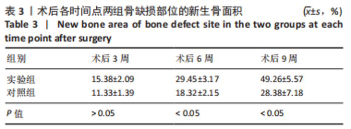
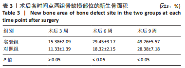
通过对骨缺损部位的成骨情况进行统计分析,结果显示:实验组术后6,9周的新生骨面积大于对照组(P < 0.05),见表3。"
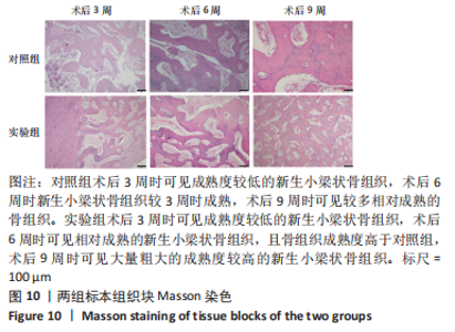
2.5.3 标本Masson染色结果 两组标本Masson染色结果见图10(呈现蓝绿色的为新生骨组织,红染越深代表骨质成熟度越高)。对照组术后3周时可见成熟度较低的新生小梁状骨组织,术后6周时新生小梁状骨组织较3周时成熟,术后9周时可见较多相对成熟的骨组织,但骨小梁排列稀疏。实验组术后3周时可见成熟度较低的新生小梁状骨组织,术后6周时可见相对成熟的新生小梁状骨组织,且骨组织成熟度高于对照组,术后9周时可见大量粗大的成熟度较高的新生小梁状骨组织。"

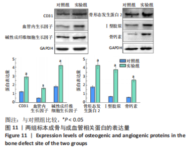
2.5.4 标本Western blot检测结果 与对照组比较,实验组术后9周骨缺损部位的CD31、血管内皮生长因子、碱性成纤维细胞生长因子、骨形态发生蛋白2、Ⅰ型胶原、骨钙素的蛋白表达量显著升高(P < 0.05),见图11。"


2.6 可注射水凝胶的生物相容性 由体外细胞实验及动物体内应用实验可知,负载外泌体的可注射葡萄糖酸内酯-海藻酸钠/β-磷酸三钙-聚乙二醇水凝胶具有良好的生物相容性。"

| [1] AGHALOO T, PI-ANFRUNS J, MOSHAVERINIA A, et al. The Effects of Systemic Diseases and Medications on Implant Osseointegration: A Systematic Review. Int J Oral Maxillofac Implants. 2019 Suppl;34: s35-s49. [2] OVERMANN AL, APARICIO C, RICHARDS JT, et al. Orthopaedic osseointegration: Implantology and future directions. J Orthop Res. 2020;38(7):1445-1454. [3] TAŞDEMIR U, KIRTAY M, KELEŞ A, et al. Autogenous Tooth Bone Graft and Simvastatin Combination Effect on Bone Healing. J Craniofac Surg. 2020;31(8):2350-2354. [4] TASDEMIR U, IYILIKÇI B, AKTÜRK MC, et al. The Effect of Autogenous Bone Graft Mixed With Recombinant Human Vascular Endothelial Growth Factor on Bone Regeneration. J Craniofac Surg. 2021;32(6):2233-2237. [5] BALDWIN P, LI DJ, AUSTON DA, et al. Autograft, Allograft, and Bone Graft Substitutes: Clinical Evidence and Indications for Use in the Setting of Orthopaedic Trauma Surgery. J Orthop Trauma. 2019;33(4):203-213. [6] SUHARDI JV, MORGAN DFA, MURATOGLU OK, et al. Radioprotection and cross-linking of allograft bone in the presence of vitamin E. J Biomed Mater Res B Appl Biomater. 2020;108(5):2354-2367. [7] CHEN MY, FANG JJ, LEE JN, et al. Supercritical Carbon Dioxide Decellularized Xenograft-3D CAD/CAM Carved Bone Matrix Personalized for Human Bone Defect Repair. Genes (Basel). 2022;13(5):755. [8] XIAO Y, GU Y, QIN L, et al. Injectable thermosensitive hydrogel-based drug delivery system for local cancer therapy. Colloids Surf B Biointerfaces. 2021;200:111581. [9] WANG Z, ZHANG Y, YIN Y, et al. High-Strength and Injectable Supramolecular Hydrogel Self-Assembled by Monomeric Nucleoside for Tooth-Extraction Wound Healing. Adv Mater. 2022;34(13): e2108300. [10] SEO JW, SHIN SR, LEE MY, et al. Injectable hydrogel derived from chitosan with tunable mechanical properties via hybrid-crosslinking system. Carbohydr Polym. 2021;251:117036. [11] BHATTACHARJEE M, ESCOBAR IVIRICO JL, KAN HM, et al. Injectable amnion hydrogel-mediated delivery of adipose-derived stem cells for osteoarthritis treatment. Proc Natl Acad Sci U S A. 2022;119(4): e2120968119. [12] ZHANG L, JIAO G, REN S, et al. Exosomes from bone marrow mesenchymal stem cells enhance fracture healing through the promotion of osteogenesis and angiogenesis in a rat model of nonunion. Stem Cell Res Ther. 2020;11(1):38. [13] YU L, SUI B, FAN W, et al. Exosomes derived from osteogenic tumor activate osteoclast differentiation and concurrently inhibit osteogenesis by transferring COL1A1-targeting miRNA-92a-1-5p. J Extracell Vesicles. 2021;10(3):e12056. [14] LI Z, WANG Y, LI S, et al. Exosomes Derived From M2 Macrophages Facilitate Osteogenesis and Reduce Adipogenesis of BMSCs. Front Endocrinol (Lausanne). 2021;12:680328. [15] 刘 印,刘琴,陈娇,等.可注射葡萄糖酸内酯-海藻酸钠/β-磷酸三钙-聚乙二醇复合水凝胶支架的性能评价[J].中国组织工程研究, 2022,26(27):4308-4313. [16] SEVERINO P, DA SILVA CF, ANDRADE LN, et al. Alginate Nanoparticles for Drug Delivery and Targeting. Curr Pharm Des. 2019;25(11):1312-1334. [17] JI J, CHEN G, LIU Z, et al. Preparation of PEG-modified wool keratin/sodium alginate porous scaffolds with elasticity recovery and good biocompatibility. J Biomed Mater Res B Appl Biomater. 2021;109(9): 1303-1312. [18] DING Z, XI W, JI M, et al. Developing a biodegradable tricalcium silicate/glucono-delta-lactone/calcium sulfate dihydrate composite cement with high preliminary mechanical property for bone filling. Mater Sci Eng C Mater Biol Appl. 2021;119:111621. [19] WU S, KAI Z, WANG D, et al. Allogenic chondrocyte/osteoblast-loaded β-tricalcium phosphate bioceramic scaffolds for articular cartilage defect treatment. Artif Cells Nanomed Biotechnol. 2019;47(1): 1570-1576. [20] VAHABZADEH S, BOSE S. Effects of Iron on Physical and Mechanical Properties, and Osteoblast Cell Interaction in β-Tricalcium Phosphate .Ann Biomed Eng. 2017;45(3):819-828. [21] Kardan T, Mohammadi R, TAGHAVIFAR S, et al. Polyethylene Glycol-Based Nanocerium Improves Healing Responses in Excisional and Incisional Wound Models in Rats. Int J Low Extrem Wounds. 2021;20(3):263-271. [22] MAZLOOM-JALALI A, SHARIATINIA Z, TAMAI IA, et al. Fabrication of chitosan-polyethylene glycol nanocomposite films containing ZIF-8 nanoparticles for application as wound dressing materials. Int J Biol Macromol. 2020;153:421-432. [23] SHI L, ZHANG J, ZHAO M, et al. Effects of polyethylene glycol on the surface of nanoparticles for targeted drug delivery. Nanoscale. 2021;13(24):10748-10764. [24] TAKEUCHI R, KATAGIRI W, ENDO S, et al. Exosomes from conditioned media of bone marrow-derived mesenchymal stem cells promote bone regeneration by enhancing angiogenesis. PLoS One. 2019;14(11): e0225472. [25] WU D, CHANG X, TIAN J, et al. Bone mesenchymal stem cells stimulation by magnetic nanoparticles and a static magnetic field: release of exosomal miR-1260a improves osteogenesis and angiogenesis. J Nanobiotechnology. 2021;19(1):209. [26] 管鹏飞,崔瑞文,王其友,等.负载骨髓干细胞来源外泌体的3D水凝胶通过调节免疫促进损伤软骨的修复[J].南方医科大学学报, 2022(4):528-537. [27] 周欣,王颖,张馨欣.间充质干细胞外泌体-温敏水凝胶的制备及其在表皮创伤修复中的应用[J].中国医药工业杂志,2021,52(9): 1199-1207. [28] AHN J, YOON MJ, HONG SH, et al. Three-dimensional microengineered vascularised endometrium-on-a-chip. Hum Reprod. 2021;36(10):2720-2731. [29] KAMAT P, FRUEH FS, MCLUCKIE M, et al. Adipose tissue and the vascularization of biomaterials: Stem cells, microvascular fragments and nanofat-a review. Cytotherapy. 2020;22(8):400-411. [30] LEI ZY, LI J, LIU T, et al. Autologous Vascularization: A Method to Enhance the Antibacterial Adhesion Properties of ePTFE. J Surg Res. 2019;236:352-358. [31] TIAN Y, ZHU P, LIU S, et al. IL-4-polarized BV2 microglia cells promote angiogenesis by secreting exosomes.Adv Clin Exp Med. 2019;28(4): 421-430. [32] YAN C, CHEN J, WANG C, et al. Milk exosomes-mediated miR-31-5p delivery accelerates diabetic wound healing through promoting angiogenesis.Drug Deliv. 2022;29(1):214-228. [33] QI X, ZHANG J, YUAN H, et al. Exosomes Secreted by Human-Induced Pluripotent Stem Cell-Derived Mesenchymal Stem Cells Repair Critical-Sized Bone Defects through Enhanced Angiogenesis and Osteogenesis in Osteoporotic Rats.Int J Biol Sci. 2016;12(7):836-849. [34] ZHA Y, LI Y, LIN T, et al. Progenitor cell-derived exosomes endowed with VEGF plasmids enhance osteogenic induction and vascular remodeling in large segmental bone defects. Theranostics. 2021;11(1):397-409. |
| [1] | Cao Sheng, Kong Lingwei, Xu Kun, Sun Zhijie. Effect of gelatin methacryloyl hydrogel loaded with salvianolic acid B on intervertebral disc degeneration [J]. Chinese Journal of Tissue Engineering Research, 2024, 28(3): 380-386. |
| [2] | Bi Yujie, Ma Dujun, Peng Liping, Zhou Ziqiong, Zhao Jing, Zhu Houjun, Zhong Qiuhui, Yang Yuxin. Strategy and significance of Chinese medicine combined with medical hydrogel for disease treatment [J]. Chinese Journal of Tissue Engineering Research, 2024, 28(3): 419-425. |
| [3] | Chen Pinrui, Pei Xibo, Xue Yiyuan. Function and advantages of magnetically responsive hydrogel in bone tissue engineering [J]. Chinese Journal of Tissue Engineering Research, 2024, 28(3): 452-457. |
| [4] | Long Zhirui, Huang Lei, Xiao Fang, Wang Lin, Wang Xiaobei. Characteristics of hydrogel microspheres in bone tissue engineering [J]. Chinese Journal of Tissue Engineering Research, 2024, 28(3): 472-478. |
| [5] | Xu Jing, Lyu Huixin, Bao Xin, Zhang Yi, Wang Yihan, Zhou Yanmin. Application of near infrared responsive hydrogels in tissue engineering [J]. Chinese Journal of Tissue Engineering Research, 2024, 28(3): 486-492. |
| [6] | Long Yi, Yang Jiaming, Ye Hua, Zhong Yanbiao, Wang Maoyuan. Extracellular vesicles in sarcopenic obesity: roles and mechanisms [J]. Chinese Journal of Tissue Engineering Research, 2024, 28(2): 315-320. |
| [7] | Wang Xianfeng, Wang Kun, Sun Han, Sun Xiaoliang, Yan Litao. Mechanism underlying exosomal lncRNA H19 derived from umbilical cord mesenchymal stem cells promotes cartilage injury repair [J]. Chinese Journal of Tissue Engineering Research, 2024, 28(1): 20-25. |
| [8] | Zheng Rongjiong, Deng Zerun, Han Dan, Sun Lihua. Mechanism underlying rat hepatocyte apoptosis regulated by exosomes derived from bone marrow mesenchymal stem cells [J]. Chinese Journal of Tissue Engineering Research, 2024, 28(1): 44-49. |
| [9] | Zheng Mingkui, Xue Chenhui, Guan Xiaoming, Ma Xun. Human umbilical cord mesenchymal stem cell-derived exosomes reduce the permeability of blood-spinal cord barrier after spinal cord injury [J]. Chinese Journal of Tissue Engineering Research, 2024, 28(1): 50-55. |
| [10] | Chen Guanting, Zhang Linqi, Li Qingru. Research hot spots and trends of exosomes in theranostic application for chronic kidney disease [J]. Chinese Journal of Tissue Engineering Research, 2024, 28(1): 86-92. |
| [11] | Huang Yongbin, Wang Tao, Lou Yuanyi, Pang Jingqun, Chen Guanghua. Application prospect of mesenchymal stem cells in promoting muscle tissue repair [J]. Chinese Journal of Tissue Engineering Research, 2024, 28(1): 107-112. |
| [12] | Nong Fuxiang, Jiang Zhixiong, Li Yinghao, Xu Wencong, Shi Zhilan, Luo Hui, Zhang Qinglang, Zhong Shuang, Tang Meiwen. Bone cement augmented proximal femoral nail antirotation for type A3.3 intertrochanteric femoral fracturalysis [J]. Chinese Journal of Tissue Engineering Research, 2023, 27(在线): 1-10. |
| [13] | Pan Zhongjie, Qin Zhihong, Zheng Tiejun, Ding Xiaofei, Liao Shijie. Targeting of non-coding RNAs in the pathogenesis of the osteonecrosis of the femoral head [J]. Chinese Journal of Tissue Engineering Research, 2023, 27(9): 1441-1447. |
| [14] | Liu Xiaolin, Mu Xinyue, Ma Ziyu, Liu Shutai, Wang Wenlong, Han Xiaoqian, Dong Zhiheng. Effect of hydrogel-loaded simvastatin microspheres on osteoblast proliferation and differentiation [J]. Chinese Journal of Tissue Engineering Research, 2023, 27(7): 998-1003. |
| [15] | Li Cheng, Zheng Guoshuang, Kuai Xiandong, Yu Weiting. Alginate scaffold in articular cartilage repair [J]. Chinese Journal of Tissue Engineering Research, 2023, 27(7): 1080-1088. |
| Viewed | ||||||
|
Full text |
|
|||||
|
Abstract |
|
|||||