Chinese Journal of Tissue Engineering Research ›› 2024, Vol. 28 ›› Issue (6): 891-899.doi: 10.12307/2023.710
Previous Articles Next Articles
Finite element analysis on correction effect of varus foot orthosis based on the three-point force principle
Ning Tianliang1, Wang Kun1, Wang Lingbiao2, Han Pengfei1
- 1School of Mechanical Engineering, Inner Mongolia University of Technology, Hohhot 010000, Inner Mongolia Autonomous Region, China; 2Rehabilitation Aids Center of Inner Mongolia Autonomous Region, Hohhot 010000, Inner Mongolia Autonomous Region, China
-
Received:2022-09-26Accepted:2022-11-16Online:2024-02-28Published:2023-07-12 -
Contact:Wang Kun, PhD, Associate professor, School of Mechanical Engineering, Inner Mongolia University of Technology, Hohhot 010000, Inner Mongolia Autonomous Region, China -
About author:Ning Tianliang, Master candidate, School of Mechanical Engineering, Inner Mongolia University of Technology, Hohhot 010000, Inner Mongolia Autonomous Region, China -
Supported by:Scientific Research Start-Up Fund Project of Inner Mongolia University of Technology in 2021, No. DC2200000931 (to WK); Basic Scientific Research Fund Project of Universities Directly under the Inner Mongolia Autonomous Region in 2022, No. JY20220026 (to WK)
CLC Number:
Cite this article
Ning Tianliang, Wang Kun, Wang Lingbiao, Han Pengfei. Finite element analysis on correction effect of varus foot orthosis based on the three-point force principle[J]. Chinese Journal of Tissue Engineering Research, 2024, 28(6): 891-899.
share this article
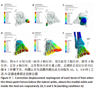
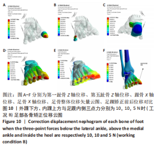
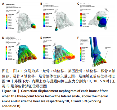
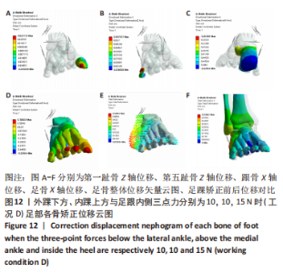
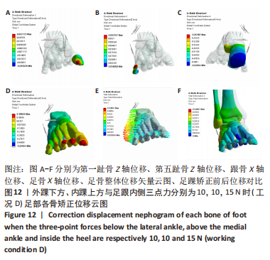
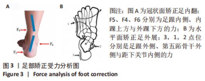
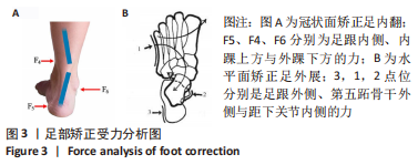
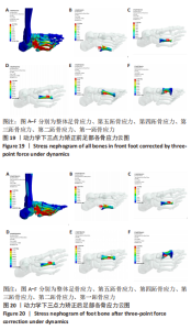
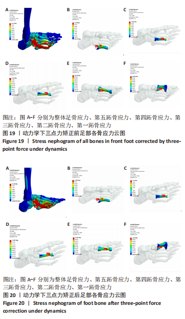
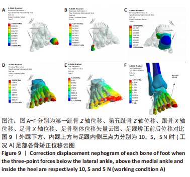
2.5 冠状面穿戴踝足矫形器三点力静力学分析 足部穿戴踝足矫形器时,三点力的施加主要依靠绑带力的调整,而软组织与矫形器的力作用是相互的,故在穿戴踝足矫形器时将三点力施加踝足矫形器内表面,作者在此部分进行2次分析。 (1)矫形效果的评定:对于前足与地面内翻角,以冠状面第一趾骨和第五趾骨上的两节点分别连线为A,与地面水平线B形成夹角,即前足内翻角,两节点分别设为(X2,Z2),(X1,Z1),依据公式(1)与公式(2)并以A线斜率变化评估前足内翻角。对于后足内翻角,以跟骨的中线相对于垂直方向的偏斜角度作为矫正评估标准[27]。 公式(1)中,K1为矫正前前足内翻角对应斜率,z2与z1分别为第一趾骨、第五趾骨Z轴节点坐标,x2与x1则为两节点X轴坐标。 公式(2)中,K2为矫正后前足内翻角对应斜率,横纵坐标与上述一致,Δ1与Δ2分别为第一趾骨与第五趾骨节点沿Z轴位移变化,Δ3与Δ4分别为两趾骨节点沿X轴位移变化量。 (2)将外踝下方、内踝上方与足跟内侧三点力分别加载10,5,5 N与10,10,5 N,分别记为工况A与B,进行初次分析。依据结果调整跟骨内侧与内踝上方矫正力大小,以对皮肤施力区产生适当压迫,计算结果得到的位移太小,为方便观察,截取变形放大后的云图[28],变形比例调整为自动比例。工况A与B计算结果见图9,10。 "
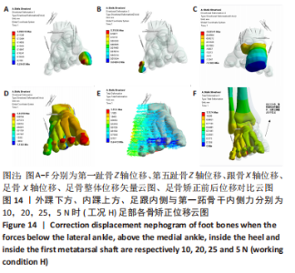
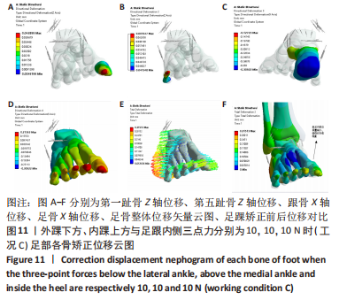
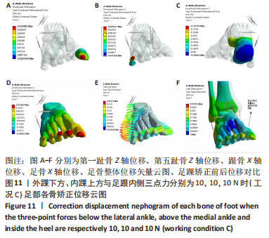
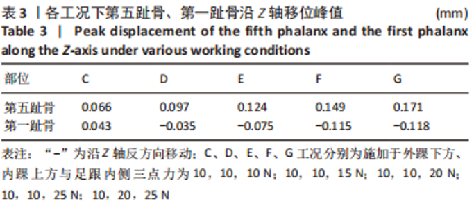
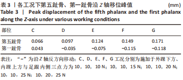
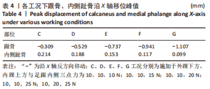
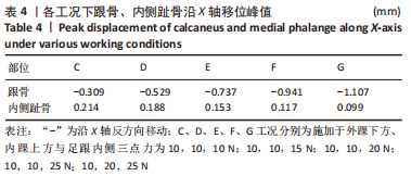
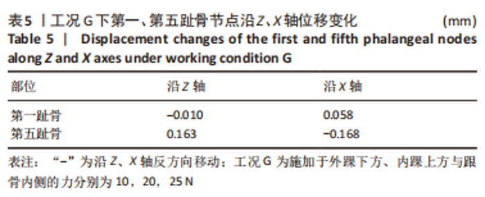
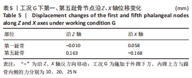
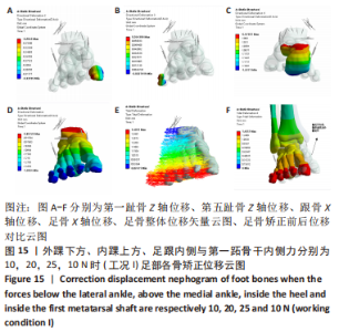
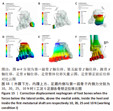
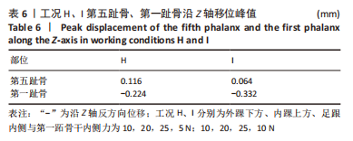
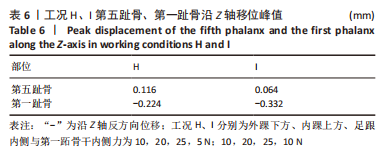
由图11和表3,4数据分析工况C,即在三点矫正载荷下,第一趾骨与第五趾骨均沿Z轴上移,且第五趾骨Z轴位移大于第一趾骨0.023 mm,依据公式(2),可推断趾骨区与地面偏斜角减小,跟骨沿X轴反向移动,即外翻移位0.309 mm,在冠状面对后足内翻有一定矫正效果,但内侧趾骨沿X轴最大移位0.214 mm,内侧趾骨区内收移位会进一步加剧患者前足部内翻与内收畸形。数据表明,单靠冠状面三力系统虽可恢复跟骨移位,但前足矫正效果并不理想。 由表3,4数据分析可知,工况C-F力系区别在于不断增加跟骨内侧的矫正力,跟骨内侧矫正力增大后,第五趾骨与第一趾骨Z轴垂直矫正位移会更明显,跟骨外翻移位量也随之增加,即矫正跟骨内翻位。对工况G依据公式(2)得: 不难看出,斜率值的减小可推断出趾骨区与地面间的前足内翻角得到矫正。 工况G在工况F基础上,固定跟骨与外踝力系,将内踝上方力系增至20 N,结果发现,跟骨矫正移位量更优于前4组工况,两趾骨间的垂直距离变化更大,且足部内侧趾骨沿X轴内翻、内收仅为0.099 mm,冠状面上,工况G力系显然更利于矫正足内翻。 2.6 冠状面、水平面穿戴踝足矫形器静力学分析 在进行冠状面三点力生物力学分析后,无论怎样调整三点力系大小,发现前足沿X轴内侧均有不同程度的内翻与内收移位,而内翻患者主要病症表现为内侧趾骨朝向内侧靠拢,内侧趾骨如再向内侧移位可能会进一步恶化患者前足内翻程度。为此添加水平面的三点力系,分析两平面三力系统联合作用下足部矫形效果,基于冠状面最优三点力即G工况下,在踝足矫形器内壁靠近第一跖骨干内侧分别施加5,10 N的矫正力,方向向外,分别记为工况H、I。计算结果见图14,15,表6-9。"

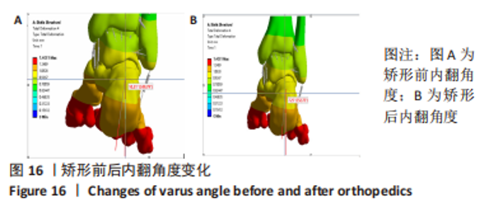
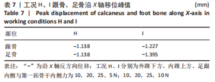
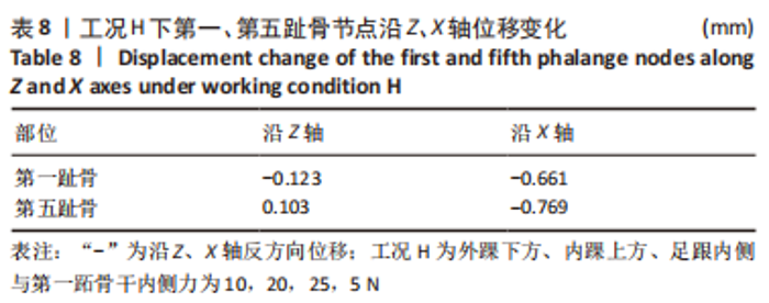
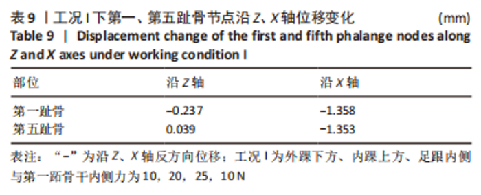
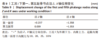
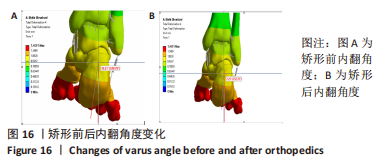
由数据分析可知,在水平面第一跖骨干施加5 N与10 N矫正力后,在X轴方向前足不再有内侧偏移,而是向阻抗内翻方向移位。第一跖骨干加载5 N时,足骨X轴位移聚集在跟骨,跟骨外翻移位1.138 mm,第一跖骨干加载10 N时,足骨X轴位移主要聚集在前足,前足外翻移位1.395 mm。依据公式(2)与表8,9可推出两工况下均有良好的矫正效果,但侧重点位不同,在矫形时应依据患者前足、后足内翻严重程度调整施力点位矫正力。 以工况I为例对矫正效果进行评估,依据公式(2)得: 斜率倾斜程度减缓推测出前足内翻角得到一定的矫正。结合跟骨的中线相对于垂直方向的偏斜角度作为矫正评估标准,在放大变形比例下的工况I后足位移角度测量结果见图16,在变形比例放大云图下跟骨内翻角由10.21°矫正至7.25°,内翻角改善28.9%。"
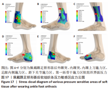
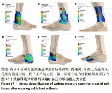
2.7 施加三点力后人体痛阈分析 患者在佩戴矫形器后,可能由于绑带的作用力长时间挤压足部而感到疼痛,文中对上述冠状面与水平面联合作用下矫正效果最佳且矫正力较大的三点力进行分析,即施加于跟骨内侧、内踝上方、外踝下方和第一跖骨干内侧的力分别为25,20,10,10 N。人体穿戴矫形器长时间能够承受而不被破坏的压力为2.5 N/cm2,文中施加在上述矫正部位的面积分别为10.93,13.80,5.39,5.15 cm2,依据公式(3),在矫正力F一定时,增加面积S,压强P即可减小。增加施力面积后,4个矫正区域长时间最大耐受压力分别为27.3,34.5,13.5,12.9 N。故文中施加的矫正力皆可满足人体皮肤疼痛阈值要求[29]。佩戴矫形器并施加冠状面与水平面三点力系后的软组织表面应力云图见图17。 P=F/S (3) 公式(3)中;P为压强,F为矫正力,S为施力面积。"
| [1] KIRIAKI T, CHRISTOS I, PETROS O, et al. Forefoot Varus (FV) in Children. International Journal of Engineering and Applied Sciences. 2016;3(4):102-104. [2] GÖK H, KÜÇÜKDEVECI A, ALTINKAYNAK H, et al. Effects of ankle-foot orthoses on hemiparetic gait. Clin Rehabil. 2003;17(2):137-139. [3] ZHANG Y, LIU HW, FU CH, et al. The biomechanical effect of acupuncture for poststroke cavovarus foot: study protocol for a randomized controlled pilot trial. Trials. 2016;17(1):146. [4] WARD AB. A literature review of the pathophysiology and onset of post-stroke spasticity. Eur J Neurol. 2012;19(1):21-27. [5] 胡金鲁,陈彦.中西医康复治疗中风后足内翻研究进展[J].中医药临床杂志, 2020,32(2):383-386. [6] MENG X, REN M, ZHUANG Y, et al. Application Experience and Patient Feedback Analysis of 3D Printed AFO with Different Materials: A Random Crossover Study. Biomed Res Int. 2021;2021:8493505. [7] KHAN SF, RADZMI I. Design and Analysis of Various Thermoplastic for Optimized Ankle Foot Orthosis. J Phys Conf Ser. 2021;2051(1): 012034. [8] 刘震,张盘德,容小川,等.脑卒中踝足矫形器的3D打印研究[J].中国康复医学杂志,2017,32(8):874-878. [9] KEMAL SH, ZIYA AY. Evaluation of various design concepts in passive ankle-foot orthoses using finite element analysis. Eng Sci Technol. 2021;24(6):1301-1307. [10] 王岩,张明.足踝矫形器及其生物力学研究进展[J].科技导报,2019,37(22): 60-68. [11] 路鹏程.3D打印踝足紧密接触型外固定支具的数字化设计、计算机仿真分析及临床应用[D].广州:南方医科大学,2021. [12] LIU X, YUE Y, WU X, et al. Analysis of transient response of the human foot based on the finite element method.Technol Health Care. 2022;30(1):79-92. [13] GU YD, REN XJ, LI JS, et al. Computer simulation of stress distribution in the metatarsals at different inversion landing angles using the finite element method. Int Orthop. 2010;34(5):669-676. [14] 彭志鑫,闫文刚,王坤,等.3D打印前臂外固定支具的有限元分析与结构优化设计[J].中国组织工程研究,2023,27(9):1340-1345. [15] CHO JR, LEE DY, AHN YJ. Finite element investigation of the biomechanical responses of human foot to the heel height and a rigid hemisphere cleat. J Mech Sci Technol. 2016;30(9):4269-4274. [16] 何晓宇,王朝强,周之平,等.三维有限元方法构建足部健康骨骼与常见疾病模型及生物力学分析[J].中国组织工程研究,2020,24(9):1410-1415. [17] DARWICH A, NAZHA H, NAZHA A, et al. Bio-Numerical Analysis of the Human Ankle-Foot Model Corresponding to Neutral Standing Condition. J Biomed Phys Eng. 2020;10(5):645-650. [18] WANG D, CAI P. Finite Element Analysis of the Expression of Plantar Pressure Distribution in the Injury of the Lateral Ligament of the Ankle. Nano Biomed Eng. 2019;11(3):290-296. [19] 弓太生,康路平,李姝,等.足部有限元模型静态分析及验证[J].中国皮革, 2022,51(4):61-66. [20] 刘清华.数字化人体足踝部三维有限元模型的建立及分析[D].广州:南方医科大学,2010. [21] KATHIRGAMANATHAN B, SILVA P, FERNANDEZ J. Implication of obesity on motion, posture and internal stress of the foot: an experimental and finite element analysis. Comput Methods Biomech Biomed Engin. 2019;22(1):47-54. [22] CHEUNG JT, ZHANG M, AN KN. Effect of Achilles tendon loading on plantar fascia tension in the standing foot. Clin Biomech (Bristol, Avon). 2006;21(2):194-203. [23] 胡航帆.基于有限元分析的3D打印踝关节矫形器的设计与制作[D].昆明:昆明医科大学,2019. [24] 赵辉三.假肢与矫形器学[M].北京:华夏出版社,2005. [25] 喻洪流.假肢矫形器原理与应用[M].南京:东南大学出版社,2011. [26] WANG CQ, HE XY, ZHANG ZN, et al. Three-Dimensional Finite Element Analysis and Biomechanical Analysis of Midfoot von Mises Stress Levels in Flatfoot, Clubfoot, and Lisfranc Joint Injury. Med Sci Monit. 2021;27:e931969. [27] 樊瑜波,张明.康复工程与生物力学[M].上海:上海交通大学出版社,2017. [28] 吴云成,许苑晶,鲁德志,等.增材制造脊柱侧弯矫形器治疗效果的有限元分析[J].医用生物力学,2022,37(3):492-497. [29] 王晓辉,王坤,胡志勇,等.假肢接受腔设计及界面应力的有限元分析[J].中国组织工程研究,2020,24(6):862-868. [30] BANGA HK, KALRA P, BELOKAR RM, et al. Customized design and additive manufacturing of kids’ ankle foot orthosis. Rapid Prototyping J. 2020;26(10): 1677-1685. [31] FU JC, CHEN YJ, LI CF, et al. The effect of three dimensional printing hinged ankle foot orthosis for equinovarus control in stroke patients. Clin Biomech (Bristol, Avon). 2022;94:105622. [32] SANAD DA. Moderate effect of ankle foot orthosis versus ground reaction ankle foot orthosis on balance in children with diplegic cerebral palsy. Prosthet Orthot Int. 2022;46(1):2-6. [33] FAHAD MOHANAD KADHIM, MUHAMMAD SAFA AL-DIN TAHIR. Design and Analysis of Three-Point Pressure for Varus Foot Deformity. J Biomim Biomater BI. 2020;4739: 1-11. [34] SHINICHI E, JUNG HC, TSUTAO K. Design/Manufacturing System for Composite Ankle Foot Orthosis. Key Eng Mater. 2014;627:261-264. [35] TELFER S, PALLARI J, MUNGUIA J, et al. Embracing additive manufacture: implications for foot and ankle orthosis design. BMC Musculoskelet Disord. 2012;13:84. [36] MASO AD, COSMI F. 3D-printed ankle-foot orthosis: a design method. Mater Today: Proc. 2019;12:252-261. [37] PANDEY R, SINGH R. Experimental Analysis of Various Materials on Custom-Fit Ankle Foot Orthosis. J Phys Conf Ser. 2021;1950(1):1-7. [38] 王坤,张振江,赵卫国.3D打印外固定支具参数化反求建模方法[J].机械设计,2021,38(5):121-126. |
| [1] | Kang Zhijie, Cao Zhenhua, Xu Yangyang, Zhang Yunfeng, Jin Feng, Su Baoke, Wang Lidong, Tong Ling, Liu Qinghua, Fang Yuan, Sha Lirong, Liang Liang, Li Mengmeng, Du Yifei, Lin Lin, Wang Haiyan, Li Xiaohe, Li Zhijun. Finite element model establishment and stress analysis of lumbar-sacral intervertebral disc in ankylosing spondylitis [J]. Chinese Journal of Tissue Engineering Research, 2024, 28(6): 840-846. |
| [2] | Zhang Min, Peng Jing, Zhang Qiang, Chen Dewang. Mechanical properties of L3/4 laminar decompression and intervertebral fusion in elderly osteoporosis patients analyzed by finite element method [J]. Chinese Journal of Tissue Engineering Research, 2024, 28(6): 847-851. |
| [3] | Xue Xiaofeng, Wei Yongkang, Qiao Xiaohong, Du Yuyong, Niu Jianjun, Ren Lixin, Yang Huifeng, Zhang Zhimin, Guo Yuan, Chen Weiyi. Finite element analysis of osteoporosis in proximal femur after cannulated screw fixation for femoral neck fracture [J]. Chinese Journal of Tissue Engineering Research, 2024, 28(6): 862-867. |
| [4] | Huang Peizhen, Dong Hang, Cai Qunbin, Lin Ziling, Huang Feng. Finite element analysis of anterograde and retrograde intramedullary nail for different areas of femoral shaft fractures [J]. Chinese Journal of Tissue Engineering Research, 2024, 28(6): 868-872. |
| [5] | Wang Mingming, Zhang Zhong, Sun Jianhua, Zhao Gang, Song Hua, Yan Huadong, Lyu Bin. Finite element analysis of three different minimally invasive fixation methods for distal tibial fractures with soft tissue injury [J]. Chinese Journal of Tissue Engineering Research, 2024, 28(6): 879-885. |
| [6] | Hou Zexin, Xu Benke, Dai Yuan, He Chuan, Zhang Chaoju, Li Xiaolin. Finite element analysis of the mechanism of dorsiflexion injury of wrist joint in elderly people after falls [J]. Chinese Journal of Tissue Engineering Research, 2024, 28(6): 886-890. |
| [7] | Zhang Lili, Zhang Xinglai, Zhang Jie, Zheng Jiejiao. Effect of visual feedback on landing biomechanics in chronic ankle instability patients [J]. Chinese Journal of Tissue Engineering Research, 2024, 28(6): 900-904. |
| [8] | Wang Qiang, Li Shiyun, Xiong Ying, Li Tiantian. Biomechanical changes of the cervical spine in internal fixation with different anterior cervical interbody fusion systems [J]. Chinese Journal of Tissue Engineering Research, 2024, 28(6): 821-826. |
| [9] | Wei Yuanbiao, Lin Zhan, Chen Yanmei, Yang Tenghui, Zhao Xiao, Chen Yangsheng, Zhou Yanhui, Yang Minchao, Huang Feiqi. Finite element analysis of effects of sagittal cervical manipulation on intervertebral disc and facet joints [J]. Chinese Journal of Tissue Engineering Research, 2024, 28(6): 827-832. |
| [10] | Zhang Rui, Wang Kun, Shen Zicong, Mao Lu, Wu Xiaotao. Effects of endoscopic foraminoplasty and laminoplasty on biomechanical properties of intervertebral disc and isthmus [J]. Chinese Journal of Tissue Engineering Research, 2024, 28(6): 833-839. |
| [11] | Zhang Qianlong, Maihemuti•Yakufu, Song Chenhui, Liu Xiuxin, Ren Zheng, Liu Yuzhe, Muyashaer•Abudushalamu, Sajidan•Aikebaier, Ran Jian. Finite element analysis of the effect of the distribution position and content of bone cement on the stress and displacement of reverse femoral intertrochanteric fracture [J]. Chinese Journal of Tissue Engineering Research, 2024, 28(3): 336-340. |
| [12] | Xu Xiaodong, Zhou Jiping, Zhang Qi, Feng Chen, Zhu Mianshun, Shi Hongcan. 3D printing process of gelatin/oxidized nanocellulose skin scaffold with high elastic modulus and high porosity [J]. Chinese Journal of Tissue Engineering Research, 2024, 28(3): 398-403. |
| [13] | Chen Yuanyuan, Wang Wei, Zhao Lu, Annikaer·Aniwaer, Nijati·Turson. Finite element analysis of the influence of scaffold materials on the fixed restoration of edentulous maxillary implants under two designs [J]. Chinese Journal of Tissue Engineering Research, 2024, 28(3): 411-418. |
| [14] | Yang Jie, Hu Haolei, Li Shuo, Yue Wei, Xu Tao, Li Yi. Application of bio-inks for 3D printing in tissue repair and regenerative medicine [J]. Chinese Journal of Tissue Engineering Research, 2024, 28(3): 445-451. |
| [15] | Kong Xiangyu, Wang Xing, Pei Zhiwei, Chang Jiale, Li Siqin, Hao Ting, He Wanxiong, Zhang Baoxin, Jia Yanfei. Biological scaffold materials and printing technology for repairing bone defects [J]. Chinese Journal of Tissue Engineering Research, 2024, 28(3): 479-485. |
| Viewed | ||||||||||||||||||||||||||||||||||||||||||||||||||
|
Full text 324
|
|
|||||||||||||||||||||||||||||||||||||||||||||||||
|
Abstract 549
|
|
|||||||||||||||||||||||||||||||||||||||||||||||||
