Chinese Journal of Tissue Engineering Research ›› 2024, Vol. 28 ›› Issue (22): 3509-3516.doi: 10.12307/2024.488
Previous Articles Next Articles
Repair of infected osteochondral defect with sustained release vancomycin three-dimensional scaffold in rabbits
Li Xingyu1, Zhou Jie1, Li Shasha1, Zhang Tianxi1, Guo Guoning1, Yu Anyong1, Deng Jiang2, Ye Peng1
- 1Emergency Department of Affiliated Hospital of Zunyi Medical University (First Aid Trauma Ward), Zunyi 563003, Guizhou Province, China; 2Department of Orthopedics, Zunyi First People’s Hospital, Zunyi 563003, Guizhou Province, China
-
Received:2023-04-04Accepted:2023-09-28Online:2024-08-08Published:2024-01-20 -
Contact:Ye Peng, MD, Associate chief physician, Emergency Department of Affiliated Hospital of Zunyi Medical University (First Aid Trauma Ward), Zunyi 563003, Guizhou Province, China -
About author:Li Xingyu, Master, Emergency Department of Affiliated Hospital of Zunyi Medical University (First Aid Trauma Ward), Zunyi 563003, Guizhou Province, China -
Supported by:National Natural Science Foundation of China (Regional Science Foundation), No. H0606 (to DJ); Guizhou Science and Technology Plan Project, No. [2021]074 (to YP); Guizhou Science and Technology Plan Project, No. [2022]034 (to GGN); Outstanding Young Talents Training Program of Affiliated Hospital of Zunyi Medical University, No. RC220220911 (to YP)
CLC Number:
Cite this article
Li Xingyu, Zhou Jie, Li Shasha, Zhang Tianxi, Guo Guoning, Yu Anyong, Deng Jiang, Ye Peng. Repair of infected osteochondral defect with sustained release vancomycin three-dimensional scaffold in rabbits[J]. Chinese Journal of Tissue Engineering Research, 2024, 28(22): 3509-3516.
share this article
Add to citation manager EndNote|Reference Manager|ProCite|BibTeX|RefWorks
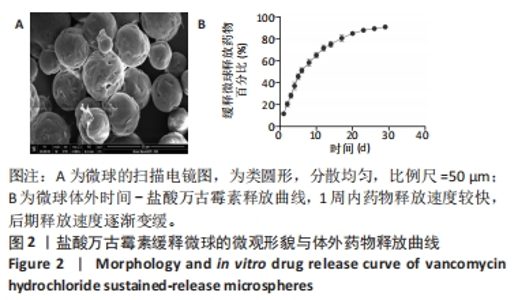
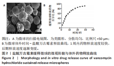
2.1 VANCO缓释微球表征结果 大体可见VANCO缓释微球呈白色粉末,扫描电镜下为类圆形,分散均匀(图2A),微球粒径平均为(21.45±6.09) μm,载药率为(23.86±1.35)%,包封率为 (46.21±11.78)%。 体外浸泡于PBS中第1天,微球释放VANCO的量为(11.62±1.85)%,第8天为(58.29±3.39)%,1周内药物释放速度较快,后期释放速度逐渐变缓(图2B)。"
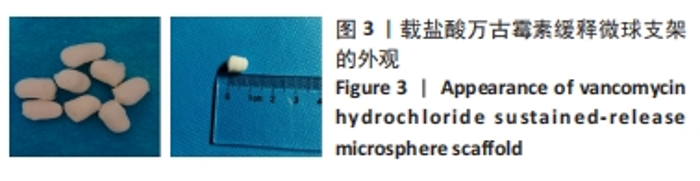
2.2 载VANCO缓释微球支架物理性能表征结果 2.2.1 支架大体形态 VANCO缓释微球丝素蛋白-壳聚糖-纳米羟基磷灰石支架呈白色,形似子弹,无异味,质量轻,支架高约7 mm,直径4.0-5.0 mm,具有较好的弹性和韧性见图3。"

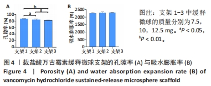
2.2.2 支架的孔隙率、吸水膨胀率及热水溶失率 支架1-3的孔隙率分别为(87.79±1.03)%,(84.37±3.92)%,(82.14±1.36)%,3组间两两比较差异有显著性意义(P < 0.05,P < 0.01)(图4A)。以上支架孔隙率均可满足实验细胞生长的空间需求。 支架1-3的吸水膨胀率分别为(2 267±38)%,(2 292±57)%,(2 302±14)%,3组间比较差异无显著性意义(P > 0.05)(图4B)。以上各组支架吸水膨胀率均达到骨软骨组织工程支架要求。"

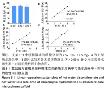
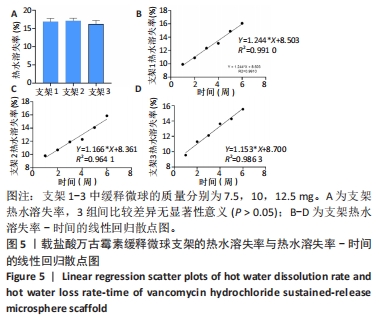
支架1-3的热水溶失率分别为(16.86±0.89)%,(17.11±0.69)%,(16.24±0.96)%,3组间比较差异无显著性意义(P > 0.05)(图5A)。线性回归分析结果显示,支架1-3的R2分别是0.991 0,0.964 1,0.986 3,根据回归曲线计算支架1-3完全被溶蚀的时间分别是56.15,59.47,65.21周(图5B-D),各组支架热水溶失率结均达到实验支架要求标准。"
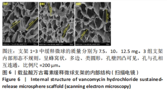
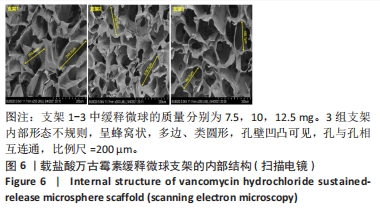
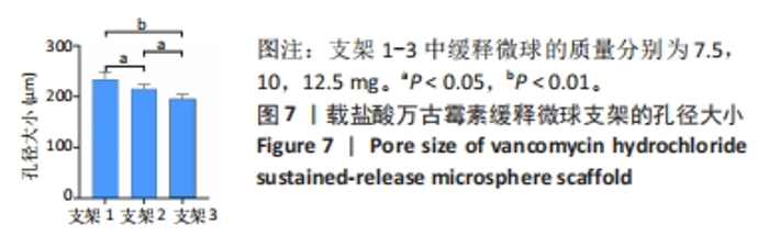
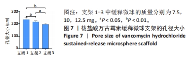
2.2.3 支架形态及孔径大小 扫描电镜下见支架内部形态不规则,呈蜂窝状,多边、类圆形,孔壁凹凸可见,孔与孔相互连通,内部可见VANCO缓释微球和纳米羟基磷灰石沉着(图6)。支架1-3的平均孔径分别为(233.8±15.0),(215.3±8.3),(196.0±8.2) μm,3组间两两比较差异有显著性意义(P < 0.05,P < 0.01),见图7。各组支架孔径大小均达到实验要求(100-300 μm)。"
"

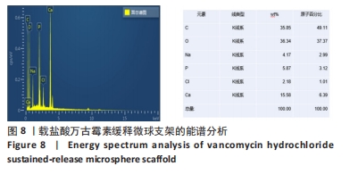
2.2.4 支架的能谱分析及离子分布 载盐酸万古霉素缓释微球支架的组成成分中以 Ca、O和 P元素为主体(图8),研究表明,在该支架表面存在羟基磷灰石沉淀,可以为骨组织的矿化形成提供原料,同时还具备了很好的生物活性,均符合骨软骨组织工程支架要求。"
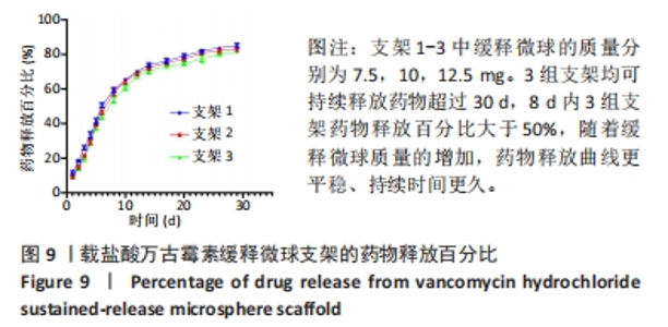
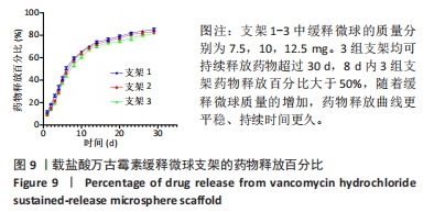
2.2.5 支架中VANCO累积缓释率 根据各组支架缓释曲线可知VANCO持续释放超过30 d,所有支架8 d内ANCO释放百分比大于50%,随着缓释微球质量的增加,VANCO释放曲线更平稳、持续时间更久(图9)。"

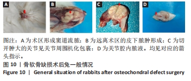
尽管增加载药微球的量可以提高支架的药物释放效率,但是太多的载药微球也会导致支架的孔隙率和孔径减小,只有加入适当的载药微球才能为细胞的生长、增殖、分化等提供更好的空间和环境。综合以上结果,在支架中加入10 mg VANCO缓释微球最符合细胞生长需求,故选择支架2完成动物实验部分。 2.3 兔软骨缺损修复效果 2.3.1 实验动物数量分析 45只兔全部进入结果分析。 2.3.2 术后兔一般情况 3组兔术后第1-3天精神饮食差,饮食自如。在术后1个月内,空白组有12只兔术区肿胀,红肿面积大,周围组织坏死,见窦道形成,并伴随大量脓液的排出周围形成窦道并流脓;对照组有10只兔术区红肿明显,周围形成窦道并流脓;实验组有2只兔膝关节周围形成窦道并流脓。1个月以后,空白组、对照组兔术后术区及关节处肿胀明显,关节畸形,步态不稳,活动明显受限,切开肿大的关节见机化包裹,暴露关节腔见黄色脓性关节液,部分兔在远离术区的颈背部可见皮下脓肿;实验组兔术后关节活动及饮食自如,部分兔关节活动稍受限,暴露关节腔见关节液透明。见图10。"
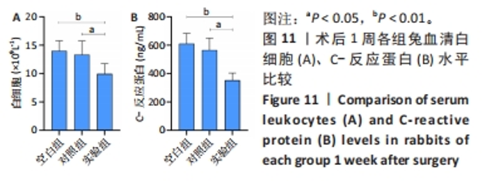
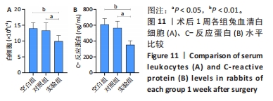
2.3.3 各组兔血清炎性指标检测 空白组、对照组、实验组兔血清白细胞水平分别为(14.02±1.78)×109 L-1,(13.33±2.42)×109 L-1,(9.98±1.78)×109 L-1,实验组白细胞水平低于空白组、对照组(P < 0.05,P < 0.01),见图11A。 空白组、对照组、实验组兔血清C-反应蛋白质量浓度分别为(610.7±73.01),(566.20±84.31),(354.20±50.17) ng/mL,实验组C-反应蛋白质量浓度低于空白组、对照组(P < 0.05,P < 0.01),见图11B。"

2.3.4 各组兔关节软骨大体观察及ICRS软骨修复评分 空白组、对照组术区均有明显粘连,其中一些液体为黄色,轻微红肿;实验组2只兔术区有轻微粘连,其余兔关节和关节囊均无异常,无明显的滑膜炎和骨赘。空白组术后4-12周缺损部位没有愈合,缺损部位和正常软骨之间的界限清楚。对照组术后4周缺损区无明显修复,8周后术区已被局部软组织覆盖,并出现了局部凹陷;术后12周,术区周边被透明软骨和纤维组织包裹,但中心凹痕依然清晰可见且边缘清晰。实验组术后4周修复区域仍呈明显的凹形,并有一层新的纤维层包裹在其表面;术后8周,术区内充盈纤维及透明软骨;第12周,术区被新的软骨细胞替代,表面平整、光滑,缺损处与关节面边界模糊。各组兔关节软骨大体观察见图12。"


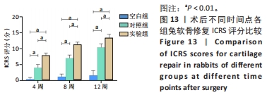
随着术后时间的延长,各组兔软骨修复ICRS评分增加,相同时间点下,实验组ICRS评分高于空白组、对照组(P < 0.01),对照组ICRS评分高于空白组(P < 0.01),见图13。"
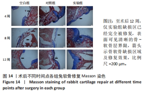
2.3.5 各组兔骨软骨修复组织学观察及Wakitani病理评分 Masson染色:空白组至术后12周缺损区仍然没有明显的软骨化现象;对照组至术后12周时缺损区已由新的组织逐步充满,但软骨修复效果差,表面欠光滑,缺损区组织与周围软骨分界明显;实验组术后4-12周时缺损区被新的组织填满,表面平滑,颜色从浅染到暗蓝色,术后12周缺损区已经完全被修复,表面可见清晰的骨-软骨层界限,见图14。"

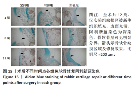
阿利新蓝染色:空白组至术后12周缺损区仍可见明显软骨缺失,阿利新蓝染色阴性;对照组术后4-12周缺损区逐渐被新生组织填充,12周时可见关节面光滑,缺损区与周围软骨分界明显,阿利新蓝染色较淡;实验组术后4-12周缺损区被新生组织填充,表面光滑,阿利新蓝染色由淡染逐渐变为深染色,骨软骨层可见明显分界,见图15。"


苏木精-伊红染色:空白组与对照组术后各时间点可见大量炎性细胞浸润,实验组可见相对较少的炎性细胞浸润。空白组术后4-12周缺损区均未见明显组织再生;对照组术后4周缺损区支架表面未见明显纤维组织,术后8周缺损区软骨层纤维组织增生,软骨下骨层出现部分骨小梁结构,分界不清,至12周缺损区修复组织见少量纤维软骨,软骨下骨未完全重建,骨软骨层分界不清晰;实验组术后4周缺损区可见纤维软骨修复,8周缺损区透明软骨增生明显且可见较多骨小梁结构,至12周软骨层与软骨下骨层修复比较完整、分界明显,可见潮线,见图16。"

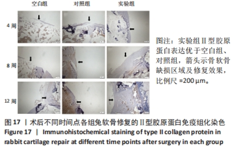
免疫组化染色:空白组至术后12周仍未见明显Ⅱ型胶原蛋白表达;对照组至术后12周修复组织中可见Ⅱ型胶原蛋白少量表达;实验组术后4周修复区见有少量Ⅱ型胶原蛋白均有表达,术后8,12周修复组织中Ⅱ型胶原蛋白表达增加,骨软骨分界明显,见图17。"

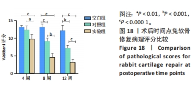
Wakitani评分:随着术后时间的延长,对照组、实验组Wakitani评分降低,相同时间点下,实验组Wakitani评分低于空白组、对照组(P < 0.01,P < 0.001,P < 0.000 1),见图18。"

| [1] DING H, CHENG Y, NIU X, et al. Application of electrospun nanofibers in bone, cartilage and osteochondral tissue engineering. J Biomater Sci Polym Ed. 2021;32(4):536-561. [2] VAISH A, SHANMUGASUNDARAM S, KIM SA, et al. Biological reconstruction of the joint: Concepts of articular cartilage regeneration and their scientific basis. J Clin Orthop Trauma. 2022;24:101718. [3] LIU Y, LI M, YIN Z, et al. SUMO-modified bone marrow mesenchymal stem cells promoted the repair of articular cartilage in rats. Cell Biol Int. 2020;44(2):560-568. [4] LI H, CAO X, WANG J, et al. [Redintegration of articular surface and alignment with tibia type III Pilon fracture]. Zhongguo Xiu Fu Chong Jian Wai Ke Za Zhi. 2012;26(8):926-929. [5] LEE CH, KWON Y, JUNG IY, et al. Effect of the Articular Surface Incongruency on Surgical Outcome of the Distal Radius Fracture. Biomed Res Int. 2022;2022:8357675. [6] COOK JL, RUCINSKI K, CRECELIUS CR, et al. Initial Outcomes After Unicompartmental Tibiofemoral Bipolar Osteochondral and Meniscal Allograft Transplantation in the Knee Using MOPS-Preserved Fresh (Viable) Tissues. Am J Sports Med. 2023;51(3):596-604. [7] FLETCHER AN, JOHNSON LG, EASLEY ME, et al. Midterm Prospective Evaluation of Structural Allograft Transplantation for Osteochondral Lesions of the Talar Shoulder. Foot Ankle Int. 2022;43(7):899-912. [8] DHILLON J, DECILVEO AP, KRAEUTLER MJ, et al. Third-Generation Autologous Chondrocyte Implantation (Cells Cultured Within Collagen Membrane) Is Superior to Microfracture for Focal Chondral Defects of the Knee Joint: Systematic Review and Meta-analysis. Arthroscopy. 2022;38(8):2579-2586. [9] SCHUETTE HB, KRAEUTLER MJ, SCHROCK JB, et al. Primary Autologous Chondrocyte Implantation of the Knee Versus Autologous Chondrocyte Implantation After Failed Marrow Stimulation: A Systematic Review. Am J Sports Med. 2021;49(9):2536-2541. [10] LEE H, KIM HT, JO S. Femoral head cartilage reconstruction using autologous osteochondral mosaicplasty: A case report. Medicine (Baltimore). 2023;102(6):e32913. [11] YU JR, NAVARRO J, COBURN JC, et al. Current and Future Perspectives on Skin Tissue Engineering: Key Features of Biomedical Research, Translational Assessment, and Clinical Application. Adv Healthc Mater. 2019;8(5):e1801471. [12] YANG Z, WANG B, LIU W, et al. In situ self-assembled organoid for osteochondral tissue regeneration with dual functional units. Bioact Mater. 2023;27:200-215. [13] SHI Y, HAN X, PAN S, et al. Gold Nanomaterials and Bone/Cartilage Tissue Engineering: Biomedical Applications and Molecular Mechanisms. Front Chem. 2021;9:724188. [14] OMID H, ABDOLLAHI S, BONAKDAR S, et al. Biomimetic vascular tissue engineering by decellularized scaffold and concurrent cyclic tensile and shear stresses. J Mater Sci Mater Med. 2023;34(3):12. [15] YU H, FENG M, MAO G, et al. Implementation of Photosensitive, Injectable, Interpenetrating, and Kartogenin-Modified GELMA/PEDGA Biomimetic Scaffolds to Restore Cartilage Integrity in a Full-Thickness Osteochondral Defect Model. ACS Biomater Sci Eng. 2022;8(10):4474-4485. [16] HE T, LI B, COLOMBANI T, et al. Hyaluronic Acid-Based Shape-Memory Cryogel Scaffolds for Focal Cartilage Defect Repair. Tissue Eng Part A. 2021;27(11-12):748-760. [17] WANG T, XU W, ZHAO X, et al. Repair of osteochondral defects mediated by double-layer scaffolds with natural osteochondral-biomimetic microenvironment and interface. Mater Today Bio. 2022;14:100234. [18] KIM JW, LEE KJ. Development of a Single-nucleotide Polymorphism Genotyping Assay for the Rapid Detection of Vancomycin-intermediate Resistance in Staphylococcus aureus Epidemic Lineage ST5. Ann Lab Med. 2023;43(4):355-363. [19] LANDERSDORFER CB, LEE WL, NATION RL, et al. Penetration of Vancomycin into Noninfected Bone in Patients Undergoing Total Joint Arthroplasty Evaluated by a Minimal Physiologically Based Population Pharmacokinetic Modeling Approach. Mol Pharm. 2023;20(3):1509-1518. [20] MOSTAFA AA, EL-SAYED M, MAHMOUD AA, et al. Bioactive/Natural Polymeric Scaffolds Loaded with Ciprofloxacin for Treatment of Osteomyelitis. AAPS PharmSciTech. 2017;18(4):1056-1069. [21] WANG G, CUI Y, LIU H, et al. Antibacterial peptides-loaded bioactive materials for the treatment of bone infection. Colloids Surf B Biointerfaces. 2023;225:113255. [22] KHAYAT KASHANI HR. Answer to the letter to the editor of M. Kataria et al. concerning “Local vancomycin therapy to reduce surgical site infection in adult spine surgery: a randomized prospective study” by Salimi S, et al. (Eur Spine J [2022];31:454-460). Eur Spine J. 2023;32(3):1090. [23] LI S, SHI X, XU B, et al. In vitro drug release and antibacterial activity evaluation of silk fibroin coated vancomycin hydrochloride loaded poly (lactic-co-glycolic acid) (PLGA) sustained release microspheres. J Biomater Appl. 2022;36(9):1676-1688. [24] LI J, LI K, DU Y, et al. Dual-Nozzle 3D Printed Nano-Hydroxyapatite Scaffold Loaded with Vancomycin Sustained-Release Microspheres for Enhancing Bone Regeneration. Int J Nanomedicine. 2023;18:307-322. [25] 肖红利,邓江,韩子冀,等.渐进性梯度孔径骨软骨支架的制备及细胞相容性研究[J].中华老年医学杂志,2020,39(4):456-461. [26] SALTZMAN BM, RIBOH JC. Subchondral Bone and the Osteochondral Unit: Basic Science and Clinical Implications in Sports Medicine. Sports Health. 2018;10(5):412-418. [27] ZHANG Y, YU J, REN K, et al. Thermosensitive Hydrogels as Scaffolds for Cartilage Tissue Engineering. Biomacromolecules. 2019;20(4):1478-1492. [28] DONELL S. Subchondral bone remodelling in osteoarthritis. EFORT Open Rev. 2019;4(6):221-229. [29] LI B, DING T, CHEN H, et al. CircStrn3 targeting microRNA-9-5p is involved in the regulation of cartilage degeneration and subchondral bone remodelling in osteoarthritis. Bone Joint Res. 2023;12(1):33-45. [30] JACOB G, SHIMOMURA K, NAKAMURA N. Osteochondral Injury, Management and Tissue Engineering Approaches. Front Cell Dev Biol. 2020;8:580868. [31] FU L, YANG Z, GAO C, et al. Advances and prospects in biomimetic multilayered scaffolds for articular cartilage regeneration. Regen Biomater. 2020;7(6):527-542. [32] LE TM, VU NB, HUYNH PD, et al. Treatment of Osteochondral Femoral Head Defect by Human Umbilical Cord Mesenchymal Stem Cell Sheet Transplantation: An Experimental Study in Rats. Adv Exp Med Biol. 2021. doi: 10.1007/5584_2021_671. [33] VAN TUIJN IM, EMANUEL KS, VAN HUGTEN P, et al. Prognostic Factors for the Clinical Outcome after Microfracture Treatment of Chondral and Osteochondral Defects in the Knee Joint: A Systematic Review. Cartilage. 2023;14(1):5-16. [34] OTLANS P, LATTERMANN C, SHERMAN SL, et al. Cartilage Disease of the Patellofemoral Joint: Realignment, Restoration, Replacement. Instr Course Lect. 2021;70:289-308. [35] MICHALIK R, PAUER T, BRILL N, et al. Quantitative articular cartilage sub-surface defect assessment using optical coherence tomography: An in-vitro study. Ann Anat. 2019;221:125-134. [36] HIGA K, KITAMURA N, GOTO K, et al. Effects of osteochondral defect size on cartilage regeneration using a double-network hydrogel. BMC Musculoskelet Disord. 2017;18(1):210. [37] LUO M, CHEN M, BAI J, et al. A bionic composite hydrogel with dual regulatory functions for the osteochondral repair. Colloids Surf B Biointerfaces. 2022;219:112821. [38] ALOMAR AZ, SOMILY AM, ALRAIYES TM, et al. Quantification Analysis of the Intraoperative Bacterial Contamination Rate and Level in Osteochondral Autografts. Am J Sports Med. 2016; 44(3):761-766. [39] WEI J, XIA X, XIAO S, et al. Sequential Dual-Biofactor Release from the Scaffold of Mesoporous HA Microspheres and PLGA Matrix for Boosting Endogenous Bone Regeneration. Adv Healthc Mater. 2023;12(20):e2300624. [40] WANG W, SONG Y, TIAN Y, et al. TCPP/MgO-loaded PLGA microspheres combining photodynamic antibacterial therapy with PBM-assisted fibroblast activation to treat periodontitis. Biomater Sci. 2023;11(8):2828-2844. [41] GANDOMKARZADEH M, MOGHIMI HR, MAHBOUBI A. Evaluation of the Effect of Ciprofloxacin and Vancomycin on Mechanical Properties of PMMA Cement; a Preliminary Study on Molecular Weight. Sci Rep. 2020;10(1):3981. [42] LIU N, HUANG S, GUO F, et al. Calcium phosphate cement with icariin-loaded gelatin microspheres as a local drug delivery system for bone regeneration. Biomed Eng Online. 2022;21(1):89. [43] GHAREH SM, SHABANI RN, BEHROUZI M, et al. Engineered PLGA microspheres for extended-release of naltrexone: in vitro, in vivo, and IVIVR. Pharm Dev Technol. 2023;28(2):190-199. [44] 刘华蔚.壳聚糖导管复合NGF缓释微球修复兔面神经缺损的实验研究[D].北京:解放军医学院,军医进修学院,解放军总医院,2011. [45] FAN L, TENG W, HE J, et al. Value of 3D Printed PLGA Scaffolds for Cartilage Defects in Terms of Repair. Evid Based Complement Alternat Med. 2022;2022:3561430. [46] LUO M, CHEN M, BAI J, et al. A bionic composite hydrogel with dual regulatory functions for the osteochondral repair. Colloids Surf B Biointerfaces. 2022;219:112821. [47] 高泽,石志良,李锋,等.材料和孔隙率对可降解支架内骨形成的影响[J].医用生物力学, 2021,36(4):582-588. [48] PRANANINGRUM W, NAITO Y, GALLI S, et al. Bone ingrowth of various porous titanium scaffolds produced by a moldless and space holder technique: an in vivo study in rabbits. Biomed Mater. 2016;11(1):15012. [49] MOURA CS, SILVA JC, FARIA S, et al. Chondrogenic differentiation of mesenchymal stem/stromal cells on 3D porous poly (epsilon-caprolactone) scaffolds: Effects of material alkaline treatment and chondroitin sulfate supplementation. J Biosci Bioeng. 2020;129(6):756-764. [50] MARTINS EA, MICHELACCI YM, BACCARIN RY, et al. Evaluation of chitosan-GP hydrogel biocompatibility in osteochondral defects: an experimental approach. BMC Vet Res. 2014;10:197. [51] ZHU D, WANG H, TRINH P, et al. Elastin-like protein-hyaluronic acid (ELP-HA) hydrogels with decoupled mechanical and biochemical cues for cartilage regeneration. Biomaterials. 2017; 127:132-140. [52] 向柄彦,李鹏,柏帆,等.载万古霉素缓释微球纳米羟基磷灰石/壳聚糖支架联合自体红骨髓可修复慢性骨髓炎兔的骨缺损[J].中国组织工程研究,2019,23(6):843-848. [53] LI J, LI K, DU Y, et al. Dual-Nozzle 3D Printed Nano-Hydroxyapatite Scaffold Loaded with Vancomycin Sustained-Release Microspheres for Enhancing Bone Regeneration. Int J Nanomedicine. 2023;18:307-322. [54] 马涛,尚北城,陈庆华,等.万古霉素阳离子脂质体复合纳米羟基磷灰石/壳聚糖/魔芋葡苷聚糖支架的制备与降解及药物的体外释放研究[J].中华创伤骨科杂志,2014,16(10):891-897. [55] 冯建波,李陈诚,刘金月,等.金黄色葡萄球菌生物膜克氏针置入建立创伤性大鼠骨髓炎模型[J].中国组织工程研究,2022,26(5):700-705. [56] 唐洪.自体软骨微粒复合仿生凝胶修复局灶性关节软骨缺损研究[D].重庆:第三军医大学,2016. |
| [1] | Yang Yufang, Yang Zhishan, Duan Mianmian, Liu Yiheng, Tang Zhenglong, Wang Yu. Application and prospects of erythropoietin in bone tissue engineering [J]. Chinese Journal of Tissue Engineering Research, 2024, 28(9): 1443-1449. |
| [2] | Chen Kaijia, Liu Jingyun, Cao Ning, Sun Jianbo, Zhou Yan, Mei Jianguo, Ren Qiang. Application and prospect of tissue engineering in treatment of osteonecrosis of the femoral head [J]. Chinese Journal of Tissue Engineering Research, 2024, 28(9): 1450-1456. |
| [3] | Wang Shanshan, Shu Qing, Tian Jun. Physical factors promote osteogenic differentiation of stem cells [J]. Chinese Journal of Tissue Engineering Research, 2024, 28(7): 1083-1090. |
| [4] | Mei Jingyi, Liu Jiang, Xiao Cong, Liu Peng, Zhou Haohao, Lin Zhanyi. Proliferation and metabolic patterns of smooth muscle cells during construction of tissue-engineered blood vessels [J]. Chinese Journal of Tissue Engineering Research, 2024, 28(7): 1043-1049. |
| [5] | Chen Xiaofang, Zheng Guoshuang, Li Maoyuan, Yu Weiting. Preparation and application of injectable sodium alginate hydrogels [J]. Chinese Journal of Tissue Engineering Research, 2024, 28(5): 789-794. |
| [6] | Wang Jiani, Chen Junyu. Angiogenesis mechanism of metal ions and their application in bone tissue engineering [J]. Chinese Journal of Tissue Engineering Research, 2024, 28(5): 804-812. |
| [7] | Wang Wu, Fan Xiaolei, Xie Jie, Hu Yihe, Zeng Min. Hydroxyapatite-polyvinyl alcohol/collagen-chitosan-gelatin composite hydrogel for repairing rabbit osteochondral defect [J]. Chinese Journal of Tissue Engineering Research, 2024, 28(5): 682-689. |
| [8] | Shen Ziqing, Xia Tian, Shan Yibo, Zhu Ruijun, Wan Haoxin, Ding Hao, Pan Shu, Zhao Jun. Vascularized tracheal substitutes constructed by exosome-load hydrogel-modified 3D printed scaffolds [J]. Chinese Journal of Tissue Engineering Research, 2024, 28(5): 697-705. |
| [9] | Wang Jianchun, Yang Shuqing, Su Xin, Wang Hongyuan. Different contents of B2O3 affect mechanical properties and bioactivity of bioactive glass scaffolds [J]. Chinese Journal of Tissue Engineering Research, 2024, 28(5): 712-716. |
| [10] | Zhang Yihai, Shang Peng, Ma Benyuan, Hou Guanghui, Cui Lunxu, Song Wanzhen, Qi Dexuan, Liu Yancheng. Structural design and mechanical property analysis of trabecular scaffold of triply periodic minimal surface with a radial gradient [J]. Chinese Journal of Tissue Engineering Research, 2024, 28(5): 741-746. |
| [11] | Zhu Liwei, Wang Jiangyue, Bai Ding. Application value of nanocomposite gelatin methacryloyl hydrogels in different bone defect environments [J]. Chinese Journal of Tissue Engineering Research, 2024, 28(5): 753-758. |
| [12] | Yang Yuqing, Chen Zhiyu. Role and application of early transient presence of M1 macrophages in bone tissue engineering [J]. Chinese Journal of Tissue Engineering Research, 2024, 28(4): 594-601. |
| [13] | Li Yulin, Yu Haipeng, Tang Huajing, Zhang Zitong, Lin Xingnan. The mechanism, safety and application of berberine in promoting bone regeneration [J]. Chinese Journal of Tissue Engineering Research, 2024, 28(35): 5702-5708. |
| [14] | Yu Pengxin, Han Yuqiu, Guo Lina, Wang Xiuli. The construction of rat intestinal smooth muscle collagen band and evaluation of periodic stretching culture in vitro [J]. Chinese Journal of Tissue Engineering Research, 2024, 28(35): 5630-5635. |
| [15] | Shang Yonghui, Li Shuai, Liu Yicong, Zhao Qihang, Liu Wen. Three-dimensional finite element study on the effect of posterior tooth forward movement on temporomandibular joint stress in orthodontic reduction patients [J]. Chinese Journal of Tissue Engineering Research, 2024, 28(34): 5516-5520. |
| Viewed | ||||||
|
Full text |
|
|||||
|
Abstract |
|
|||||