Chinese Journal of Tissue Engineering Research ›› 2024, Vol. 28 ›› Issue (15): 2302-2306.doi: 10.12307/2024.402
Previous Articles Next Articles
Effect of nano-modified titanium surface with alkali heat treatment on early adhesion and growth of osteoblasts
Gao Yan1, Lin Xi1, Liu Ying2
- 1Implant Center, 2Department of Dentistry and Endodontics, Stomatological Hospital, School of Stomatology, Southern Medical University, Guangzhou 510280, Guangdong Province, China
-
Received:2023-05-13Accepted:2023-07-08Online:2024-05-28Published:2023-09-19 -
Contact:Liu Ying, MD, Associate chief physician, Department of Dentistry and Endodontics, Stomatological Hospital, School of Stomatology, Southern Medical University, Guangzhou 510280, Guangdong Province, China -
About author:Gao Yan, MD, Associate chief physician, Implant Center, Stomatological Hospital, School of Stomatology, Southern Medical University, Guangzhou 510280, Guangdong Province, China -
Supported by:Natural Science Foundation of Guangdong Province, No. 2018A030310439 (to GY); Guangdong Medical Science and Technology Research Fund Project, No. A2021480 (to GY); Guangdong Medical Science and Technology Research Fund Project, No. B2021061 (to LX)
CLC Number:
Cite this article
Gao Yan, Lin Xi, Liu Ying. Effect of nano-modified titanium surface with alkali heat treatment on early adhesion and growth of osteoblasts[J]. Chinese Journal of Tissue Engineering Research, 2024, 28(15): 2302-2306.
share this article
Add to citation manager EndNote|Reference Manager|ProCite|BibTeX|RefWorks

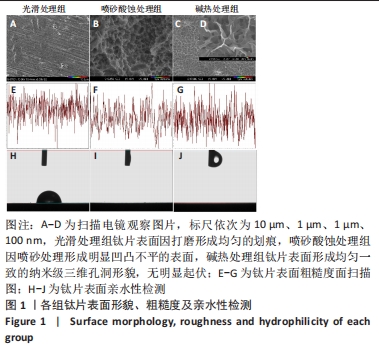
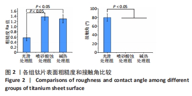
2.1 各组钛片表面形貌、粗糙度及亲水性检测结果 各组钛片表面的扫描电镜观察结果见图1A-D,光滑处理组钛片表面因打磨形成带有均匀的划痕,喷砂酸蚀处理组因喷砂处理形成明显凹凸不平的表面,碱热处理组钛片表面形成均匀一致的纳米级三维孔洞形貌,无明显起伏。 图1E-G为3组钛片表面粗糙度面扫描图;图1H-J为3组钛片表面亲水性检测图片,当水滴接触钛片表面时,喷砂酸蚀处理组和碱热处理组钛片表面水滴迅速散开,而光滑处理组水滴与钛片表面成一定角度。比较3组钛片表面的粗糙度值与接触角,结果显示:喷砂酸蚀处理组、碱热处理组钛片表面的粗糙度值大于光滑组(P < 0.05),接触角低于光滑处理组(P < 0.05);喷砂酸蚀处理组、碱热处理组钛片表面的粗糙度值与接触角比较差异均无显著性意义(P > 0.05),见图2。"

"
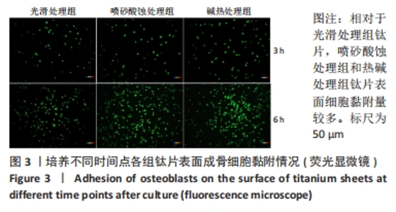
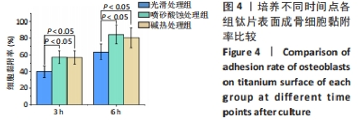
2.2 各组钛片表面成骨细胞黏附情况 图3为各组钛片表面接种MG63细胞分别培养3 h和6 h后的细胞黏附情况,可以明显观察到相对于光滑处理组钛片,喷砂酸蚀处理组和热碱处理组钛片表面细胞黏附量较多。计算细胞黏附率结果显示,喷砂酸蚀处理组、碱热处理组钛片表面培养3,6 h的黏附细胞数量均高于光滑处理组(P < 0.05),喷砂酸蚀处理组、碱热处理组钛片表面黏附细胞数量比较差异无显著性意义(P > 0.05),见图4。"
"

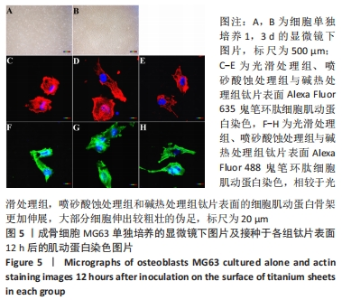
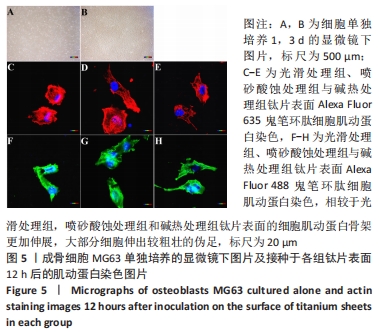
图5为成骨细胞MG63单独培养的显微镜下图片及接种于各组钛片表面12 h后的肌动蛋白染色结果,接种于钛片表面12 h后,共聚焦显微镜下可见相较于光滑处理组,喷砂酸蚀处理组和碱热处理组钛片表面的细胞肌动蛋白骨架更加伸展,大部分细胞伸出较粗壮的伪足,利于后续细胞间信号传导和细胞间相互作用。"
| [1] DANIEL B, LARS S, HUGO DB. Modern implant dentistry based on osseointegr- ation: 50 years of progress, current trends and open questions. Periodontol 2000. 2017;73(1):7-21. [2] 车振家,朱正清,朱礼伟,等.钛植入物表面生物化学改性对骨整合的影响[J].中国组织工程研究,2022,26(16):2576-2583. [3] SCULEAN A, GRUBER R, BOSSHARDT DD. Soft tissue wound healing around teeth and dental implants. J Clin Periodontol. 2014;41 Suppl 15:S6-22. [4] HASEGAWA M, SARUTA J, HIROTA M, et al. A newly created meso-, micro-, and nano-scale rough titanium surface promotes bone-implant integration. Int J Mol Sci. 2020;21(3):783. [5] 王旻,姜楠,祝颂松.新型钛表面微纳米共存梯度仿生结构对骨髓间充质细胞黏附、增殖及成骨分化的影响[J].口腔疾病防治,2021,29(4):226-233. [6] PINTO TS, MARTINS BR, FERREIRA MR, et al. Nanohydroxyapatite-Blasted Bioactive Surface Drives Shear-Stressed Endothelial Cell Growth and Angiogenesis. Biomed Res Int. 2022;2022:1433221. [7] WANG L, XU C, MENG K, et al. Biomimetic Hydroxyapatite Composite Coatings with a Variable Morphology Mediated by Silk Fibroin and Its Derived Peptides Enhance the Bioactivity on Titanium. ACS Biomater Sci Eng. 2023;9(1):165-181. [8] LÓPEZ-VALVERDE N, ARAGONESES J, LÓPEZ-VALVERDE A, et al. Effectiveness of biomolecule-based bioactive surfaces, on os-seointegration of titanium dental implants: A systematic review and meta-analysis of in vivo studies. Front Bioeng Biotechnol. 2022;10:986112. [9] WU J, JIANG S, XIE W, et al. Surface modification of the Ti surface with nanoscale bio-MOF-1 for improving biocompatibility and osteointegration in vitro and in vivo. J Mater Chem B. 2022;10(41):8535-8548. [10] CHEN M, WANG D, LI M, et al. Nanocatalytic Biofunctional MOF Coating on Titanium Implants Promotes Osteoporotic Bone Regeneration through Cooperative Pro-osteoblastogenesis MSC Reprogramming. ACS Nano. 2022;16(9):15397-15412. [11] LI Y, CHEN G, HE Y, et al. Selenomethionine-Modified Polyethylenimine-Based Nanoparticles Loaded with miR-132-3p Inhibitor-Biofunctionalized Titanium Implants for Improved Osteointegration. ACS Biomater Sci Eng. 2021;7(10):4933-4945. [12] SUN T, HUANG J, ZHANG W, et al. Simvastatin-hydroxyapatite coatings prevent biofilm formation and improve bone formation in implant-associated infections. Bioact Mater. 2022;21:44-56. [13] ZHU LS, LUO D, LIU Y. Effect of the nano/microscale structure of biomaterial scaffolds on bone regeneration. Int J Oral Sci. 2020;12(1):6. [14] DIOMEDE F, MARCONI GD, FONTICOLI L, et al. Functional relationship between osteogenesis and angiogenesis in tissue regeneration. Int J Mol Sci. 2020;21(9):3242. [15] MA L, LI G, LEI J, et al. Nanotopography Sequentially Mediates Human Mesenchymal Stem Cell-Derived Small Extracellular Vesicles for Enhancing Osteogenesis. ACS Nano. 2022;16(1):415-430. [16] XUE Y, ZHANG L, LIU F, et al. Surface Bandgap Engineering of Nanostructured Implants for Rapid Photothermal Ion Therapy of Bone Defects. Adv Healthc Mater. 2022;11(22):e2200998. [17] KIM HM, MIYAJI F, KOKUBO T, et al. Preparation of bioactive Ti and its alloys via simple chemical surface treatment. J Biomed Mater Res. 1996;32(3):409-417. [18] Baek HL, YOUNG DK, JI HS, et al. Surface modification by alkali and heat treatments in titanium alloys. J Biomed Mater Res. 2002;61(3):466-473. [19] TADASHI K, SEIJI Y. Groeth of novel ceramic layers on metals via chemical and heat treatments for inducing various biological functions. Front Bioeng Biotechnol. 2015;3:176. [20] WANG XJ, LI YC, LIN JG, et al. Effect of heat-treatment atmosphere on the bond strength of apatite layer on Ti substrate. Dent Mater. 2008;24(11):1549-1555. [21] ZHOU Y, WANG YB, ZHANG EW, et al. Alkali-heat treatment of a low modulus biomedical Ti-27NB alloy. Biomed Mater. 2009;4(4):044108. [22] ZHAO LZ, HU LS, HUO KF, et al. Mechanism of cell repellence on quasi-aligned nanowires arrays on Ti alloy. Biomaterials. 2010;31(32):8341-8349. [23] DING XL, YANG XQ, ZHOU L, et al. Titanate nanowires scaffolds decorated with anatase nanocrystals show good protein adsorption and low cell adhesion capacity. Int J Nanomedicine. 2013;8:569-579. [24] SticH T, ALAGBOSO F, KŘENEK T, et al. Implant-bone-interface: Reviewing the impact of titanium surface modifications on osteogenic processes in vitro and in vivo. Bioeng Transl Med. 2021;7(1):e10239. [25] Li S, Ni J, Liu X, et al. Surface characteristics and biocompatibility of sandblasted and acid-etched titanium surface modified by ultraviolet irradiation: an in vitro study. J Biomed Mater Res B Appl Biomater. 2012;100(6):1587-1598. [26] DADFARNIA M, NOVAK P, AHN DC, et al. Recent advances in the study of structural materials compatibility with hydrogen. Adv Mater. 2010;22(10):1128-1135. [27] HU P, GAO Q, ZHENG H, et al. The Role and Activation Mechanism of TAZ in Hierarchical Microgroove/Nanopore Topography-Mediated Regulation of Stem Cell Differentiation. Int J Nanomedicine. 2021;16:1021-1036. [28] MA L, KE W, LIAO Z, et al. Small extracellular vesicles with nanomorphology memory promote osteogenesis. Bioact Mater. 2022;17:425-438. [29] YU X, XU R, ZHANG Z. Different Cell and Tissue Behavior of Micro-/Nano-Tubes and Micro-/Nano-Nets Topographies on Selective Laser Melting Titanium to Enhance Osseointegration. Int J Nanomedicine. 2021;16:3329-3342. [30] FUJIBAYASHI S, NAKAMURA T, NISHIGUCHI S, et al. Bioactive titanium: effect of sodium removal on the bone-bonding ability of bioactive titanium prepared by alkali and heat treatment. J Biomed Mater Res. 2001;56(4):562-570. [31] KIM HM, MIYAJI F, KOKUBO T, et al. Graded surface structure of bioactive titanium prepared by chemical treatment. J Biomed Mater Res. 1999;45(2):100-107. [32] BOERCKER JE, ENACHE-POMMER E, AYDIL ES. Growth mechanism of titanium dioxide nanowires for dye-sensitized solar cells. Nanotechnology. 2008;19(9): 095604. [33] WANG F, DAI HX, DENG JG, et al. Manganese oxides with rod-, wire-, tube-, and flower-like morphologies: highly effective catalysts for the removal of toluene. Environ Sci Technol. 2012;46(7):4034-4041. [34] LIU W, LIANG L, LIU B, et al. The response of macrophages and their osteogenic potential modulated by micro/nano-structured Ti surfaces. Colloids Surf B Biointerfaces. 2021;205:111848. [35] MASAMOTO K, FUJIBAYASHI S, YAMAGUCHI S, et al. Bioactivity and antibacterial activity of strontium and silver ion releasing titanium. J Biomed Mater Res B Appl Biomater. 2021;109(2):238-245. [36] OKUZU Y, FUJIBAYASHI S, YAMAGUCHI S, et al. In vitro study of antibacterial and osteogenic activity of titanium metal releasing strontium and silver ions. J Biomater Appl. 2021;35(6):670-680. [37] DOYMUS B, KEREM G, YAZGAN KARATAS A, et al. A functional coating to enhance antibacterial and bioactivity properties of titanium implants and its performance in vitro. J Biomater Appl. 2021;35(6):655-669. [38] JEONG SJ, JEONG MJ. Effect of Thymosin beta4 on the Differentiation and Mineralization of MC3T3-E1 Cell on a Titanium Surface. J Nanosci Nanotechnol. 2016;16(2):1979-1983. [39] GAO Q, HOU Y, LI Z, et al. mTORC2 regulates hierarchical micro/nano topography-induced osteogenic differentiation via promoting cell adhesion and cytoskeletal polymerization. J Cell Mol Med. 2021;25(14):6695-6708. [40] WANG H, XU Q, HU H, et al. The Fabrication and Function of Strontium-modified Hierarchical Micro/Nano Titanium Implant. Int J Nanomedicine. 2020;15:8983-8998. [41] ALBERTINI M, FERNANDEZ-YAGUE M, LÁZARO P, et al. Advances in surfaces and osseointegration in implantology. Biomimetic surfaces. Med Oral Patol Oral Cir Bucal. 2015;20(3):e316-325. [42] DIAS CORPA TARDELLI J, LIMA DA COSTA VALENTE M, THEODORO DE OLIVEIRA T, et al. Influence of chemical composition on cell viability on titanium surfaces: A systematic review. J Prosthet Dent. 2021;125(3):421-425. [43] LIU J, RUAN J, YIN J, et al. Fabrication of multilevel porous structure networks on Nb-Ta-Ti alloy scaffolds and the effects of surface characteristics on behaviors of MC3T3-E1 cells. Biomed Mater. 2022;17(6). doi: 10.1088/1748-605X/ac9ffd. [44] SHICHMAN I, OAKLEY C, WILLEMS JH, et al. Novel metaphyseal porous titanium cones allow favorable outcomes in revision total knee arthroplasty. Arch Orthop Trauma Surg. 2023;143(3):1537-1547. [45] ZHANG Y, SUN N, ZHU M, et al. The contribution of pore size and porosity of 3D printed porous titanium scaffolds to osteogenesis. Biomater Adv. 2022;133:112651. [46] DAVIES JE. Understanding peri-implant endosseous healing. J Dent Educ. 2003; 67(8):932-949. [47] PULEO DA, NANCI A. Understanding and controlling the bone-implant interface. Biomaterials. 1999;20(23-24):2311-2321. [48] HORI N, UENO T, SUZUKI T, et al. Ultraviolet light treatment for the restoration of age-related degradation of titanium bioactivity. Int J Oral Maxillofac Implants. 2010;25(1):49-62. [49] MIYAUCHI T, YAMADA M, YAMAMOTO A, et al. The enhanced characteristics of osteoblast adhesion to photofunctionalized nanoscale TiO2 layers on biomaterials surfaces. Biomaterials. 2010;31(14):3827-3839. [50] TabUCHI M, HAMAJIMA K, TANAKA M, et al. UV Light-Generated Superhydrophilicity of a Titanium Surface Enhances the Transfer, Diffusion and Adsorption of Osteogenic Factors from a Collagen Sponge. Int J Mol Sci. 2021; 22(13):6811. |
| [1] | Yang Yufang, Yang Zhishan, Duan Mianmian, Liu Yiheng, Tang Zhenglong, Wang Yu. Application and prospects of erythropoietin in bone tissue engineering [J]. Chinese Journal of Tissue Engineering Research, 2024, 28(9): 1443-1449. |
| [2] | Bai Chen, Yang Wenqian, Meng Zhichao, Wang Yuze. Strategies for repairing injured anterior cruciate ligament and promoting graft healing [J]. Chinese Journal of Tissue Engineering Research, 2024, 28(9): 1457-1463. |
| [3] | Wang Wen, Zheng Pengpeng, Meng Haohao, Liu Hao, Yuan Changyong. Overexpression of Sema3A promotes osteogenic differentiation of dental pulp stem cells and MC3T3-E1 [J]. Chinese Journal of Tissue Engineering Research, 2024, 28(7): 993-999. |
| [4] | Wei Yuanxun, Chen Feng, Lin Zonghan, Zhang Chi, Pan Chengzhen, Wei Zongbo. The mechanism of Notch signaling pathway in osteoporosis and its prevention and treatment with traditional Chinese medicine [J]. Chinese Journal of Tissue Engineering Research, 2024, 28(4): 587-593. |
| [5] | Zhu Zhiqi, Yuan Sijie, Zhang Zilin, Ji Shijie, Meng Mingsong, Yan Anming, Han Jing. Mechanism underlying the effect of Liuwei Dihuang Pill on osteolysis and osteogenesis induced by titanium particles [J]. Chinese Journal of Tissue Engineering Research, 2024, 28(3): 392-397. |
| [6] | Gao Xueyu, Zhang Wentao, Sun Tianze, Zhang Jing, Li Zhonghai. Application of metal ions in bone tissue engineering [J]. Chinese Journal of Tissue Engineering Research, 2024, 28(3): 439-444. |
| [7] | Yang Jie, Hu Haolei, Li Shuo, Yue Wei, Xu Tao, Li Yi. Application of bio-inks for 3D printing in tissue repair and regenerative medicine [J]. Chinese Journal of Tissue Engineering Research, 2024, 28(3): 445-451. |
| [8] | Chen Junyan, Meng Qingqi, Li Siming. Cartilage targeting function in the drug delivery system by intra-articular injection for the treatment of osteoarthritis [J]. Chinese Journal of Tissue Engineering Research, 2024, 28(3): 458-463. |
| [9] | Kong Xiangyu, Wang Xing, Pei Zhiwei, Chang Jiale, Li Siqin, Hao Ting, He Wanxiong, Zhang Baoxin, Jia Yanfei. Biological scaffold materials and printing technology for repairing bone defects [J]. Chinese Journal of Tissue Engineering Research, 2024, 28(3): 479-485. |
| [10] | Chen Xiangshan, Liu Hua, Sun Weikang, Li Huanan. Mechanism of m6A methylation regulating bone metabolism for prevention and treatment of osteoporosis [J]. Chinese Journal of Tissue Engineering Research, 2024, 28(28): 4572-4577. |
| [11] | Zhang Shudong, Huang Yilin, Yao Qi. Punicalagin treats postmenopausal osteoporosis by promoting osteogenesis [J]. Chinese Journal of Tissue Engineering Research, 2024, 28(26): 4101-4105. |
| [12] | Li Jiale, Luo Dasheng, Zheng Liujie, Liu Wei, Yao Yunfeng. Human osteoarthritic chondrocytes up-regulate the expression of osteoprotegerin in osteoblasts via the Indian hedgehog signaling pathway [J]. Chinese Journal of Tissue Engineering Research, 2024, 28(26): 4194-4201. |
| [13] | Yang Qipei, Chen Feng, Cui Wei, Zhang Chi, Wu Ruiqi, Song Zhenheng, Meng Xin. Signaling pathways related to kaempferol active monomers in the treatment of osteoporosis [J]. Chinese Journal of Tissue Engineering Research, 2024, 28(26): 4242-4249. |
| [14] | Yu Yangyi, Lian Qiang, Wu Jianqun, Zhang Xuan, Ren Jinke, Li Guangheng. Cell-of-origin for heterotopic ossification induced by bone morphogenetic protein 4 in skeletal muscle [J]. Chinese Journal of Tissue Engineering Research, 2024, 28(25): 4034-4040. |
| [15] | Luo Peng, Wang Yi, Wang Ansu, Dang Yi, Ma Yaping, Zhang Yi, Wang Xin. Role and mechanism of interleukin-8 in bone regeneration [J]. Chinese Journal of Tissue Engineering Research, 2024, 28(24): 3910-3914. |
| Viewed | ||||||
|
Full text |
|
|||||
|
Abstract |
|
|||||