Chinese Journal of Tissue Engineering Research ›› 2024, Vol. 28 ›› Issue (26): 4194-4201.doi: 10.12307/2024.419
Previous Articles Next Articles
Human osteoarthritic chondrocytes up-regulate the expression of osteoprotegerin in osteoblasts via the Indian hedgehog signaling pathway
Li Jiale1, 2, Luo Dasheng1, Zheng Liujie1, Liu Wei1, Yao Yunfeng1
- 1The Second Hospital of Anhui Medical University, Hefei 230601, Anhui Province, China; 2Fuyang Hospital of Anhui Medical University, Fuyang 236000, Anhui Province, China
-
Received:2023-06-10Accepted:2023-07-24Online:2024-09-18Published:2023-10-07 -
Contact:Yao Yunfeng, MD, Chief physician, The Second Hospital of Anhui Medical University, Hefei 230601, Anhui Province, China -
About author:Li Jiale, Master, Physician, The Second Hospital of Anhui Medical University, Hefei 230601, Anhui Province, China; Fuyang Hospital of Anhui Medical University, Fuyang 236000, Anhui Province, China -
Supported by:the Natural Science Foundation of Anhui Province, No. 1608085MH167 (to YYF)
CLC Number:
Cite this article
Li Jiale, Luo Dasheng, Zheng Liujie, Liu Wei, Yao Yunfeng. Human osteoarthritic chondrocytes up-regulate the expression of osteoprotegerin in osteoblasts via the Indian hedgehog signaling pathway[J]. Chinese Journal of Tissue Engineering Research, 2024, 28(26): 4194-4201.
share this article
Add to citation manager EndNote|Reference Manager|ProCite|BibTeX|RefWorks

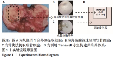
2.1 提取细胞及构建共培养系统的流程 从手术室收集标本后,快速转至实验室,无菌生理盐水、PBS反复冲洗干净标本后,从胫骨平台外侧区域(图1A)分离提取人骨关节炎成骨细胞与软骨细胞。酶解法提取软骨细胞后,除一部分细胞2D培养进行鉴定外,其余细胞采用海藻酸钠珠包埋后培养(图1B)。将骨块用胰酶、胶原酶预消化后种植在培养皿中,1周左右可在倒置显微镜下看到成骨细胞从骨块中爬出(图1C)。建立软骨细胞-成骨细胞共培养体系时,将海藻酸钠包埋的软骨细胞放在0.4 μm孔径的Transwell小室中,然后将小室放在培养有成骨细胞的6孔板中(图1D)。"

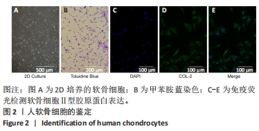
2.2 培养及鉴定人软骨细胞 从胫骨平台分离出软骨细胞后,经细胞滤网过滤、离心后,将细胞高密度种植(约2.5×10⁴/cm2) 到培养板中。待细胞贴壁后显微镜下观察拍照(图2A)。使用甲苯胺蓝对原代软骨细胞进行染色,可见软骨细胞呈紫红色(图2B)。通过免疫荧光检测软骨细胞表达分泌Ⅱ型胶原蛋白的情况,结果显示软骨细胞的细胞核呈蓝色,细胞胞浆中可见散在的Ⅱ型胶原蛋白呈绿色(图2C-E)。"

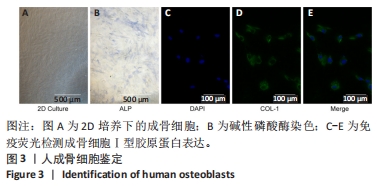
2.3 培养及鉴定人成骨细胞 待第2代成骨细胞长至80%融合时,消化收集成骨细胞,将成骨细胞种植在6孔板中(图3A)。培养一段时间后,显微镜下观察细胞并拍照。碱性磷酸酶染色可见成骨细胞成蓝紫色(图3B)。免疫荧光检测成骨细胞表达分泌Ⅰ型胶原蛋白的情况,结果显示成骨细胞核呈蓝色,细胞胞浆中可见散在的Ⅰ型胶原蛋白呈绿色(图3C-E)。"
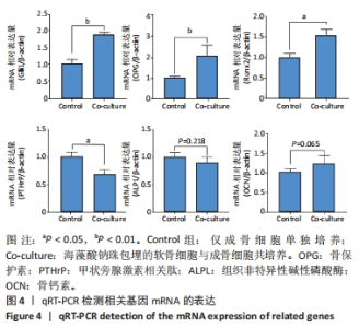
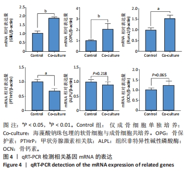
2.4 共培养下人软骨细胞促进成骨细胞OPG、Runx2等成骨相关基因表达 与软骨细胞共培养48 h后,利用qRT-PCR检测成骨细胞成骨基因的相对于内参基因β-actin的表达情况,发现成骨细胞中Gli1、OPG、Runx2的mRNA均升高(P < 0.05),PTHrP的mRNA表达相对下降(P < 0.05)。在与软骨细胞共培养时,成骨细胞中ALPL 的mRNA表达稍降低,骨钙素的mRNA表达稍增加,但差异无显著性意义(P > 0.05),见图4。"

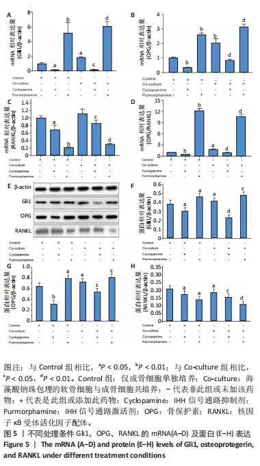
2.5 IHH信号通路对OPG/RANKL的影响 共培养时,成骨细胞中IHH信号通路激活的目的基因Gli1的表达明显升高,提示骨关节炎软骨细胞能够通过IHH信号通路影响成骨细胞。为了进一步探究IHH信号通路的作用,使用了该通路的抑制剂(环巴胺,10 nmol/L)和激活剂(Purmorphamine,10 nmol/L),两者通过作用在细胞膜受体smo蛋白,进而控制IHH下游基因的表达。在使用药物48 h后测定成骨细胞中Gli1以及成骨相关基因的表达情况,结果发现在使用激活剂后Gli1表达相较于对照组显著升高,使用抑制剂后Gli1表达显著降低,这说明加入激活剂或抑制剂能够特异性激活或抑制成骨细胞中IHH的信号通路(图5A)。在OPG、Runx2、PTHrP等相关检测基因里,仅OPG的mRNA表达趋势与Gli1完全一致,当IHH信号通路被激活时,OPG蛋白的mRNA表达显著升高;当IHH信号通路被抑制时,OPG蛋白的mRNA的表达显著降低(图5B)。另外在OPG基因的表达被抑制时RANKL的表达没有显著的升高;但OPG基因的表达增高时RANKL的表达明显降低(图5B,C),差异有显著性意义(P < 0.05)。进一步计算了OPG/RANKL的mRNA相对表达值,发现OPG/RANKL的mRNA的比值与OPG mRNA以及Gli1 mRNA的趋势相同(图5D),最后使用Western blot检测了这些mRNA对应蛋白的表达量,发现与mRNA的趋势一致(图5E-H)。这些结果表明骨关节炎软骨细胞能够通过IHH信号通路上调骨关节炎成骨细胞OPG的表达,从而进一步影响OPG/RANKL系统。"
| [1] NELSON AE. Osteoarthritis year in review 2017: clinical.Osteoarthritis Cartilage. 2018;26(3):319-325. [2] O’NEILL TW, MCCABE PS, MCBETH J. Update on the epidemiology, risk factors and disease outcomes of osteoarthritis. Best Pract Res Clin Rheumatol. 2018;32(2):312-326. [3] AHO OM, FINNILA M, THEVENOT J, et al. Subchondral bone histology and grading in osteoarthritis. PLoS One. 2017;12(3):e0173726. [4] HUGLE T, GEURTS J. What drives osteoarthritis?-synovial versus subchondral bone pathology. Rheumatology (Oxford). 2017;56(9): 1461-1471. [5] STEWART HL, KAWCAK CE. The Importance of Subchondral Bone in the Pathophysiology of Osteoarthritis. Front Vet Sci. 2018;5:178. [6] LI G, YIN J, GAO J, et al. Subchondral bone in osteoarthritis: insight into risk factors and microstructural changes. Arthritis Res Ther. 2013; 15(6):223. [7] BURR DB, GALLANT MA. Bone remodelling in osteoarthritis. Nat Rev Rheumatol. 2012;8(11):665-673. [8] SANCHEZ C, DEBERG MA, PICCARDI N, et al. Subchondral bone osteoblasts induce phenotypic changes in human osteoarthritic chondrocytes. Osteoarthritis Cartilage. 2005;13(11):988-997. [9] SANCHEZ C, DEBERG MA, PICCARDI N, et al. Osteoblasts from the sclerotic subchondral bone downregulate aggrecan but upregulate metalloproteinases expression by chondrocytes. This effect is mimicked by interleukin-6, -1beta and oncostatin M pre-treated non-sclerotic osteoblasts. Osteoarthritis Cartilage. 2005;13(11):979-987. [10] 朴星银,蒋传路.胶质瘤相关Hedgehog信号通路的研究进展[J].中国微侵袭神经外科杂志,2012,11(17):526-528. [11] LIN AC, SEETO BL, BARTOSZKO JM, et al. Modulating hedgehog signaling can attenuate the severity of osteoarthritis. Nat Med. 2009;15(12): 1421-1425. [12] MAEDA Y, SCHIPANI E, DENSMORE MJ, et al. Partial rescue of postnatal growth plate abnormalities in Ihh mutants by expression of a constitutively active PTH/PTHrP receptor. Bone. 2010;46(2):472-478. [13] HOFBAUER LC, KUHNE CA, VIERECK V. The OPG/RANKL/RANK system in metabolic bone diseases. J Musculoskelet Neuronal Interact. 2004; 4(3):268-275. [14] SCHARSTUHL A, GLANSBEEK HL, VAN BEUNINGEN HM, et al. Inhibition of endogenous TGF-beta during experimental osteoarthritis prevents osteophyte formation and impairs cartilage repair. J Immunol. 2002; 169(1):507-514. [15] TCHETINA EV, SQUIRES G, POOLE AR. Increased type II collagen degradation and very early focal cartilage degeneration is associated with upregulation of chondrocyte differentiation related genes in early human articular cartilage lesions. J Rheumatol. 2005;32(5):876-886. [16] SALTZMAN BM, RIBOH JC. Subchondral Bone and the Osteochondral Unit: Basic Science and Clinical Implications in Sports Medicine. Sports Health. 2018;10(5):412-418. [17] ANDERSON HC, CHACKO S, ABBOTT J, et al. The loss of phenotypic traits by differentiated cells in vitro. VII. Effects of 5-bromodeoxyuridine and prolonged culturing on fine structure of chondrocytes. Am J Pathol. 1970;60(2):289-312. [18] VON DER MARK K, GAUSS V, VON DER MARK H, et al. Relationship between cell shape and type of collagen synthesised as chondrocytes lose their cartilage phenotype in culture. Nature. 1977;267(5611): 531-532. [19] REICHENBERGER E, AIGNER T, VON DER MARK K,et al. In situ hybridization studies on the expression of type X collagen in fetal human cartilage. Dev Biol. 1991;148(2):562-572. [20] FUKUI N, PURPLE CR, SANDELL LJ. Cell biology of osteoarthritis: the chondrocyte’s response to injury. Curr Rheumatol Rep. 2001;3(6): 496-505. [21] SUN MM, BEIER F. Chondrocyte hypertrophy in skeletal development, growth, and disease. Birth Defects Res C Embryo Today. 2014;102(1): 74-82. [22] PITSILLIDES AA, BEIER F. Cartilage biology in osteoarthritis--lessons from developmental biology. Nat Rev Rheumatol. 2011;7(11):654-663. [23] MORT JS, BILLINGTON CJ. Articular cartilage and changes in arthritis: matrix degradation. Arthritis Res. 2001;3(6):337-341. [24] MOULIN D, SALONE V, KOUFANY M, et al. MicroRNA-29b Contributes to Collagens Imbalance in Human Osteoarthritic and Dedifferentiated Articular Chondrocytes. Biomed Res Int. 2017;2017:9792512. [25] HDUD IM, EL-SHAFEI AA, LOUGHNA P, et al. Expression of Transient Receptor Potential Vanilloid (TRPV) channels in different passages of articular chondrocytes. Int J Mol Sci. 2012;13(4):4433-4445. [26] WANG D, REN H, LI C, et al. Orexin A prevents degradation of the articular matrixes in human primary chondrocyte culture. Mol Immunol. 2018;101:102-107. [27] FINGER F, SCHORLE C, ZIEN A, et al. Molecular phenotyping of human chondrocyte cell lines T/C-28a2, T/C-28a4, and C-28/I2. Arthritis Rheum. 2003;48(12):3395-3403. [28] SAAS J, LINDAUER K, BAU B, et al. Molecular phenotyping of HCS-2/8 cells as an in vitro model of human chondrocytes. Osteoarthritis Cartilage. 2004;12(11):924-934. [29] SCHULZE S, WEHRUM D, DIETER P, et al. A supplement-free osteoclast-osteoblast co-culture for pre-clinical application. J Cell Physiol. 2018; 233(6):4391-4400. [30] SUN Q, CHOUDHARY S, MANNION C, et al. Ex vivo construction of human primary 3D-networked osteocytes. Bone. 2017;105:245-252. [31] ROSENBERG N, ROSENBERG O, WEIZMAN A, et al. In vitro Effects of the Specific Mitochondrial TSPO Ligand Ro5 4864 in Cultured Human Osteoblasts. Exp Clin Endocrinol Diabetes. 2018;126(2):77-84. [32] FLORENCIO-SILVA R, SASSO GR, SASSO-CERRI E, et al. Biology of Bone Tissue: Structure, Function, and Factors That Influence Bone Cells.Biomed Res Int. 2015;2015:421746. [33] SANCHEZ C, DEBERG MA, BELLAHCENE A, et al. Phenotypic characterization of osteoblasts from the sclerotic zones of osteoarthritic subchondral bone. Arthritis Rheum. 2008;58(2):442-455. [34] NAKAOKA R, HSIONG SX, MOONEY DJ. Regulation of chondrocyte differentiation level via co-culture with osteoblasts. Tissue Eng. 2006; 12(9):2425-2433. [35] SIMONET WS, LACEY DL, DUNSTAN CR, et al. Osteoprotegerin: a novel secreted protein involved in the regulation of bone density. Cell. 1997; 89(2):309-319. [36] YASUDA H, SHIMA N, NAKAGAWA N, et al. Identity of Osteoclastogenesis Inhibitory Factor (OCIF) and Osteoprotegerin (OPG): A Mechanism by which OPG/OCIF Inhibits Osteoclastogenesis in Vitro 1. Endocrinology. 1998;139(3):1329-1337. [37] SCHNEEWEIS LA, WILLARD D, MILLA ME. Functional Dissection of Osteoprotegerin and Its Interaction with Receptor Activator of NF-κB Ligand. J Biol Chem. 2005;280(50):41155-41164. [38] REID P, HOLEN I. Pathophysiological roles of osteoprotegerin (OPG). Eur J Cell Biol. 2009;88(1):1-17. [39] GEUSENS P. Emerging treatments for postmenopausal osteoporosis–focus on denosumab. Clin Interv Aging. 2009;4:241-250. [40] SILVA I, BRANCO JC. Rank/Rankl/opg:literature review. Acta Reumatol Port. 2011;36(3):209-218. [41] TYROVOLA JB. The “Mechanostat” Principle and the Osteoprotegerin-OPG/RANKL/RANK System PART II. The Role of the Hypothalamic-Pituitary Axis. J Cell Biochem. 2017;118(5):962-966. [42] HOFBAUER LC, KHOSLA S, DUNSTAN CR, et al. The roles of osteoprotegerin and osteoprotegerin ligand in the paracrine regulation of bone resorption. J Bone Miner Res. 2000;15(1):2-12. [43] UPTON AR, HOLDING CA, DHARMAPATNI AA, et al. The expression of RANKL and OPG in the various grades of osteoarthritic cartilage. Rheumatol Int. 2012;32(2):535-540. [44] MAK KK, BI Y, WAN C, et al. Hedgehog signaling in mature osteoblasts regulates bone formation and resorption by controlling PTHrP and RANKL expression. Developmental Cell. 2008;14(5):674-688. [45] LIANG W, LI X, GAO B, et al. Observing the development of the temporomandibular joint in embryonic and post-natal mice using various staining methods. Exp Ther Med. 2016;11(2):481-489. [46] MAEDA Y, NAKAMURA E, NGUYEN MT, et al. Indian Hedgehog produced by postnatal chondrocytes is essential for maintaining a growth plate and trabecular bone. Proc Natl Acad Sci U S A. 2007;104(15): 6382-6387. [47] HUANGFU D, ANDERSON KV. Signaling from Smo to Ci/Gli: conservation and divergence of Hedgehog pathways from Drosophila to vertebrates.Development. 2006;133(1):3-14. [48] DIDIASOVA M, SCHAEFER L, WYGRECKA M. Targeting GLI Transcription Factors in Cancer. Molecules. 2018;23(5):1003. [49] LEIJTEN JC, BOS SD, LANDMAN EB, et al. GREM1, FRZB and DKK1 mRNA levels correlate with osteoarthritis and are regulated by osteoarthritis-associated factors. Arthritis Res Ther. 2013;15(5):R126. [50] ALI SA, AL-JAZRAWE M, MA H, et al. Regulation of Cholesterol Homeostasis by Hedgehog Signaling in Osteoarthritic Cartilage. Arthritis Rheumatol. 2016;68(1):127-137. |
| [1] | Yang Yufang, Yang Zhishan, Duan Mianmian, Liu Yiheng, Tang Zhenglong, Wang Yu. Application and prospects of erythropoietin in bone tissue engineering [J]. Chinese Journal of Tissue Engineering Research, 2024, 28(9): 1443-1449. |
| [2] | Li Yongjie, Fu Shenyu, Xia Yuan, Zhang Dakuan, Liu Hongju. Correlation of knee extensor muscle strength and spatiotemporal gait parameters with peak knee flexion/adduction moment in female patients with knee osteoarthritis [J]. Chinese Journal of Tissue Engineering Research, 2024, 28(9): 1354-1358. |
| [3] | Qi Haodong, Lu Chao, Xu Hanbo, Wang Mengfei, Hao Yangquan. Effect of diabetes mellitus on perioperative blood loss and pain after primary total knee arthroplasty [J]. Chinese Journal of Tissue Engineering Research, 2024, 28(9): 1383-1387. |
| [4] | Du Changling, Shi Hui, Zhang Shoutao, Meng Tao, Liu Dong, Li Jian, Cao Heng, Xu Chuang. Efficacy and safety of different applications of tranexamic acid in high tibial osteotomy [J]. Chinese Journal of Tissue Engineering Research, 2024, 28(9): 1409-1413. |
| [5] | Huang Xiarong, Hu Lizhi, Sun Guanghua, Peng Xinke, Liao Ying, Liao Yuan, Liu Jing, Yin Linwei, Zhong Peirui, Peng Ting, Zhou Jun, Qu Mengjian. Effect of electroacupuncture on the expression of P53 and P21 in articular cartilage and subchondral bone of aged rats with knee osteoarthritis [J]. Chinese Journal of Tissue Engineering Research, 2024, 28(8): 1174-1179. |
| [6] | Zhao Garida, Ren Yizhong, Han Changxu, Kong Lingyue, Jia Yanbo. Mechanism of Mongolian Medicine Erden-uril on osteoarthritis in rats [J]. Chinese Journal of Tissue Engineering Research, 2024, 28(8): 1193-1199. |
| [7] | Li Rui, Zhang Guihong, Wang Tao, Fan Ping. Effect of ginseng polysaccharide on the expression of prostaglandin E2/6-keto-prostaglandin 1alpha in traumatic osteoarthritis model rats [J]. Chinese Journal of Tissue Engineering Research, 2024, 28(8): 1235-1240. |
| [8] | Wang Wen, Zheng Pengpeng, Meng Haohao, Liu Hao, Yuan Changyong. Overexpression of Sema3A promotes osteogenic differentiation of dental pulp stem cells and MC3T3-E1 [J]. Chinese Journal of Tissue Engineering Research, 2024, 28(7): 993-999. |
| [9] | Zhang Kefan, Shi Hui. Research status and application prospect of cytokine therapy for osteoarthritis [J]. Chinese Journal of Tissue Engineering Research, 2024, 28(6): 961-967. |
| [10] | Zhang Zeyi, Yang Yimin, Li Wenyan, Zhang Meizhen. Effect of foot progression angle on lower extremity kinetics of knee osteoarthritis patients of different ages: a systematic review and meta-analysis [J]. Chinese Journal of Tissue Engineering Research, 2024, 28(6): 968-975. |
| [11] | Shen Feiyan, Yao Jixiang, Su Shanshan, Zhao Zhongmin, Tang Weidong. Knockdown of circRNA WD repeat containing protein 1 inhibits proliferation and induces apoptosis of chondrocytes in knee osteoarthritis [J]. Chinese Journal of Tissue Engineering Research, 2024, 28(4): 499-504. |
| [12] | Maisituremu·Heilili, Zhang Wanxia, Nijiati·Nuermuhanmode, Maimaitituxun·Tuerdi. Effect of intraarticular injection of different concentrations of ozone on condylar histology of rats with early temporomandibular joint osteoarthritis [J]. Chinese Journal of Tissue Engineering Research, 2024, 28(4): 505-509. |
| [13] | Qiao Hujun, Wang Guoxiang. Evaluation of rat osteoarthritis chondrocyte models induced by interleukin-1beta [J]. Chinese Journal of Tissue Engineering Research, 2024, 28(4): 516-521. |
| [14] | Liu Yuhan, Fan Yujiang, Wang Qiguang. Comparison of protocols for constructing animal models of early traumatic knee osteoarthritis [J]. Chinese Journal of Tissue Engineering Research, 2024, 28(4): 542-549. |
| [15] | Zhang Yaru, Chen Yanjun, Zhang Xiaodong, Chen Shenghua, Huang Wenhua. Effect of ferroptosis mediated by glutathione peroxidase 4 in the occurrence and progression of synovitis in knee osteoarthritis [J]. Chinese Journal of Tissue Engineering Research, 2024, 28(4): 550-555. |
| Viewed | ||||||
|
Full text |
|
|||||
|
Abstract |
|
|||||