Chinese Journal of Tissue Engineering Research ›› 2024, Vol. 28 ›› Issue (4): 516-521.doi: 10.12307/2024.226
Previous Articles Next Articles
Evaluation of rat osteoarthritis chondrocyte models induced by interleukin-1beta
Qiao Hujun1, 2, Wang Guoxiang1
- 1School of Physical Education, Soochow University, Suzhou 215021, Jiangsu Province, China; 2Changzhi University, Changzhi 046000, Shanxi Province, China
-
Received:2023-01-03Accepted:2023-02-24Online:2024-02-08Published:2023-07-13 -
Contact:Wang Guoxiang, Professor, Doctoral supervisor, School of Physical Education, Soochow University, Suzhou 215021, Jiangsu Province, China -
About author:Qiao Hujun, MD, Lecturer, School of Physical Education, Soochow University, Suzhou 215021, Jiangsu Province, China; Changzhi University, Changzhi 046000, Shanxi Province, China -
Supported by:Postgraduate Research & Practice Innovation Program of Jiangsu Province, No. KYCX18-2485 (to QHJ)
CLC Number:
Cite this article
Qiao Hujun, Wang Guoxiang. Evaluation of rat osteoarthritis chondrocyte models induced by interleukin-1beta[J]. Chinese Journal of Tissue Engineering Research, 2024, 28(4): 516-521.
share this article
Add to citation manager EndNote|Reference Manager|ProCite|BibTeX|RefWorks
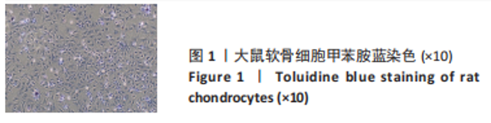
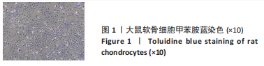
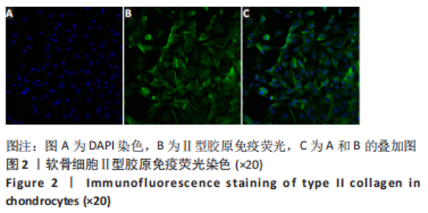
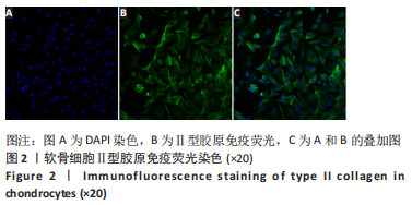
2.1 软骨细胞鉴定 软骨细胞经甲苯胺蓝染色,显微镜下可见细胞为梭形或多角形,轮廓明显,排列紧密;细胞核呈圆形或椭圆形,呈深蓝色,可见一两个核仁,见图1。软骨细胞经Ⅱ型胶原免疫荧光染色,可见细胞质呈绿色荧光,细胞核经DAPI染色呈深蓝色,胞质和胞核图叠加可见典型软骨细胞形态,见图2。由此证明,此次实验培养细胞为软骨细胞。"
"

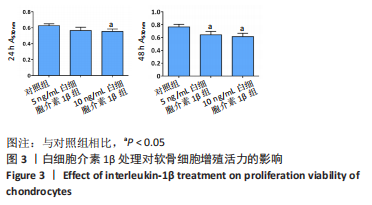
2.2 软骨细胞活性 MTT检测结果发现,白细胞介素1β处理软骨细胞24 h,10 ng/mL白细胞介素1β组软骨细胞活性较对照组显著降低(P < 0.05);白细胞介素1β处理软骨细胞48 h,同对照组相比,5 ng/mL 和10 ng/mL白细胞介素1β组软骨细胞活性均显著降低(P < 0.05),见图3。"
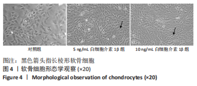
2.3 软骨细胞形态学变化 软骨细胞形态学变化见图4,对照组软骨细胞呈梭形、多角形和椭圆形,细胞形态结构完整,白细胞介素1β处理后软骨细胞呈长梭形。"

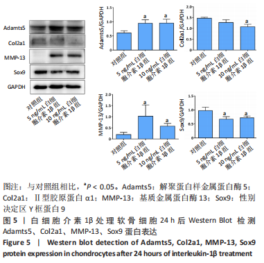
2.4 Western Blot检测结果 白细胞介素1β处理软骨细胞24 h,同对照组相比,5 ng/mL白细胞介素1β组MMP-13、Adamts5蛋白表达增加(P < 0.05),Sox9蛋白表达下降(P < 0.05),Ⅱ型胶原蛋白表达差异不明显;10 ng/mL白细胞介素1β组MMP-13、Adamts5蛋白表达增加(P < 0.05),Ⅱ型胶原蛋白、Sox9蛋白表达下降(P < 0.05),见图5。"
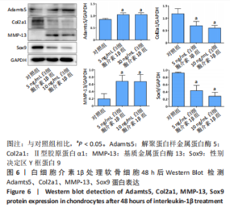
白细胞介素1β处理软骨细胞48 h,同对照组相比,5 ng/mL和10 ng/mL白细胞介素1β组MMP-13、Adamts5蛋白表达均增加(P < 0.05),Sox9、Ⅱ型胶原蛋白表达均下降(P < 0.05),见图6。"

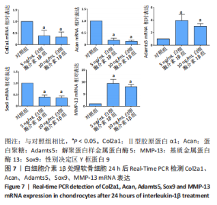
2.5 Real-Time PCR检测结果 白细胞介素1β处理软骨细胞24 h,同对照组相比,5 ng/mL和10 ng/mL白细胞介素1β组Ⅱ胶原蛋白、蛋白聚糖、Sox9 mRNA表达均显著下降(P < 0.05),Adamts5、MMP-13 mRNA表达均显著增加(P < 0.05),见图7。"

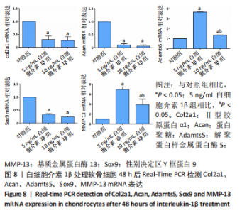
白细胞介素1β处理软骨细胞48 h,同对照组相比,5 ng/mL和10 ng/mL白细胞介素1β组Ⅱ型胶原蛋白、蛋白聚糖、Sox9 mRNA表达均显著下降(P < 0.05),Adamts5、MMP-13 mRNA表达均显著增加(P < 0.05),见图8。"
| [1] LOESER RF, GOLDRING SR, SCANZELLO CR, et al. Osteoarthritis: a disease of the joint as an organ. Arthritis Rheum. 2012;64(6):1697-1707. [2] ROBINSON WH, LEPUS CM, WANG Q, et al. Low-grade inflammation as a key mediator of the pathogenesis of osteoarthritis. Nat Rev Rheumatol. 2016; 12(10):580-592. [3] SHENTU CY, YAN G, XU DC, et al.Emerging pharmaceutical therapeutics and delivery technologies for osteoarthritis therapy. Front Pharmacol. 2022;13: 1-16. [4] ANSARI MY, AHMAD N, HAQQI TM.Oxidative stress and inflammation in osteoarthritis pathogenesis: Role of polyphenols. Biomed Pharmacother. 2020;129:1-9. [5] KAPOOR M, MARTEL-PELLETIER J, LAJEUNESSE D, et al. Role of proinflammatory cytokines in the pathophysiology of osteoarthritis. Nat Rev Rheumatol. 2011;7(1):33-42. [6] WOJDASIEWICZ P, PONIATOWSKI LA, SZUKIEWICZ D. The role of inflammatory and anti-inflammatory cytokines in the pathogenesis of osteoarthritis. Mediators Inflamm. 2014;2014:1-19. [7] CHEN J, GU YT, XIE JJ, et al. Gastrodin reduces IL-1beta-induced apoptosis, inflammation, and matrix catabolism in osteoarthritis chondrocytes and attenuates rat cartilage degeneration in vivo. Biomed Pharmacother. 2018; 97:642-651. [8] SUN FF, HU PF, XIONG Y, et al. Tricetin Protects Rat Chondrocytes against IL-1beta-Induced Inflammation and Apoptosis. Oxid Med Cell Longev. 2019; 2019:1-10. [9] GUO Z, LIN J, SUN K, et al. Deferoxamine Alleviates Osteoarthritis by Inhibiting Chondrocyte Ferroptosis and Activating the Nrf2 Pathway. Front Pharmacol. 2022;13:1-15. [10] JIANG Z, QI G, LU W, et al. Omaveloxolone inhibits IL-1beta-induced chondrocyte apoptosis through the Nrf2/ARE and NF-kappaB signalling pathways in vitro and attenuates osteoarthritis in vivo. Front Pharmacol. 2022;13:1-16. [11] ZHOU Q, WANG W, WU J, et al. Ubiquitin-specific protease 3 attenuates interleukin-1beta-mediated chondrocyte senescence by deacetylating forkhead box O-3 via sirtuin-3. Bioengineered. 2022;13(2):2017-2027. [12] KONG J, WANG J, GONG X, et al. Punicalagin Inhibits Tert-Butyl Hydroperoxide-Induced Apoptosis and Extracellular Matrix Degradation in Chondrocytes by Activating Autophagy and Ameliorates Murine Osteoarthritis. Drug Des Devel Ther. 2020;14:5521-5533. [13] VARELA-EIRIN M, LOUREIRO J, FONSECA E, et al. Cartilage regeneration and ageing: Targeting cellular plasticity in osteoarthritis. Ageing Res Rev. 2018; 42:56-71. [14] 杨帆, 刘保一, 刘家河,等.体外培养SD大鼠关节软骨细胞原代至第3代的形态学特点[J].中国组织工程研究,2021,25(14):2161-2165. [15] LI Z, MENG D, LIU Y, et al. Circular RNA VMA21 ameliorates IL-1beta-engendered chondrocyte injury through the miR-495-3p/FBWX7 signaling axis. Clin Immunol. 2022;238:1-10. [16] MU Y, WANG L, FU L, et al.K nockdown of LMX1B Suppressed Cell Apoptosis and Inflammatory Response in IL-1β-Induced Human Osteoarthritis Chondrocytes through NF-κB and NLRP3 Signal Pathway. Mediators Inflamm. 2022;2022:1-15. [17] FU G, YIN F, ZHAO J. Depletion of circ_0128846 ameliorates interleukin-1beta-induced human chondrocyte apoptosis and inflammation through the miR-940/PTPN12 pathway. Int Immunopharmacol. 2022;110:1-10. [18] LI X, FENG K, LI J, et al. Curcumin Inhibits Apoptosis of Chondrocytes through Activation ERK1/2 Signaling Pathways Induced Autophagy. Nutrients. 2017; 9(4):1-14. [19] FU S, FAN Q, XU J, et al. Circ_0008956 contributes to IL-1beta-induced osteoarthritis progression via miR-149-5p/NAMPT axis. Int Immunopharmacol. 2021;98:1-9. [20] MA Y, TU C, LIU W, et al. Isorhapontigenin Suppresses Interleukin-1beta-Induced Inflammation and Cartilage Matrix Damage in Rat Chondrocytes. Inflammation. 2019;42(6):2278-2285. [21] HAO L, MA C, LI Z, et al. Effects of type II collagen hydrolysates on osteoarthritis through the NF-kappaB, Wnt/beta-catenin and MAPK pathways. Food Funct. 2022;13(3):1192-1205. [22] JORGENSEN AEM, KJAER M, HEINEMEIER KM. The Effect of Aging and Mechanical Loading on the Metabolism of Articular Cartilage.J Rheumatol. 2017;44(4):410-417. [23] LEFEBVRE V, DVIR-GINZBERG M. SOX9 and the many facets of its regulation in the chondrocyte lineage. Connect Tissue Res. 2017;58(1):1-22. [24] LIU CF, LEFEBVRE V. The transcription factors SOX9 and SOX5/SOX6 cooperate genome-wide through super-enhancers to drive chondrogenesis.Nucleic Acids Res. 2015;43(17):8183-8203. [25] HATTORI T, M LLER C, GEBHARD S. SOX9 is a major negative regulator of cartilage vascularization, bone marrow formation and endochondral ossification. Development. 2010;137(6):901-911. [26] HASEEB A, KC R, ANGELOZZI M, et al. SOX9 keeps growth plates and articular cartilage healthy by inhibiting chondrocyte dedifferentiation/osteoblastic redifferentiation. PNAS. 2021;118(8):1-11. [27] BREW CJ, CLEGG PD, BOOT-HANDFORD RP, et al. Gene expression in human chondrocytes in late osteoarthritis is changed in both fibrillated and intact cartilage without evidence of generalised chondrocyte hypertrophy. Ann Rheum Dis. 2010;69(1):234-240. [28] GOLDRING MB, OTERO M, PLUMB DA, et al. Roles of inflammatory and anabolic cytokines in cartilage metabolism signals and multiple effectors converge upon MMP-13 regulation in osteoarthritis. Eur Cell Mater. 2011; 21:1-29. [29] RAHMATI M, MOBASHERI A, MOZAFARI M. Inflammatory mediators in osteoarthritis: A critical review of the state-of-the-art, current prospects, and future challenges. Bone. 2016;85:81-90. [30] ZREIQAT H, BELLUOCCIO D, SMITH MM. S100A8 and S100A9 in experimental osteoarthritis. Arthritis Res Ther. 2010;12(1):1-13. [31] POZGAN U, CAGLIC D, ROZMAN B, et al. Expression and activity profiling of selected cysteine cathepsins and matrix metalloproteinases in synovial fluids from patients with rheumatoid arthritis and osteoarthritis. Biol Chem. 2010;391(5):571-579. [32] GOLDRING MB, OTERO M. Inflammation in osteoarthritis.Curr Opin Rheumatol. 2011;23(5):471-478. [33] APTE SS. Anti-ADAMTS5 monoclonal antibodies: implications for aggrecanase inhibition in osteoarthritis. Biochem J. 2016;473(1):e1-e4. |
| [1] | Tan Nengxian, Wu Wenzheng, Zheng Churong, Luo Lieliang, Gu Peng, Ouyang Chongzhi, Zheng Xiaohui. Finite element analysis of different fixation methods of partially threaded cannulated screws for treating vertical femoral neck fractures [J]. Chinese Journal of Tissue Engineering Research, 2024, 28(6): 873-878. |
| [2] | Chen Zan, Lei Fei, Ye Fei, Zhou Qingzhong, Yuan Hao, Zheng Lipeng, Zha Xian, Feng Daxiong. Relationship between drainage time and early efficacy after short-segment lumbar fusion [J]. Chinese Journal of Tissue Engineering Research, 2024, 28(6): 927-933. |
| [3] | Kaiyisaier•Abudukelimu, Maimaitimin•Abulimiti, Li Lei, Yang Xiaokai, Zhang Yukun, Liu Shuai. Effect of lumbar CT values in the diagnosis of osteoporosis in women patients with lumbar degenerative diseases [J]. Chinese Journal of Tissue Engineering Research, 2024, 28(6): 945-949. |
| [4] | Zhang Kefan, Shi Hui. Research status and application prospect of cytokine therapy for osteoarthritis [J]. Chinese Journal of Tissue Engineering Research, 2024, 28(6): 961-967. |
| [5] | Zhang Zeyi, Yang Yimin, Li Wenyan, Zhang Meizhen. Effect of foot progression angle on lower extremity kinetics of knee osteoarthritis patients of different ages: a systematic review and meta-analysis [J]. Chinese Journal of Tissue Engineering Research, 2024, 28(6): 968-975. |
| [6] | Jiang Zihao, Wang Guanglan, Chen Peng, Sun Xianghong, Wang Ting, Jia Shaohui, Zheng Cheng. Effect of eccentric training combined with different frequency whole body vibration training on patellar tendinopathy [J]. Chinese Journal of Tissue Engineering Research, 2024, 28(4): 493-498. |
| [7] | Shen Feiyan, Yao Jixiang, Su Shanshan, Zhao Zhongmin, Tang Weidong. Knockdown of circRNA WD repeat containing protein 1 inhibits proliferation and induces apoptosis of chondrocytes in knee osteoarthritis [J]. Chinese Journal of Tissue Engineering Research, 2024, 28(4): 499-504. |
| [8] | Maisituremu·Heilili, Zhang Wanxia, Nijiati·Nuermuhanmode, Maimaitituxun·Tuerdi. Effect of intraarticular injection of different concentrations of ozone on condylar histology of rats with early temporomandibular joint osteoarthritis [J]. Chinese Journal of Tissue Engineering Research, 2024, 28(4): 505-509. |
| [9] | Liu Yuhan, Fan Yujiang, Wang Qiguang. Comparison of protocols for constructing animal models of early traumatic knee osteoarthritis [J]. Chinese Journal of Tissue Engineering Research, 2024, 28(4): 542-549. |
| [10] | Zhang Yaru, Chen Yanjun, Zhang Xiaodong, Chen Shenghua, Huang Wenhua. Effect of ferroptosis mediated by glutathione peroxidase 4 in the occurrence and progression of synovitis in knee osteoarthritis [J]. Chinese Journal of Tissue Engineering Research, 2024, 28(4): 550-555. |
| [11] | Yang Yuqing, Chen Zhiyu. Role and application of early transient presence of M1 macrophages in bone tissue engineering [J]. Chinese Journal of Tissue Engineering Research, 2024, 28(4): 594-601. |
| [12] | Liu Luxing, Di Mingyuan, Yang Qiang. Signaling pathways in the mechanism underlying active ingredients of Chinese medicine in the treatment of osteoarthritis [J]. Chinese Journal of Tissue Engineering Research, 2024, 28(4): 609-614. |
| [13] | Ma Sicong, Chen Jing, Li Yunqing. Functions and roles of connective tissue growth factor in nervous systems [J]. Chinese Journal of Tissue Engineering Research, 2024, 28(4): 615-620. |
| [14] | Yan Binghan, Li Zhichao, Su Hui, Xue Haipeng, Xu Zhanwang, Tan Guoqing. Mechanisms of traditional Chinese medicine monomers in the treatment of osteoarthritis by targeting autophagy [J]. Chinese Journal of Tissue Engineering Research, 2024, 28(4): 627-632. |
| [15] | Abuduwupuer·Haibier, Alimujiang·Yusufu, Maihemuti·Yakufu, Maimaitimin·Abulimiti, Tuerhongjiang·Abudurexiti. Meta-analysis of efficacy and safety of terlipatide and bisphosphate in the treatment of postmenopausal osteoporosis fractures [J]. Chinese Journal of Tissue Engineering Research, 2024, 28(4): 639-645. |
| Viewed | ||||||
|
Full text |
|
|||||
|
Abstract |
|
|||||