Chinese Journal of Tissue Engineering Research ›› 2014, Vol. 18 ›› Issue (3): 335-340.doi: 10.3969/j.issn.2095-4344.2014.03.002
Previous Articles Next Articles
Biocompatibility of hydroxyapatite/TiO2 nanotube composites
Zhang Hang-zhou1, Sun Yu1, Wang Lin1, Tian Ang2, Xue Xiang-xin2, Bai Xi-zhuang1
- 1 Department of Sports Medicine and Joint Surgery, First Affiliated Hospital of China Medical University, Shenyang 110001, Liaoning Province, China; 2 School of Materials and Metallurgy, Northeastern University, Shenyang 110001, Liaoning Province, China
-
Online:2014-01-15Published:2014-01-15 -
Contact:Bai Xi-zhuang, M.D., Doctoral supervisor, Chief physician, Department of Sports Medicine and Joint Surgery, First Affiliated Hospital of China Medical University, Shenyang 110001, Liaoning Province, China -
About author:Zhang Hang-zhou, Studying for master’s degree, Department of Sports Medicine and Joint Surgery, First Affiliated Hospital of China Medical University, Shenyang 110001, Liaoning Province, China -
Supported by:the National Natural Science Foundation of China, No. 81071449, 50872019, 51002027; the Scientific Research Project of Liaoning Educational Bureau, No. L2010645
CLC Number:
Cite this article
Zhang Hang-zhou, Sun Yu, Wang Lin, Tian Ang, Xue Xiang-xin, Bai Xi-zhuang. Biocompatibility of hydroxyapatite/TiO2 nanotube composites[J]. Chinese Journal of Tissue Engineering Research, 2014, 18(3): 335-340.
share this article
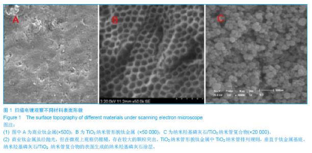
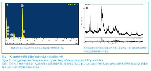
2.1 扫描电镜观察商业钛金属、TiO2纳米管形貌钛金属、纳米羟基磷灰石/TiO2 纳米管复合物的表面形貌 见图1。 图1为商业钛金属、TiO2纳米管形貌钛金属与纳米羟基磷灰石/TiO2 纳米管复合载体表面形貌的扫描电镜图片,可见商业钛金属虽经抛光,但在微观上观察仍粗糙,存在较大的颗粒突出;经阳极氧化技术生成的TiO2纳米管排列规则,垂直于钛金属基底。EDX 分析示TiO2 纳米管的主要化学成分为TiO2(图2A)。图1C为TiO2纳米管经过电沉积处理后生成的纳米羟基磷灰石/TiO2纳米管复合物,经X射线衍射分析涂层证实涂层成分基本成分为羟基磷灰石(图2B)。"
"
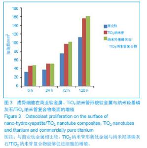
2.2 倒置显微镜观察不同材料周围小鼠成骨细胞MC-3T3-E1的生长情况 TiO2纳米管形貌钛金属、纳米羟基磷灰石/TiO2纳米管复合物与小鼠成骨细胞MC-3T3-E1共培养3 d后,经倒置显微镜观察可见复合物周围的细胞生长状态良好,细胞的形态良好,未见复合物抑制细胞的生长,与商业钛金属相比较可见纳米羟基磷灰石/TiO2纳米管复合物能够促进细胞的增殖(图3)。"

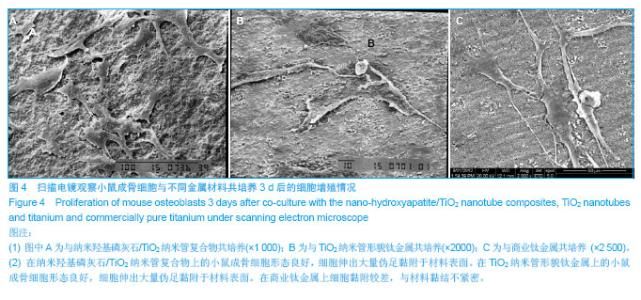
2.3 细胞在不同材料上的黏附与增殖 图4A显示为小鼠成骨细胞MC-3T3-E1在纳米羟基磷灰石/TiO2纳米管复合物上的增殖情况,图4B显示为小鼠成骨细胞MC-3T3-E1在TiO2纳米管形貌钛金属上的增殖情况,可见两组细胞在复合物上的生长状态良好,细胞形态良好,可见细胞的大量伪足附着于复合物上,说明纳米羟基磷灰石/TiO2纳米管复合体具有良好的生物相容性。图4C显示小鼠成骨细胞在商业钛金属上的生长情况,可见细胞伸出的伪足少于TiO2纳米管形貌钛金属及纳米羟基磷灰石/TiO2纳米管复合物组。"

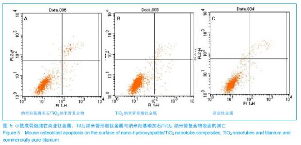
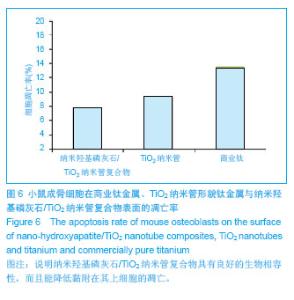
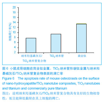
2.4 细胞在不同材料上的凋亡情况 将小鼠成骨细胞MC-3T3-E1与纳米羟基磷灰石/TiO2纳米管复合物共培养3 d后,经胰酶消化材料表面的细胞,使用Annexin-V与PI双染后流式细胞术检测凋亡情况(图5),结果显示纳米羟基磷灰石/TiO2纳米管复合物上的细胞凋亡率(早期凋亡与晚期凋亡)为7.8%,TiO2纳米管形貌钛金属表面细胞的凋亡率是9.4%,而商业钛金属表面细胞的凋亡率为13.5%,说明纳米羟基磷灰石/TiO2纳米管复合物不仅具有良好的生物相容性,而且能降低黏附在其上细胞的凋亡(图6)。"
"
| [1]Lentino JR.Prosthetic joint infections: Bane of orthopedists, challenge for infectious disease specialists.Clin Infect Dis. 2003;36:1157-1161.[2]Darouiche RO. Treatment of infections associated with surgical implants. New Engl J Med.2004;350:1422-1429.[3]Li S,Huang B,Chen Y,et al.Hydroxyapatite-coated femoral stems in primary total hip arthroplasty: a meta-analysis of randomized controlled trials. Int J Surg.2013;11(6):477-482. [4]Oonishi H.A long term histological analysis of effect of interposed hydroxyapatite between bone and bone cement in THA and TKA.J Long Term Eff Med Implants.2012;22(2): 165-176.[5]Munzinger U,Guggi T,Kaptein B,et al.A titanium plasma-sprayed cup with and without hydroxyapatite-coating: a randomised radiostereometric study of stability and osseointegration.Hip Int.2013;23(1):33-39. [6]Cossetto DJ.Mid-term outcome of a modular, cementless, proximally hydroxyapatite-coated, anatomic femoral stem.J Orthop Surg. 2012;20(3):322-326.[7]Herrera A, Mateo J, Lobo-Escolar A et al. Long-term outcomes of a new model of anatomical hydroxyapatite-coated hip prosthesis.J Arthroplasty. 2013; 28(7):1160-1166. [8]Eschen J,Kring S,Brix M,et al.Outcome of an uncemented hydroxyapatite coated hemiarthroplasty for displaced femoral neck fractures: a clinical and radiographic 2-year follow-up study.Hip Int.2012;22(5):574-579.[9]Lazarinis S,Kärrholm J,Hailer NP.Effects of hydroxyapatite coating of cups used in hip revision arthroplasty.Acta Orthop. 2012;83(5):427-435.[10]Melton JT,Mayahi R,Baxter SE,et al.Long-term outcome in an uncemented, hydroxyapatite-coated total knee replacement: a 15- to 18-year survivorship analysis.J Bone Joint Surg Br. 2012;94(8):1067-1070.[11]Martin JY,Schwartz Z,Hummert TW,et al.Effect of titanium surface roughness on proliferation, differentiation, and protein synthesis of human osteoblast-like cells (MG63).J Biomed Mater Res.1995;29(3):389-401.[12]Del Curto B,Brunella MF,Giordano C,et al.Decreased bacterial adhesion to surface-treated titanium.Int J Artif Organs.2005;28(7):718-730.[13]Rajesh P,Muraleedharan CV,Komath M,et al.Laser surface modification of titanium substrate for pulsed laser deposition of highly adherent hydroxyapatite. J Mater Sci Mater Med. 2011;22(7):1671-1679.[14]Popat KC,Eltgroth M,Latempa TJ,et al.Decreased Staphylococcus epidermis adhesion and increased osteoblast functionality on antibiotic-loaded titania nanotubes. Biomaterials. 2007;28(32):4880-4888.[15]George EA,Chang Y,Thomas JW.Enhanced osteoblast adhesion to drug-coated anodized nanotular titanium safaces. Int J Nanomed.2008;3:257-264.[16]Peng L,Mendelsohn AD,Latempa TJ,et al.Long-term small molecule and protein elution from TiO(2) nanotubes.Nano Lett.2009;9(5):1932-1936.[17]田昂,王超,管格非,等.Nano-HA 涂层与HA/ CTS 复合涂层抑制胶质母细胞瘤细胞活力作用的比较[J].功能材料,2010,2(41): 300-303.[18]Tian A,Xue XX,Liu C,et al.Electrodeposited hydroxyapatite coatings in static magnetic field.MaterLett.2010;64(10): 1197-1199.[19]Tian A,Wang C,Xue XX,et al.Inhibitory Effect of Nano-HA Particles on Human U87 Glioblastoma Cells Viability. J Inorganic Mater.2010;25(1): 101-106.[20]Piao Z,Qiu J,Wu Y,et al.Effects of the nano-tubular anodic TiO2 buffer layer on bioactive hydroxyapatite coating.J Nanosci Nanotechnol.2011;11(1):286-290.[21]Tsuchiya H, Macak JM, Müller L,et al.Hydroxyapatite growth on anodic TiO2 nanotubes.J Biomed Mater Res A.2006; 77(3):534-541.[22]Oh SH,Finõnes RR,Daraio C,et al.Growth of nano-scale hydroxyapatite using chemically treated titanium oxide nanotubes.Biomaterials.2005;26(24):4938-4943.[23]Li W, Kabius B,Auciello O.Science and technology of biocompatible thin films for implantable biomedical devices.Conf Proc IEEE Eng Med Biol Soc. 2010;2010: 6237-6242.[24]Bose S,Roy M,Das K,et al.Surface modification of titanium for load-bearing applications.J Mater Sci Mater Med.2009;20 Suppl 1:S19-24.[25]Xie J,Luan BL.Nanometer-scale surface modification of Ti6Al4V alloy for orthopedic applications.J Biomed Mater Res A.2008;84(1):63-72.[26]Yang BC,Uchida M,Kim HM,et al.Preparation of bioactive titanium metal via anodic oxidation treatment.Biomaterials. 2004;25(6):1003-1010.[27]Cooper LF.A role for surface topography in creating and maintaining bone at titanium endosseous implants.J Prosthet Dent.2000;84(5):522-534.[28]Webster TJ,Ejiofor JU.Increased osteoblast adhesion on nanophase metals: Ti, Ti6Al4V, and CoCrMo.Biomaterials. 2004;25:4731-4739.[29]Shinto Y,Uchida A,Korkusuz F,et al.Calcium hydroxyapatite ceramic used as a delivery system for antibiotics.J Bone Joint Surg Br.1992;74(4):600-604.[30]Brammer KS,Seunghan O,Cobb CJ,et al.Improved bone-forming functionality on diameter-controlled TiO2 nanotube surface. Acta biomater.2009;5:3215-3223.[31]von Wilmowsky C,Bauer S,Lutz R,et al.In vivo evaluation of anodic TiO2 nanotubes: an experimental study in the pig.J Biomed Mater Res B. 2009;89(1):165-171.[32]Bjursten LM,Rasmusson L,Oh S,et al.Titanium dioxide nanotubes enhance bone bonding in vivo.2010;92(3): 1218-1224. |
| [1] | Chen Song, He Yuanli, Xie Wenjia, Zhong Linna, Wang Jian. Advantages of calcium phosphate nanoparticles for drug delivery in bone tissue engineering research and application [J]. Chinese Journal of Tissue Engineering Research, 2021, 25(22): 3565-3570. |
| [2] | Gan Zhoujie, Pei Xibo. Enzyme-responsive nanoparticles in tumor therapy: superiority of nanoparticles in accumulation and drug release [J]. Chinese Journal of Tissue Engineering Research, 2021, 25(16): 2562-2568. |
| [3] | Li Xuan, Lu Min, Li Mingxing, Ao Meng, Tang Linmei, Zeng Zhen, Hu Jingwei, Huang Zhiqiang, Xuan Jiqing. In vitro multi-modal imaging of magnetic targeted nanoparticles and their targeting effect on hepatic stellate cells [J]. Chinese Journal of Tissue Engineering Research, 2020, 24(4): 566-571. |
| [4] | Xiang Haibin, Li Xinxia, Liang Qiuzhen, Song Xinghua. Specific bone-targeting nanoscale drug delivery system: advantages and clinical applicability [J]. Chinese Journal of Tissue Engineering Research, 2020, 24(4): 612-618. |
| [5] | Gao Jianbo, Xia Bing, Li Shengyou, Yang Yujie, Ma Teng, Yu Peng, Luo Zhuojing, Huang Jinghui. Effect of nanoparticles carrying chondroitin sulfate ABC on the migration of Schwann cells in a magnetic field [J]. Chinese Journal of Tissue Engineering Research, 2020, 24(28): 4526-4532. |
| [6] | Li Hongcai, Ma Zhuang, Guo Youling. Biological behaviors of fibroblasts on the surface of TiO2 nanotubes with different diameters after annealing treatment [J]. Chinese Journal of Tissue Engineering Research, 2020, 24(22): 3504-3509. |
| [7] | Zhang Zhongyan, Li Yubo, Qi Tongning, Chang Tao. Three-dimensional printed polylactic acid resin humerus combined with bioactive coating promotes osteoblast adhesion and increases antibacterial ability [J]. Chinese Journal of Tissue Engineering Research, 2020, 24(16): 2485-2492. |
| [8] | Yuan Bo, Wang Zhiwei, Tang Yifan, Zhou Shengyuan, Chen Xiongsheng, Jia Lianshun. Construction of polycaprolactone-tricalcium phosphate with different mixture ratios using three-dimensional printing technology and its osteoinductivity in vitro [J]. Chinese Journal of Tissue Engineering Research, 2019, 23(6): 821-826. |
| [9] | Li Yuling, Jiang Ke, Chen Lu, Yu Peng, Chen Qian, Qiao Bo, Jiang Dianming. Preparation and in vitro biocompatibility of a novel ternary biomaterial, yttria-stabilized zirconia reinforced nano-hydroxyapatite/polyamide 66 [J]. Chinese Journal of Tissue Engineering Research, 2019, 23(6): 930-935. |
| [10] | Li Wuyuntana, Zhao Lingyan, Zhang Wei, Xia Yuan, Yang Liqun. In vitro enzymatic degradation of poly(trimethylene carbonate): influencing factors and their effects [J]. Chinese Journal of Tissue Engineering Research, 2019, 23(6): 945-950. |
| [11] | Zong Qiang, Xu Yanan, Qu Tianyi, Li Lijun, Hai Miti·Abuduaini, Ni Dongkui. Magnetic verapamil nanoparticles promote peripheral nerve regeneration [J]. Chinese Journal of Tissue Engineering Research, 2019, 23(34): 5425-5429. |
| [12] | Li Yongheng, Cui Yan, Zhang Zhiyu. Inhibitory effects of doxorubicin-loaded chitosan nanoparticles on osteosarcoma in mice [J]. Chinese Journal of Tissue Engineering Research, 2019, 23(26): 4194-4199. |
| [13] | Du Jianhong, Fan Chunshui, Du Yuelian, Sun Xiaoyu, Wang Qingfeng. Preparation, sustained release and anti-tumor characteristics of hydroxypropyl chitosan/heparin nano-drug system [J]. Chinese Journal of Tissue Engineering Research, 2019, 23(22): 3536-3541. |
| [14] | Wei Wei, Liu Yanfei, Zhang Ling, Xiong Na . Self-assembling peptide hydrogel: hemostatic effect and mechanism [J]. Chinese Journal of Tissue Engineering Research, 2019, 23(2): 310-316. |
| [15] | Zou Qiang, Sun Yu, Li Xuanze, Wu Zhanyu, Yang Long, Wang Jianji, Liu Qin, Ma Minxian, Ye Chuan. Bioprinting of 3D structure with micro-network fluidic channels [J]. Chinese Journal of Tissue Engineering Research, 2019, 23(2): 265-271. |
| Viewed | ||||||
|
Full text |
|
|||||
|
Abstract |
|
|||||