Chinese Journal of Tissue Engineering Research ›› 2020, Vol. 24 ›› Issue (16): 2485-2492.doi: 10.3969/j.issn.2095-4344.2255
Previous Articles Next Articles
Three-dimensional printed polylactic acid resin humerus combined with bioactive coating promotes osteoblast adhesion and increases antibacterial ability
Zhang Zhongyan1, Li Yubo1, Qi Tongning2, Chang Tao2
- 1Department of Traumatic Orthopedics, Affiliated Hospital of Chengde Medical University, Chengde 067000, Hebei Province, China; 2Department of Traumatic Orthopedics, Chengde City Hospital, Chengde 067000, Hebei Province, China
-
Received:2019-06-11Revised:2019-06-12Accepted:2019-07-13Online:2020-06-08Published:2020-03-24 -
Contact:Zhang Zhongyan, Department of Traumatic Orthopedics, Affiliated Hospital of Chengde Medical University, Chengde 067000, Hebei Province, China -
About author:Zhang Zhongyan, Master, Attending physician, Department of Traumatic Orthopedics, Affiliated Hospital of Chengde Medical University, Chengde 067000, Hebei Province, China -
Supported by:the Self-Financing Project of Chengde Science and Technology Program, No. 201707A008
CLC Number:
Cite this article
Zhang Zhongyan, Li Yubo, Qi Tongning, Chang Tao. Three-dimensional printed polylactic acid resin humerus combined with bioactive coating promotes osteoblast adhesion and increases antibacterial ability [J]. Chinese Journal of Tissue Engineering Research, 2020, 24(16): 2485-2492.
share this article
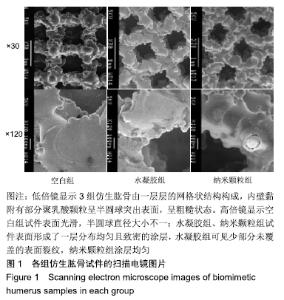
2.1 各组试件表征结果 打印试件为5 cm的正方形结构,表面粗糙,有少许凸起,内部有少许网状孔径。扫描电镜观低倍镜显示仿生肱骨由一层层的网格状结构构成,内壁黏附有部分聚乳酸颗粒呈半圆球突出表面,呈粗糙状态;高倍镜显示仿生肱骨表面光滑,半圆球直径大小不一,水凝胶组、纳米颗粒组试件表面形成了一层分布均匀且致密的涂层,水凝胶组可见少部分未覆盖的表面裂纹,纳米颗粒组涂层均匀,见图1。 "

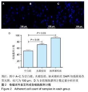
2.2 各组细胞黏附计数结果 细胞接种后8 h,荧光显微镜显示成骨细胞在各组试件上呈单层黏附生长,见图2。纳米颗粒组细胞黏附数显著高于水凝胶组(P < 0.05),水凝胶组细胞黏附数显著高于空白组(P < 0.05),定量统计分析显示纳米颗粒组细胞黏附数为水凝胶组的1.1倍,水凝胶组为空白组的1.2倍。 "
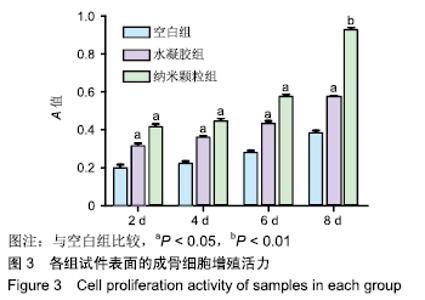
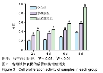
2.3 各组细胞增殖能力测定结果 细胞接种在试件上后培养2,4,6,8 d后,纳米颗粒组、水凝胶组的细胞增殖能力显著高于空白组(P < 0.05),纳米颗粒组与水凝胶组的细胞增殖能力的细胞增殖能力比较差异无显著性意义(P > 0.05),见图3。 "
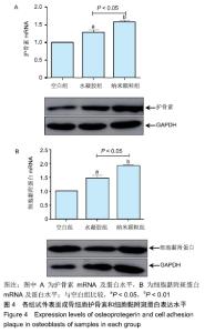
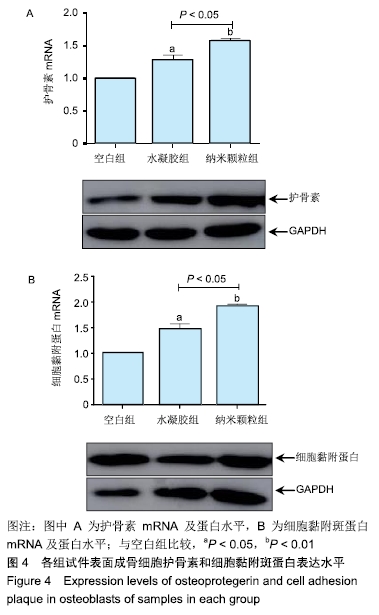
2.4 各组护骨素和细胞黏附斑蛋白表达水平 纳米颗粒组护骨素及细胞黏附斑蛋白mRNA和蛋白水平均显著高于空白组、水凝胶组(P < 0.05或P < 0.01),水凝胶组高于空白组(P < 0.05),见图4。 "
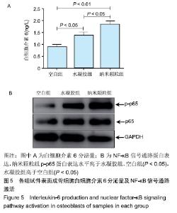
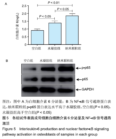
2.5 抗菌因子分泌及NF-κB信号通路激活情况 培养细胞8 d后,纳米颗粒组白细胞介素6分泌量显著高于水凝胶组、空白组(P < 0.05),可见复合生物涂层促进细胞分泌抗炎症因子提高抗菌能力;同时细胞中NF-κB信号通路激活检测结果显示纳米颗粒组p-p65蛋白表达水平高于水凝胶组、空白组(P < 0.05),温敏水凝胶组高于空白组(P < 0.05),见图5。NF-κB信号通路的激活会导致细胞分泌抗炎因子,提高细胞免疫抵抗能力,该结果和白细胞介素6分泌结果一致。 "
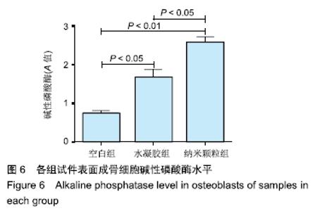
2.6 各组碱性磷酸酶测定结果 培养细胞8 d后,纳米颗粒组碱性磷酸酶活性显著高于水凝胶组、空白组(P < 0.05,P < 0.01),水凝胶组显著高于空白组(P < 0.05),见图6。 "
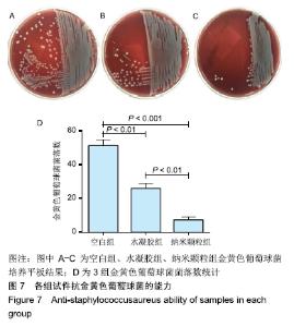
2.7 抗金黄色葡萄球菌能力检测结果 纳米颗粒组抗菌能力显著高于水凝胶组、空白组(P < 0.01,P < 0.001),水凝胶组显著高于空白组(P < 0.01),见图7。可见聚乳酸结合复合生物涂层以后能显著提高抗菌能力,这也符合前面白细胞介素6的检测结果,聚乳酸复合生物涂层后促进细胞的黏附、增殖,间接促进细胞的抗菌能力。 "


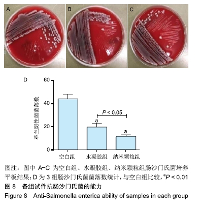
2.8 抗肠沙门氏菌能力检测结果 纳米颗粒组抗沙门氏菌能力显著高于水凝胶组、空白组(P < 0.05,P < 0.01),水凝胶组显著高于空白组(P < 0.01),见图8。可见聚乳酸结合复合生物涂层以后能显著提高细胞的抗肠沙门氏菌能力。 "
| [1] 施庆锋.基于聚乳酸的生物可降解复合材料的制备和研究[D].上海:华东理工大学,2011. [2] 邹俊.聚乳酸及其纳米复合材料的研究[D].上海:华东理工大学, 2011. [3] 桂宗彦.聚乳酸材料的改性研究[D].上海:华东理工大学,2012. [4] 王鑫众.PLA/MSM缓释微球的制备及骨关节炎治疗[D].长春:吉林大学,2013. [5] 张薷文,周延民,赵静辉.种植体骨结合相关检测手段研究现状[J].实用口腔医学杂志,2013,29(5):730-734. [6] 周弘.种植体表面不同形貌对骨质疏松症骨结合的影响[D].西安:第四军医大学,2011. [7] 石磊.种植体表面改性—光诱导超亲水性表面骨结合机制研究[D].西安:第四军医大学, 2013. [8] 唐刚.聚乳酸/次磷酸盐复合材料的制备、阻燃机理以及烟气毒性研究[D].合肥:中国科学技术大学,2013. [9] 宋亚男.聚乳酸基复合材料的性能与结构研究[D].大连:大连理工大学,2013. [10] 张绍清,蔡洪桢,王飞翔,等.钛基钽涂层人工种植体植入不同部位的骨结合对比研究[J].口腔颌面修复学杂志, 2018,19(2): 106-112. [11] 王方辉,张姗姗,舒静媛,等.纯钛种植体表面改性对骨结合的影响[J].中国组织工程研究,2014,18(52):8491-8497. [12] 叶川,马敏先,张弢,等.Ⅰ型胶原修饰纯钛片促进人脂肪间充质干细胞增殖[J].中国组织工程研究,2014,18(25):4032-4037. [13] RICARD-BLUM S, RUGGIERO F.The collagen superfamily: From the extracellular matrix to the cell membrane.Pathol Biol(Paris).2005;53(7):430-442. [14] MORRA M, CASSINELLI C, MEDA L, et al.Surface analysis and effects on interfacial bone microhardness of collagen-coated titanium implants: A rabbit model.Int J Oral Maxillofac Implants.2005;20(1):23-30. [15] ZHANG H, SUN ZQ, ZHOU LY, et al.Polylactic acid / polycaprolactone / poly (ε-caprolactone-L-lactide) copolymerstructure and performance of ternary blend system.Chin J Appl Chem.2016;32(9):1027-1031. [16] GUO XJ, ZHANG JW, HUANG JJ, et al.Poly(lactic acid)/polyoxymethylene blends:Morphology,crystallization, rheology, and thermal mechanical properties.Polymer. 2015; 69:103-109. [17] SHAHMORADI S, YAZDIAN F, TABANDEH F, et al.Controlled surface morphology and hydrophilicity of polycaprolactone toward human retinal pigment epithelium cells.Mater Sci Eng C Mater Biol Appl.2017;73:300-309. [18] YANG FW, LIN YL, TIAN XM, et al.Preparation of PLA/PCL Blend and Its Application on Medical Materials.Eng Plastic Appl.2014;2(42):1-5. [19] WU A, CHEN L, XIONG ZC, et al.Preparation and Characterization Study of Poly (ε-caprolactone-co-L-lactide)/ Poly L-lactic Acid Composites.China Plastic Industry. 2014; 42(2):47-51. [20] LI B, LI Y, LI J, et al.Influence of nanostructures on the biologicalproperties of ti implants after anodic oxidation.J Mater Sci Mater Med.2014;25(1):199-205. [21] 李赛娜,康跻耀,高建萍,等.胶原涂层对3d 打印种植体表面生物相容性的影响[J].中国组织工程研究,2017,21(10): 1558-1564. [22] TAKAYAMA T, TODO M, ARAKAWA K, et al.Characterization of Impact Fracture Behavior of BiodegradablePLA/PCL Polymer Blend.Key Eng Mater.2006;328:1569-1572. [23] GAAL G, MENDES M, DE ALMEIDA TP, et al.Simplified fabrication of integrated microfluidic devices using fused deposition modeling 3D printing.Sensor Actuat B-Chem. 2017;242:35-40. [24] 王素军.基于聚乳酸的靶向药物释放载体材料的研究[D].重庆:重庆大学,2012. [25] JIN Y, WAN Y, ZHANG B, et al.Modeling of the chemical finishing process for polylactic acid parts infused deposition modeling and investigation of its tensile properties.J Mater Proc Technol.2017;240:233-239. [26] CARRICO1 JD, TRAEDEN NW, AURELI M, et al.Fused filament 3D printing of ionic polymer-metal composites (IPMCs).Smart Mater Struct.2015;24(12):1-12. [27] 田玲玲.聚乳酸—己内酯乳液静电纺组织工程支架的制备及性能评价[D].上海:东华大学, 2014. [28] 邵俊东.聚乳酸纳米纤维支架的构建与改性及其纳米力学性能研究[D].广州:华南理工大学,2013. [29] 赵永青.生物降解聚乳酸基复合材料的制备与性能研究[D].兰州:兰州大学,2009. [30] ROCHA CR, TORRADO PEREZ AR, ROBERSON DA. Novel ABS-based binary and ternary polymer blends for materialextrusion 3D printing.J Mater Res.2014;29(17): 1859-1866. [31] SHIN BY, HAN DH. Compatibilization of immisciblepoly(lactic acid)/poly(e-caprolactone)blend through electron-beam irradiation with the addition of acompatibilizing agent.Radiat Phys Chem.2013;83:98-104. [32] ZHAN X, ZHANG C, DISSANAYAKA WL, et al.Storage media enhance osteoclastogenic potential of human periodontal ligament cells via rankl-independent signaling. Dent Traumatol. 2013;29(1):59-65. [33] DOUGLAS P, ALBADARIN AB, AL-MUHTASEB AH, et al. Al-Muhtase.Thermo-mechanical properties of polyε-caprolactone/poly L-lactic acidblends: Addition of nalidixic acidand polyethylene glycol additives.J Mech Behav Biomed Mater.2015;45:154-165. [34] 刘鹏.抗结核药物缓释微球在兔椎体中释放及分布规律的研究[D].广州:南方医科大学,2013. [35] 向琳,陈晖璐,袁影,等.降钙素基因相关肽对种植体周围神经、血管再生及骨结合的作用[J].国际口腔医学杂志, 2018,45(5): 509-515. [36] 吴倩,李德超,张慧明,等.钛合金微弧氧化载抗菌肽涂层的动物体内骨结合行为[J]. 中国体视学与图像分析,2018,23(1):1-8. [37] 陈江,陈旭晞,周麟.骨免疫调节机制对种植体骨结合及骨生物材料引导骨再生的影响[J].口腔疾病防治,2018,26(10):613-620. [38] VON WILMOWSKY C, BAUER S, ROEDL S, et al.The diameter of anodic tio2 nanotubes affects bone formation and correlates with the bone morphogenetic protein-2 expression in vivo.Clin Oral Implants Res.2012;23(3):359-366. [39] 李养军.ILK在糖尿病视网膜病变中作用的研究[D].西安:第四军医大学,2007. [40] 刘佩,彭晓燕,逄明杰,等.Ghrelin,内脂素和护骨素在Ⅱ型糖尿病发病机制中的作用研究[J].现代生物医学进展, 2016,16(18): 3423-3426. [41] KOWALCZYK M, PIORKOWSKA E, DUTKIEWICZ S, et al. Toughening of polylactide by blending with a novel randomaliphatic–aromatic copolyester.Eur Polym J.2014;59: 59-68. [42] 鲍启忠,朱康,马明.护骨素和κB-受体激活因子水平变化对缺血性股骨头坏死保护作用效果及作用机制分析[J].临床和实验医学杂志,2018,17(13):1410-1412. [43] 邓云龙,李德超,李慕勤,等.纯钛表面GRGDSP多肽对骨结合能力的影响[J].中国口腔颌面外科杂志,2018,16(3):199-204. [44] SUL YT.Electrochemical growth behavior, surface properties, and enhanced in vivo bone response of tio2 nanotubes on microstructured surfaces of blasted, screw-shaped titanium implants.Int J Nanomedicine.2010;5:87-100. [45] WANG Y, WEI Z, LI Y. Highlytoughened polylactide/epoxidized poly(styrene-bbutadiene-b-styrene) blends with excellent tensile performance. Eur Polym J.2016;85:92104. |
| [1] | Wang Debin, Bi Zhenggang. Related problems in anatomy mechanics, injury characteristics, fixed repair and three-dimensional technology application for olecranon fracture-dislocations [J]. Chinese Journal of Tissue Engineering Research, 2021, 25(9): 1446-1451. |
| [2] | Li Cai, Zhao Ting, Tan Ge, Zheng Yulin, Zhang Ruonan, Wu Yan, Tang Junming. Platelet-derived growth factor-BB promotes proliferation, differentiation and migration of skeletal muscle myoblast [J]. Chinese Journal of Tissue Engineering Research, 2021, 25(7): 1050-1055. |
| [3] | Liu Cong, Liu Su. Molecular mechanism of miR-17-5p regulation of hypoxia inducible factor-1α mediated adipocyte differentiation and angiogenesis [J]. Chinese Journal of Tissue Engineering Research, 2021, 25(7): 1069-1074. |
| [4] | Ma Zetao, Zeng Hui, Wang Deli, Weng Jian, Feng Song. MicroRNA-138-5p regulates chondrocyte proliferation and autophagy [J]. Chinese Journal of Tissue Engineering Research, 2021, 25(5): 674-678. |
| [5] | Wang Yujiao, Liu Dan, Sun Song, Sun Yong. Biphasic calcium phosphate loaded with advanced platelet rich fibrin can promote the activity of rabbit bone marrow mesenchymal stem cells [J]. Chinese Journal of Tissue Engineering Research, 2021, 25(4): 504-509. |
| [6] | Zhou Jihui, Yao Meng, Wang Yansong, Li Xinzhi, Zhou You, Huang Wei, Chen Wenyao. Influence of novel nanoscaffolds on biological behaviors of neural stem cells and the related gene expression [J]. Chinese Journal of Tissue Engineering Research, 2021, 25(4): 532-536. |
| [7] | Li Li, Ma Li. Immobilization of lactase on magnetic chitosan microspheres and its effect on enzymatic properties [J]. Chinese Journal of Tissue Engineering Research, 2021, 25(4): 576-581. |
| [8] | Chen Yang, Huang Denggao, Gao Yuanhui, Wang Shunlan, Cao Hui, Zheng Linlin, He Haowei, Luo Siqin, Xiao Jingchuan, Zhang Yingai, Zhang Shufang. Low-intensity pulsed ultrasound promotes the proliferation and adhesion of human adipose-derived mesenchymal stem cells [J]. Chinese Journal of Tissue Engineering Research, 2021, 25(25): 3949-3955. |
| [9] | Zhou Wu, Wang Binping, Wang Yawen, Cheng Yanan, Huang Xieshan. Transforming growth factor beta combined with bone morphogenetic protein-2 induces the proliferation and differentiation of mouse MC3T3-E1 cells [J]. Chinese Journal of Tissue Engineering Research, 2021, 25(23): 3630-3635. |
| [10] | Li Xinping, Cui Qiuju, Zeng Shuguang, Ran Gaoying, Zhang Zhaoqiang, Liu Xianwen, Fang Wei, Xu Shuaimei. Effect of modification of β-tricalcium phosphate/chitosan hydrogel on growth and mineralization of dental pulp stem cells [J]. Chinese Journal of Tissue Engineering Research, 2021, 25(22): 3493-3499. |
| [11] | Yu Chengshuai, Du Gang, Pang Shenning, Lao Shan. Chemerin, a pro-inflammatory adipokine, regulates chondrocyte proliferation and metabolism by increasing production of nitric oxide [J]. Chinese Journal of Tissue Engineering Research, 2021, 25(2): 258-263. |
| [12] | Dai Yaling, Chen Lewen, He Xiaojun, Lin Huawei, Jia Weiwei, Chen Lidian, Tao Jing, Liu Weilin. Construction of miR-146b overexpression lentiviral vector and the effect on the proliferation of hippocampal neural stem cells [J]. Chinese Journal of Tissue Engineering Research, 2021, 25(19): 3024-3030. |
| [13] | Ailimaierdan·Ainiwaer, Wang Ling, Gu Li, Dilidaer•Taxifulati, Wang Shan, Yin Hongbin. Effect of transforming growth factor-beta3 on the proliferation and osteogenic capability of osteoblasts [J]. Chinese Journal of Tissue Engineering Research, 2021, 25(17): 2664-2669. |
| [14] | Chen Siyu, Li Yannan, Xie Liying, Liu Siqi, Fan Yurong, Fang Changxing, Zhang Xin, Quan Jiayu, Zuo Lin. Thermosensitive chitosan-collagen composite hydrogel loaded with basic fibroblast growth factor retards ventricular remodeling after myocardial infarction in mice [J]. Chinese Journal of Tissue Engineering Research, 2021, 25(16): 2472-2478. |
| [15] | Liu Feng, Zhang Yu, Wang Yanli, Luo Wei, Han Chaoshan, Li Yangxin. Application of temperature-sensitive chitosan hydrogel encapsulated exosomes in ischemic diseases [J]. Chinese Journal of Tissue Engineering Research, 2021, 25(16): 2479-2487. |
| Viewed | ||||||
|
Full text |
|
|||||
|
Abstract |
|
|||||