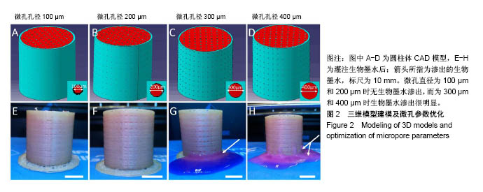
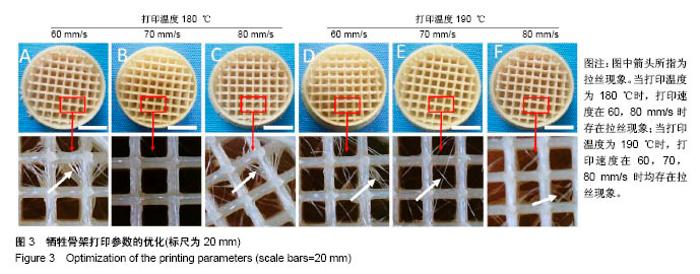
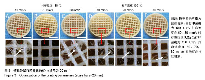
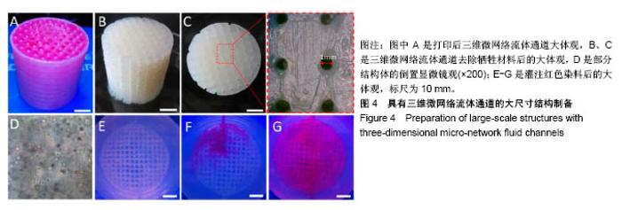
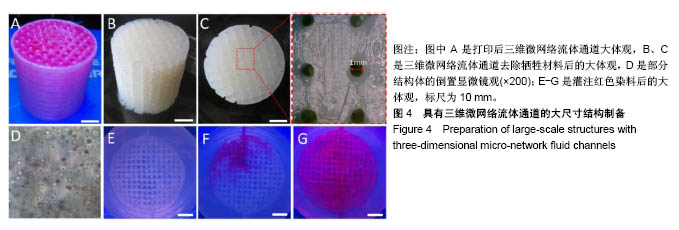
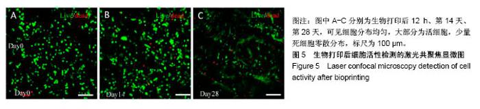
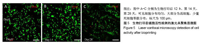
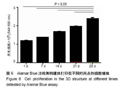
| [1] Langer R,Vacanti J.Tissue engineering.Science. 1993;260: 5110:920-926.[2] Peltola SM,Melchels FP,Grijpma DW,et al.A review of rapid prototyping techniques for tissue engineering purposes.Ann Med.2008;40(4):268-280.[3] Mironov V,Trusk T,Kasyanov V,et al.Biofabrication: a 21st century manufacturing paradigm. Biofabrication. 2009;1(2): 1-16.[4] Konig G,McAllister TN,Dusserre N,et al.Mechanical properties of completely autologous human tissue engineered blood vessels compared to human saphenous vein and mammary artery.Biomaterials. 2009;30(8:1542-1550.[5] Wang X,Yan Y,Zhang R.Recent trends and challenges in complex organ manufacturing. Tissue Eng Part B Rev. 2010; 16:2:189-197.[6] Wüst S,Müller R,Hofmann S.3D Bioprinting of complex channels-Effects of material, orientation, geometry, and cell embedding.J Biomed Mater Res A.2015;103(8):2558-2570.[7] Pati F,Ha DH,Jang J,et al.Biomimetic 3D tissue printing for soft tissue regeneration. Biomaterials.2015;62:164-175. [8] Das S,Pati F,Choi YJ,et al.Bioprintable, cell-laden silk fibroin-gelatin hydrogel supporting multilineage differentiation of stem cells for fabrication of three-dimensional tissue constructs. Acta Biomater.2015;11:233-246.[9] Gao Q,He Y,Fu JZ,et al.Coaxial nozzle-assisted 3D bioprinting with built-in microchannels for nutrients delivery. Biomaterials.2015;61:203-215.[10] Duarte CDF,Blaeser A,Buellesbach K,et al.Bioprinting Organotypic Hydrogels with Improved Mesenchymal Stem Cell Remodeling and Mineralization Properties for Bone Tissue Engineering.Adv Healthc Mater.2016;5:11:1336-45.[11] Tan YJ,Tan X,Yeong WY,et al.Hybrid microscaffold-based 3D bioprinting of multi-cellular constructs with high compressive strength: A new biofabrication strategy.Sci Rep.2016;6:39140.[12] Colosi C,Shin SR,Manoharan V,et al.Microfluidic Bioprinting of Heterogeneous 3D Tissue Constructs Using Low-Viscosity Bioink.Adv Mater.2016;28:4:677-684.[13] Blanchette CD,Knipe JM,Stolaroff JK,et al.Printable enzyme-embedded materials for methane to methanol conversion.Nat Commun.2016;7:11900.[14] He Y,Yang F,Zhao H,et al.Research on the printability of hydrogels in 3D bioprinting.Sci Rep. 2016;6:29977.[15] Yu Y,Moncal KK,Li J,et al.Three-dimensional bioprinting using self-assembling scalable scaffold-free “tissue strands” as a new bioink.Sci Rep.2016;6:28714.[16] 臧晓龙,孙健,李亚莉,等.3D生物打印构建聚乳酸羟基乙酸/纳米羟基磷灰石支架骨形态发生蛋白2缓释复合体的实验研究[J].中国组织工程研究,2016,20(16):2405-2411.[17] 宋杨,王晓飞,王宇光,等.人脂肪间充质干细胞与生物材料共混物三维打印体的体内成骨[J].北京大学学报(医学版), 2016,48(1): 45-50.[18] Jang J,Park HJ,Kim SW,et al.3D printed complex tissue construct using stem cell-laden decellularized extracellular matrix bioinks for cardiac repair. Biomaterials. 2017;112: 264-274.[19] Carvalho AF, Gasperini L,Ribeiro RS,et al.Control of osmotic pressure to improve cell viability in cell-laden tissue engineering constructs.J Tissue Eng Regen Med. 2018;12(2): e1063-e1067. [20] Costantini M,Testa S,Mozetic P,et al.Microfluidic-enhanced 3D bioprinting of aligned myoblast-laden hydrogels leads to functionally organized myofibers in vitro and in vivo. Biomaterials.2017;131:98-110. [21] Ong CS,Fukunishi T,Zhang H,et al.Biomaterial-Free Three-Dimensional Bioprinting of Cardiac Tissue using Human Induced Pluripotent Stem Cell Derived Cardiomyocytes.Sci Rep.2017;7(1):4566.[22] Chrobak KM,Potter DR,Tien J.Formation of perfused, functional microvascular tubes in vitro. Microvasc Res. 2006;71(3):185-196.[23] Ozbolat IT,Yu Y.Bioprinting toward organ fabrication: challenges and future trends.IEEE Trans Biomed Eng. 2013;60(3):691-699.[24] Paulsen SJ,Miller JS.Tissue vascularization through 3D printing: Will technology bring us flow?Dev Dyn. 2015;244(5): 629-640. [25] Lee VK,Kim DY,Ngo H,et al.Creating perfused functional vascular channels using 3D bio-printing technology. Biomaterials.2014;35(28):8092-8102.[26] 杨道朋,夏旭.3D打印生物材料研究及其临床应用优势[J].中国组织工程研究,2017,21(18):2927-2933.[27] Kundu J,Shim JH,Jang J,et al.An additive manufacturing-based PCL-alginate-chondrocyte bioprinted scaffold for cartilage tissue engineering.J Tissue Eng Regen Med.2015;9(11):1286-1297.[28] 刘伟,刘萌,祝劲松,等.人骨髓间充质干细胞的体外培养、鉴定及成骨分化[J].中国组织工程究,2012,16(14):2515-2519.[29] Ling Y,Rubin J,Deng Y,et al.A cell-laden microfluidic hydrogel. Lab Chip.2007;7(6):756-762.[30] Novosel EC,Kleinhans C,Kluger PJ.Vascularization is the key challenge in tissue engineering. Adv Drug Deliv Rev. 2011; 63:(4-5):300-311.[31] Bertassoni LE,Cecconi M,Manoharan V,et al.Hydrogel bioprinted microchannel networks for vascularization of tissue engineering constructs.Lab Chip.2014;14(13):2202-2211.[32] Kolesky DB,Homan KA,Skylar-Scott MA,et al. Three-dimensional bioprinting of thick vascularized tissues. Proc Natl Acad Sci U S A.2016;113(12):3179-3184.[33] Richards D,Jia J,Yost M,et al.3D Bioprinting for Vascularized Tissue Fabrication.Ann Biomed Eng.2017;45(1):132-147.[34] Fedorovich NE,Swennen I,Girones J,et al.Evaluation of photo crosslinked lutrol hydrogel for tissue printing applications.Biomacromolecules.2009;10(7):1689-1696.[35] 宋杨,王晓飞,王宇光,等.人脂肪间充质干细胞与生物材料共混物三维打印体的体内成骨[J].北京大学学报(医学版), 2016,48(1): 45-50.[36] 薛世华,吕培军,王勇,等.人牙髓细胞共混物三维生物打印技术[J].北京大学学报(医学版),2013,45(1):105-108.[37] Köpf M,Campos DF,Blaeser A,et al.A tailored three-dimensionally printable agarose-collagen blend allows encapsulation, spreading, and attachment of human umbilical artery smooth muscle cells. Biofabrication.2016;8(2):025011.[38] Wu Z,Xin S,Xu Y,et al.Bioprinting three-dimensional cell-laden tissue constructs with controllable degradation.Sci Rep. 2016; 6:24474. |