Chinese Journal of Tissue Engineering Research ›› 2020, Vol. 24 ›› Issue (35): 5708-5714.doi: 10.3969/j.issn.2095-4344.2876
Previous Articles Next Articles
Advantages of three-dimensional printing technology in neurosurgery
Wen Xichao, Zeng Zhaomu, Tang Yushan, Fu Meijuan, Wu Wensong, Zheng Kebin
Affiliated Hospital of Hebei University, Baoding 071000, Hebei Province, China
-
Received:2020-02-28Revised:2020-03-06Accepted:2020-03-24Online:2020-12-18Published:2020-10-19 -
Contact:Zheng Kebin, MD, Chief physician, Affiliated Hospital of Hebei University, Baoding 071000, Hebei Province, China -
About author:Wen Xichao, Master candidate, Affiliated Hospital of Hebei University, Baoding 071000, Hebei Province, China -
Supported by:Graduate Innovation Fund of Hebei University in 2020, No. hbu2020ss057
CLC Number:
Cite this article
Wen Xichao, Zeng Zhaomu, Tang Yushan, Fu Meijuan, Wu Wensong, Zheng Kebin. Advantages of three-dimensional printing technology in neurosurgery[J]. Chinese Journal of Tissue Engineering Research, 2020, 24(35): 5708-5714.
share this article
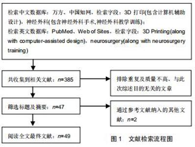
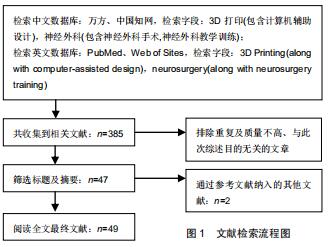
1.3 文章筛选流程 共检索、收集到相关文献385篇,排除与研究目的无关及重复研究336篇,最终纳入49篇符合标准的中英文文献进行综述,见图1。 "
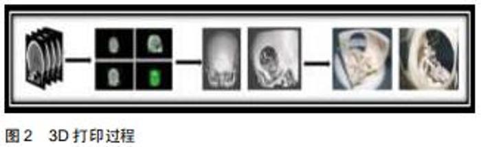
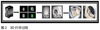
2.1.1 3D打印技术的概念及原理 3D打印又被称为增材制造、快速原型制造或者实体自由制造,是一种在计算机精确控制下,将三维数字模型用多种可黏性材料逐步堆积为实物模型的快速成型技术[2]。3D打印所用的原材料种类广泛,可以是陶瓷、塑料、金属,甚至可以是活的细胞[3]。其打印的完整过程可以分为3个步骤:①收集目标部位影像学资料;②依据影像学数据,通过计算机建模软件进行三维图像建模;③将三维图形的数据资料导入3D打印机,指导打印机逐层打印。具体过程如下:首先获取目标人体部位的MRI或CT薄层二维图像(DICOM)的数据,然后将影像学数据导入Mimics、CAD 等三维立体化软件,在各软件中构建目标区域的三维数字化模型,将得到的三维图形转化为STL(Standard Template Library)格式文件,最后将STL导入3D打印机进行分层制造,再利用激光束或热熔喷嘴等方法精确堆积材料、逐层累积,最终形成所需结构的1∶1实体三维结构[4],见图2[5]。 "
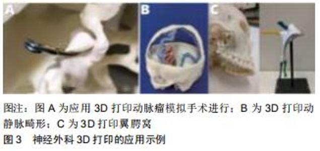
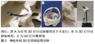
(3)脑血管病:颅内动脉瘤是颅内动脉壁局限或弥漫性扩张、膨出,颅内动脉瘤破裂导致的脑出血为神经外科急重症之一,其死亡率高,入院后需尽快干预,目前常采用的手术方式包括开颅动脉瘤夹闭、开颅动脉瘤包裹及动脉瘤介入栓塞[18]。选择合理的手术方案是保证手术效果的关键,但颅底动脉分支较多且动脉瘤生长形态各异,因而手术难度较高。SULLIVAN等[19]为1例梭形颅内动脉瘤患儿3D打印个体化模拟器,并于术前在该模型上进行神经血管内预演,练习分流器的放置,提高了复杂脑动脉瘤的术中准确性,降低了手术风险。LAN 等[20]收集动脉瘤患者模型,建立了脑动脉、动脉瘤的3D打印模型,并对模型进行模拟手术,评估不同锁孔入路的可行性,并选择合适的动脉瘤夹,为锁孔手术提供了有效的手术依据,术后数字血管造影显示全部动脉瘤完全夹闭。ALBA等[21]创建了复杂动脉瘤三维1∶1模型,帮助术者选择最佳手术入路及手术方式,可更好地观察和控制手术过程,避免对周围血管造成损伤,达到最佳手术效果。LEAL等[5]对3D打印动脉瘤进行了新尝试,制作三维空心弹性动脉瘤,较实心模型而言,空心模型具有更逼真的生物行为,并在颅骨基础上增加了血管区域作为手术参照系,而且这种模型制作时间更短,可更逼真地模拟实际手术中夹闭动脉瘤操作(图3A),更好地应用于动脉瘤破裂急诊手术。同时ANDERSON等[22]开发并验证了通过融合沉积创建脑动脉瘤模型的方法,证实该模型解剖学精度良好,并可作为MRI流动模型,用于流体力学研究。在此基础上,KANEKO等[23]以聚二甲基硅氧为材料运用3D打印技术建立了具有内皮细胞特性的体外基底动脉尖端动脉瘤,将复杂的血液动力学与动脉瘤生长或破裂结合,指导临床治疗。证实3D打印技术依据患者数据建立个性化模型,可以准确、真实地显示动脉瘤的解剖形态,更好地指导手术进行。 颅内动静脉畸形是一种先天性血管畸形,又称脑血管瘤,可采用手术全切除、供血动脉结扎、血管内栓塞或放射等治疗[24]。因动静脉的异常吻合,血管关系错综复杂,手术难度较大。二维影像学数据不能显示完整的血管信息,而借助3D打印技术可以让医生真实、全面地了解血管畸形团结构,更好地进行手术。墨西哥医师CONTI等[25]对10例动静脉畸形进行3D打印,完整显示了手术目标的体积轮廓,3D模型空间显示优于二维图像,模拟病变血管的解剖结构及其毗邻关系,并以此为依据制定手术方案,使术中更直接地找到病灶,缩短手术时间,提高手术准确性,减少并发症。陈光忠等[26]通过5例3D打印动静脉畸形模型,让医师全面真实地了解畸形团结构,为术者选取何种路径、何种栓塞材料提供参考,从而提高栓塞质量,减少术后并发症的发生,提高术后患者满意度。THAWANI等[27]利用影像学数据,3D打印1例巨大动静脉畸形模型,模拟动态血流变化,指导术前规划,改善了复杂血管病的治疗效果。DONG等[28]借助3D打印脑动静脉畸形模型(图3B),使病灶周围结构充分可视化,清楚识别所累及的血管及病变程度,从而对血管内治疗所需要栓塞的动脉及栓塞材料提前进行预判,减少手术时间及手术风险。SHAH等[29]为6例动静脉畸形患者构建三维模型,准确描述病灶的致密性、位置以及与颅骨的关系,清晰展现了供血动脉与引流静脉的数量、大小、走行,有助于手术的准备与进行。但是目前的研究对于病变血管的建模仍需要加工,具有一定主观性,因而建模准确性及客观性有待提高。总体来说,3D打印颅内动静脉畸形可精确显示动静脉状态,为手术提供指导作用。 2.2.2 3D打印模型用于神经外科教育 神经系统解剖结构复杂,对空间想象能力要求较高。神经外科疾病临床及影像学表现各异,手术难度较大,低年资神经外科医师手术经验较少,不易建立头颅及脊柱的三维立体结构,因而神经外科手术对于低年资医师、规范化培训医师而言具有极大挑战。将3D打印技术应用于神经外科医师教学中,可以提高神经外科医师学习兴趣,加深解剖结构理解,培养空间思维能力,快速掌握常规神经外科手术操作。翼腭窝是最复杂的解剖学区域之一,在身体解剖中可视性很差,而BANNON等[39]建立翼腭窝负空间模型(图3C),提供了复现整个解剖部位的方法,为神经外科及耳鼻喉科、眼科专业的低年资医师及研究生了解翼腭窝复杂的解剖关系提供有力帮助。TAI等[40]利用3D打印技术开发颅骨模型,构建脑室外引流模拟器,模型可显示颅内压力,可视化液体可以显示导管轨迹,获得逼真的触觉反馈,为神经外科受教育者提供新学习手段,提高脑室外引流、神经内镜脑室内肿瘤切除术等重要外科技能。相建等[41]通过3D打印带有孔道的穿刺模型,用于术前定位、术中引导穿刺,简化创伤性脑内血肿微创穿刺引流术,可提高穿刺准确性,降低手术难度。GHIZONI等[42]以聚酰胺为材料制作3例颅缝早闭模型,可以充分显示颅缝早闭的病理结构变异,同时模型可用于外科教育中额眶推进及后方牵张等手术过程的训练。这种新的聚酰胺颅缝早闭模型,允许外科医生进行重要手术步骤,提供真实的触觉反馈,是外科培训的重要辅助手段。CLIFTON等[43]使用特定的三维打印技术复植皮质松质细胞表面,创造了一种独特的C2椎板螺钉徒手放置三维打印模拟器,准确模拟活体内手术情况,从而显著提高受试者手术技能,提高住院医师、实习生徒手放置C2椎板螺钉的准确性。NAGASSA等[44]利用硅胶和弹道明胶浇铸颅骨,水溶性蜡和硅胶建立脑血管,成功构建了逼真的多组件脑动脉瘤手术模拟器,将该模拟器应用于神经外科住院医师的教育培训中,训练其对动脉瘤夹选取、动脉瘤手术方案制定、开颅手术和夹闭动脉瘤等手术技能,可显著提高对大脑中动脉动脉瘤手术的理解。LIN等[45]应用三维蝶鞍脑膜瘤模型辅助神经外科教育者进行教学培训,可使受训者深入理解肿瘤边界和周围正常组织间的毗邻关系,有助于其对脑蝶鞍部脑膜瘤的空间理解及记忆,增加对手术区域的认识,并引发学习兴趣。YI等[46]打印了三维脑室系统模型,可提高学生对复杂脑室系统解剖学的认识,提高学习兴趣与积极性,降低学习难度,提高教学效果。CLEARY等[47]建立了重复使用的开颅手术3D打印模型,应用于神经外科研究生一年级课程中,仿真模拟术中上矢状窦损伤合并空气栓塞等情况,使住院医师在动手培训方法中重现手术的压力,有效模拟手术,而不会使患者面临风险。通过3D打印神经外科常见疾病模型,作为外科手术计划的替代方案,既可提高培训学员的学习积极性,帮助其巩固解剖知识,还可以帮助他们提高阅片及手术技能,为神经外科教育教学提供新思路及新方法。 "
|
[1] CHANDEKAR A, MISHRA DK, SHARMA S, et al. 3D Printing technology: a new milestone in the development of pharmaceuticals. Curr Pharm Des. 2019;25(9):937-945.
[2] KHAN FA, NARASIMHAN K, SWATHI CSV, et al. 3D Printing technology in customized drug delivery system: current state of the art, prospective and the challenges. Curr Pharm Des. 2018; 24(42):5049-5061.
[3] MARTÍN-NOGUEROL T, PAULANO-GODINO F, RIASCOS RF, et al. Hybrid computed tomography and magnetic resonance imaging 3D printed models for neurosurgery planning. Ann Transl Med. 2019;7(22):684.
[4] OKONOGI S, KONDO K, HARADA N, et al. Operative simulation of anterior clinoidectomy using a rapid prototyping model molded by a three-dimensional printer. Acta Neurochir (Wien). 2017; 159(9):1619-1626.
[5] LEAL AG, MORI YT, NOHAMA P, et al. Three-Dimensional hollow elastic models for intracranial aneurysm clipping election - a case study. Conf Proc IEEE Eng Med Biol Soc. 2019;2019:4137-4140.
[6] QUAN H, ZHANG T, XU H, et al. Photo-curing 3D printing technique and its challenges. Bioact Mater. 2020;5(1):110-115.
[7] MAZZANTI V, MALAGUTTI L, MOLLICA F. FDM 3D Printing of polymers containing natural fillers: a review of their mechanical properties. Polymers (Basel). 2019;11(7). pii: E1094.
[8] RAMU M, ANANTHASUBRAMANIAN M, KUMARESAN T, et al. Optimization of the configuration of porous bone scaffolds made of Polyamide/Hydroxyapatite composites using Selective Laser Sintering for tissue engineering applications. Biomed Mater Eng. 2018;29(6):739-755.
[9] PARK EK, LIM JY, YUN IS, et al. Cranioplasty enhanced by three-dimensional printing: custom-made three-dimensional- printed titanium implants for skull defects. J Craniofac Surg. 2016;27(4):943-949.
[10] HUANG MT, JUAN PK, CHEN SY, et al. The potential of the three-dimensional printed titanium mesh implant for cranioplasty surgery applications: biomechanical behaviors and surface properties. Mater Sci Eng C Mater Biol Appl. 2019;97:412-419.
[11] ESSAYED WI, UNADKAT P, HOSNY A, et al. 3D printing and intraoperative neuronavigation tailoring for skull base reconstruction after extended endoscopic endonasal surgery: proof of concept. J Neurosurg. 2018;130(1):248-255.
[12] PANESAR SS, BELO JTA, D'SOUZA RN. Feasibility of clinician-facilitated three-dimensional printing of synthetic cranioplasty flaps. World Neurosurg. 2018;113:e628-e637.
[13] TAN ET, LING JM, DINESH SK. The feasibility of producing patient-specific acrylic cranioplasty implants with a low-cost 3D printer. J J Neurosurg. 2016;124(5):1531-1537.
[14] COELHO G, CHAVES TMF, GOES AF, et al. Multimaterial 3D printing preoperative planning for frontoethmoidal meningoencephalocele surgery. Childs Nerv Syst. 2018;34(4): 749-756.
[15] PIJPKER PAJ, WAGEMAKERS M, KRAEIMA J, et al. Three-Dimensional printed polymethylmethacrylate casting molds for posterior fossa reconstruction in the surgical treatment of chiari I malformation: technical note and illustrative cases. World Neurosurg. 2019;129:148-156.
[16] YUAN T, JIA G, YANG L, et al. Occipitocervical fusion combined with 3-dimensional navigation and 3-dimensional printing technology for the treatment of atlantoaxial dislocation with basilar invagination: a case report. Medicine (Baltimore). 2020;99(5): e18983.
[17] DU YQ, QIAO GY, YIN YH, et al. Usefulness of 3D printed models in the management of complex craniovertebral junction anomalies: choice of treatment strategy, design of screw trajectory, and protection of vertebral artery. World Neurosurg. 2020;133: e722-e729.
[18] FINGERLIN TJ, RYCHEN J, ROETHLISBERGER M, et al. Long-term aneurysm recurrence and de novo aneurysm formation after surgical treatment of unruptured intracranial aneurysms: a cohort study and systematic review. Neurol Res. 2020;42(4): 338-345.
[19] SULLIVAN S, AGUILAR-SALINAS P, SANTOS R, et al. Three-dimensional printing and neuroendovascular simulation for the treatment of a pediatric intracranial aneurysm: case report. J Neurosurg Pediatr. 2018;22(6):672-677.
[20] LAN Q, ZHU Q, XU L, et al. Application of 3D-Printed craniocerebral model in simulated surgery for complex intracranial lesions. World Neurosurg. 2020;134:e761-e770.
[21] ALBA S, FEDERICA T, ALESSIO A, et al. A workflow to generate physical 3D models of cerebral aneurysms applying open source freeware for CAD modeling and 3D printing. Interdiscip Neurosurg. 2019;17:1-6.
[22] ANDERSON JR, THOMPSON WL, ALKATTAN AK, et al. Three-dimensional printing of anatomically accurate, patient specific intracranial aneurysm models. J Neurointerv Surg. 2016; 8(5):517-520.
[23] KANEKO N, MASHIKO T, NAMBA K, et al. A patient-specific intracranial aneurysm model with endothelial lining: a novel in vitro approach to bridge the gap between biology and flow dynamics. J Neurointerv Surg. 2018;10(3):306-309.
[24] UNNITHAN A. Overview of the current concepts in the management of arteriovenous malformations of the brain. Postgrad Med J. 2020;96(1134):212-220.
[25] CONTI A, PONTORIERO A, IATÌ G, et al. 3D-Printing of Arteriovenous Malformations for Radiosurgical Treatment: Pushing Anatomy Understanding to Real Boundaries. Cureus. 2016;8(4):e594.
[26] 陈光忠,李鉴轶,秦琨,等. 3D打印技术在颅内动静脉畸形血管内介入治疗中的初步应用[J].中国脑血管病杂志,2016,13(1):25-28.
[27] THAWANI JP, PISAPIA JM, SINGH N, et al. Three-dimensional printed modeling of an arteriovenous malformation including blood flow. World Neurosurg. 2016;90:675-683.
[28] DONG M, CHEN G, LI J, et al. Three-dimensional brain arteriovenous malformation models for clinical use and resident training. Medicine (Baltimore). 2018;97(3):e9516.
[29] SHAH A, JANKHARIA B, GOEL A. Three-dimensional model printing for surgery on arteriovenous malformations. Neurol India. 2017;65(6):1350-1354.
[30] GOVSA F, KARAKAS AB, OZER MA, et al. Development of life-size patient-specific 3D-printed dural venous models for preoperative planning. World Neurosurg. 2018;110:e141-e149.
[31] ROMERO-GARCIA R, EREZ Y, OLIVER G, et al. Practical application of networks in neurosurgery: combined 3D printing, neuronavigation, and preoperative surgical planning. World Neurosurg. 2020. pii: S1878-8750(20)30103-0.
[32] KOSTERHON M, NEUFURTH M, NEULEN A, et al. Multicolor 3D printing of complex intracranial tumors in neurosurgery. J Vis Exp. 2020;155: e60471.
[33] ZHANG H, LIU G, TONG XG, et al. Application of three-dimensional printing technology in the surgical treatment of nasal skull base tumor. Zhonghua Er Bi Yan Hou Tou Jing Wai Ke Za Zhi. 2018;53(10):780-784.
[34] ZHAO S, DENG M, CAI H, et al. Clinical efficacy evaluation for treating trigeminal neuralgia using a personalized digital guide plate-assisted temperature-controlled radiofrequency. J Craniofac Surg. 2018;29(5):1322-1326.
[35] MOBBS RJ, COUGHLAN M, THOMPSON R, et al. The utility of 3D printing for surgical planning and patient-specific implant design for complex spinal pathologies: case report. J Neurosurg Spine. 2017;26(4):513-518.
[36] ZHANG YW, DENG L, ZHANG XX, et al. Three-Dimensional printing-assisted cervical anterior bilateral pedicle screw fixation of artificial vertebral body for cervical tuberculosis. World Neurosurg. 2019;127:25-30.
[37] SUGAWARA T, HIGASHIYAMA N, KANEYAMA S, et al. Accurate and simple screw insertion procedure with patient-specific screw guide templates for posterior C1- C2 fixation. Spine (Phila Pa 1976). 2017;42(6):E340-E346.
[38] LI F, HUANG X, WANG K, et al. Preparation and assessment of an individualized navigation template for lower cervical anterior transpedicular screw insertion using a three-dimensional printing technique. Spine (Phila Pa 1976). 2018;43(6):E348-E356.
[39] BANNON R, PARIHAR S, SKARPARIS Y, et al. 3D printing the pterygopalatine fossa: a negative space model of a complex structure. Surg Radiol Anat. 2018;40(2):185-191.
[40] TAI BL, ROONEY D, STEPHENSON F, et al. Development of a 3D-printed external ventricular drain placement simulator: technical note. J Neurosurg. 2015;123(4):1070-1076.
[41] 相建,李珍珠,李泽福.3D打印引导下外伤性脑内血肿微创穿刺引流术[J].中华神经创伤外科电子杂志,2016,2(1):31-33.
[42] GHIZONI E, DE SOUZA JPSAS, RAPOSO-AMARAL CE, et al. 3D-Printed craniosynostosis model: new simulation surgical tool. World Neurosurg. 2018;109:356-361.
[43] CLIFTON W, NOTTMEIER E, EDWARDS S, et al. Development of a novel 3D printed phantom for teaching neurosurgical trainees the freehand technique of C2 laminar screw placement. World Neurosurg. 2019;129:e812-e820.
[44] NAGASSA RG, MCMENAMIN PG, ADAMS JW, et al. Advanced 3D printed model of middle cerebral artery aneurysms for neurosurgery simulation. 3D Print Med. 2019;5(1):11-16.
[45] LIN QS, LIN YX, WU XY, et al. Utility of 3-Dimensional-Printed models in enhancing the learning curve of surgery of tuberculum sellae meningioma. World Neurosurg. 2018;113:e222-e231.
[46] YI X, DING C, XU H, et al. Three-Dimensional printed models in anatomy education of the ventricular system: a randomized controlled study. World Neurosurg. 2019;125:e891-e901.
[47] CLEARY DR, SILER DA, WHITNEY N, et al. A microcontroller- based simulation of dural venous sinus injury for neurosurgical training. J Neurosurg. 2018;128(5):1553-1559.
[48] HARTLEY BR, ELOWITZ E. Barriers to the enhancement of effective communication in neurosurgery. World Neurosurg. 2020; 133:466-473.
[49] EISENMENGER LB, WIGGINS RH, FULTS DW, et al. Application of 3-Dimensional printing in a case of osteogenesis imperfecta for patient education, anatomic understanding, preoperative planning, and intraoperative evaluation. World Neurosurg. 2017;107:1049. e1-1049.e7. |
| [1] | Pu Rui, Chen Ziyang, Yuan Lingyan. Characteristics and effects of exosomes from different cell sources in cardioprotection [J]. Chinese Journal of Tissue Engineering Research, 2021, 25(在线): 1-. |
| [2] | Lu Dezhi, Mei Zhao, Li Xianglei, Wang Caiping, Sun Xin, Wang Xiaowen, Wang Jinwu. Digital design and effect evaluation of three-dimensional printing scoliosis orthosis [J]. Chinese Journal of Tissue Engineering Research, 2021, 25(9): 1329-1334. |
| [3] | Zhang Tongtong, Wang Zhonghua, Wen Jie, Song Yuxin, Liu Lin. Application of three-dimensional printing model in surgical resection and reconstruction of cervical tumor [J]. Chinese Journal of Tissue Engineering Research, 2021, 25(9): 1335-1339. |
| [4] | Zhang Chao, Lü Xin. Heterotopic ossification after acetabular fracture fixation: risk factors, prevention and treatment progress [J]. Chinese Journal of Tissue Engineering Research, 2021, 25(9): 1434-1439. |
| [5] | Zhou Jihui, Li Xinzhi, Zhou You, Huang Wei, Chen Wenyao. Multiple problems in the selection of implants for patellar fracture [J]. Chinese Journal of Tissue Engineering Research, 2021, 25(9): 1440-1445. |
| [6] | Wang Debin, Bi Zhenggang. Related problems in anatomy mechanics, injury characteristics, fixed repair and three-dimensional technology application for olecranon fracture-dislocations [J]. Chinese Journal of Tissue Engineering Research, 2021, 25(9): 1446-1451. |
| [7] | Ji Zhixiang, Lan Changgong. Polymorphism of urate transporter in gout and its correlation with gout treatment [J]. Chinese Journal of Tissue Engineering Research, 2021, 25(8): 1290-1298. |
| [8] | Yuan Mei, Zhang Xinxin, Guo Yisha, Bi Xia. Diagnostic potential of circulating microRNA in vascular cognitive impairment [J]. Chinese Journal of Tissue Engineering Research, 2021, 25(8): 1299-1304. |
| [9] | Zhang Xiumei, Zhai Yunkai, Zhao Jie, Zhao Meng. Research hotspots of organoid models in recent 10 years: a search in domestic and foreign databases [J]. Chinese Journal of Tissue Engineering Research, 2021, 25(8): 1249-1255. |
| [10] | Wang Xianyao, Guan Yalin, Liu Zhongshan. Strategies for improving the therapeutic efficacy of mesenchymal stem cells in the treatment of nonhealing wounds [J]. Chinese Journal of Tissue Engineering Research, 2021, 25(7): 1081-1087. |
| [11] | Wan Ran, Shi Xu, Liu Jingsong, Wang Yansong. Research progress in the treatment of spinal cord injury with mesenchymal stem cell secretome [J]. Chinese Journal of Tissue Engineering Research, 2021, 25(7): 1088-1095. |
| [12] | Liao Chengcheng, An Jiaxing, Tan Zhangxue, Wang Qian, Liu Jianguo. Therapeutic target and application prospects of oral squamous cell carcinoma stem cells [J]. Chinese Journal of Tissue Engineering Research, 2021, 25(7): 1096-1103. |
| [13] | Zhao Min, Feng Liuxiang, Chen Yao, Gu Xia, Wang Pingyi, Li Yimei, Li Wenhua. Exosomes as a disease marker under hypoxic conditions [J]. Chinese Journal of Tissue Engineering Research, 2021, 25(7): 1104-1108. |
| [14] | Xie Wenjia, Xia Tianjiao, Zhou Qingyun, Liu Yujia, Gu Xiaoping. Role of microglia-mediated neuronal injury in neurodegenerative diseases [J]. Chinese Journal of Tissue Engineering Research, 2021, 25(7): 1109-1115. |
| [15] | Li Shanshan, Guo Xiaoxiao, You Ran, Yang Xiufen, Zhao Lu, Chen Xi, Wang Yanling. Photoreceptor cell replacement therapy for retinal degeneration diseases [J]. Chinese Journal of Tissue Engineering Research, 2021, 25(7): 1116-1121. |
| Viewed | ||||||
|
Full text |
|
|||||
|
Abstract |
|
|||||