Chinese Journal of Tissue Engineering Research ›› 2015, Vol. 19 ›› Issue (21): 3371-3376.doi: 10.3969/j.issn.2095-4344.2015.21.017
Previous Articles Next Articles
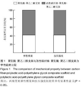
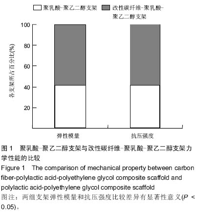
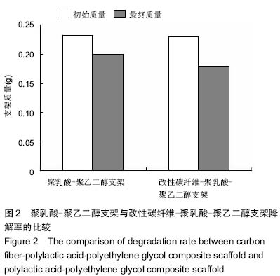
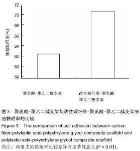
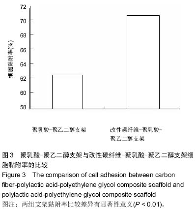
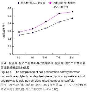
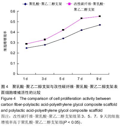
Preparation and performance detection of carbon fiber-polylactic acid-polyethylene glycol composite scaffold
Zhou Chang-yan1, Zhou Qing-huan2, Bian Jing1, Chen Ke1, Chen Wen1
- 1First Orthopedic Ward, Taihe Hospital Affiliated to Hubei University of Medicine, Shiyan 442000, Hubei Province, China;
2Nursing School, Hubei University of Medicine, Shiyan 44200, Hubei Province, China
-
Online:2015-05-21Published:2015-05-21 -
Contact:Zhou Qing-huan, Master, Senior nurse, First Orthopedic Ward, Taihe Hospital Affiliated to Hubei University of Medicine, Shiyan 442000, Hubei Province, China -
About author:Zhou Chang-yan, Nurse in charge, First Orthopedic Ward, Taihe Hospital Affiliated to Hubei University of Medicine, Shiyan 442000, Hubei Province, China
CLC Number:
Cite this article
Zhou Chang-yan, Zhou Qing-huan, Bian Jing, Chen Ke, Chen Wen. Preparation and performance detection of carbon fiber-polylactic acid-polyethylene glycol composite scaffold [J]. Chinese Journal of Tissue Engineering Research, 2015, 19(21): 3371-3376.
share this article
"

"
"
"
| [1] Kutikov AB,Song J.An amphiphilic degradable polymer/hydroxyapatite composite with enhanced handling characteristics promotes osteogenic gene expression in bone marrow stromal cells.Acta Biomater.2013;9(9):8354-8364. [2] Zhang C,Salick MR,Cordie TM,et al.Incorporation of poly(ethylene glycol) grafted cellulose nanocrystals in poly(lactic acid) electrospun nanocomposite fibers as potential scaffolds for bone tissue engineering. Mater Sci Eng C Mater Biol Appl.2015; 49:463-471.
[3] Wang D,Lin H,Jiang J,et al.Fabrication of long-acting drug release property of hierarchical porous bioglasses/polylactic acid fibre scaffolds for bone tissue engineering.IET Nanobiotechnol.2015;9(2):58-65.
[4] 田建宁,王友法,李世普.相分离/糖球沥滤复合法制备聚乳酸多孔支架[J].武汉理工大学学报,2010,32(8):30-32.
[5] Li D,Sun H,Jiang L,et al.Enhanced biocompatibility of PLGA nanofibers with gelatin/nano-hydroxyapatite bone biomimetics incorporation.ACS Appl Mater Interfaces.2014;6(12): 9402-9410.
[6] Hadinoto K,Sundaresan A,Cheow WS.Lipid-polymer hybrid nanoparticles as a new generation therapeutic delivery platform: a review.Eur J Pharm Biopharm.2013;85(3 Pt A)427-443.
[7] Xiao RZ,Zeng ZW,Zhou GL,et al.Recent advances in PEG-PLA block copolymer nanoparticles.Int J Nanomedicine. 2010;5:1057-1065.
[8] 王淑红,康非吾,潘可风.PLA-PEG复合支架材料体外细胞相容性的实验研究[J].口腔颌面外科杂志,2006,16(4):303-306.
[9] Vila A,Gill H,Mccallion O,et al.Transport of PLA-PEG particles across the nasal mucosa: effect of particle size and PEG coating density.J Control Release.2004;98(2): 231-244.
[10] 宋芹,颜军,郭晓强,等.羟基磷灰石/胶原蛋白复合支架上骨髓间充质干细胞的增殖与分化[J].中国组织工程研究与临床康复, 2010, 14(47):8795-8799.
[11] 卢剑,杨振磊,侯任.聚乳酸-聚乙二醇共聚物/表面接枝复合纤维材料与成骨细胞的黏附、增殖及活性[J].中国组织工程研究, 2014,18(39):6246-6251.
[12] Kang Y,Scully A,Young DA,et al.Enhanced mechanical performance and biological evaluation of a PLGA coated beta-TCP composite scaffold for load-bearing applications. Eur Polym J.2011;47(8):1569-1577.
[13] Jadeja H,Yeoh D,Lal M,et al.Patterns of failure with time of an artificial scaffold class ligament used for reconstruction of the human anterior cruciate ligament. Knee. 2007;14(6):439-442.
[14] Longo UG,Rizzello G,Berton A,et al.Synthetic grafts for anterior cruciate ligament reconstruction.Curr Stem Cell Res Ther.2013; 8(6):429-437.
[15] Kumar N,Sharma AK,Sharma AK,et al.Carbon fibres and plasma-preserved tendon allografts for gap repair of flexor tendon in bovines: gross, microscopic and scanning electron microscopic observations.J Vet Med A Physiol Pathol Clin Med.2002;49(5): 269-276.
[16] Li J,Yang Z,Xie H.Therapeutic effect of tissue engineered tendon in repairing old calcaneal tendon rupture and defects. Zhongguo Xiu Fu Chong Jian Wai Ke Za Zhi.2005;19(8): 639-641.
[17] Shen L,Yang H,Ying J,et al.Preparation and mechanical properties of carbon fiber reinforced hydroxyapatite/ polylactide biocomposites.J Mater Sci Mater Med.2009; 20(11):2259-2265.
[18] 徐文峰,廖晓玲.三维多孔碳纤维/聚乳酸/壳聚糖复合支架材料的体外细胞相容性评价[J].应用化学,2011,28(2):214-218.
[19] Wu Y,Hou J,Yin M,et al.Enhanced healing of rabbit segmental radius defects with surface-coated calcium phosphate cement/bone morphogenetic protein-2 scaffolds. Mater Sci Eng C Mater Biol Appl.2014;44:326-335.
[20] 史燕妮,王宁,藏永菊,等.碳纤维增强聚合物组织工程支架的制备及表征[J].高科技纤维与应用,2013,38(5):11-16.
[21] 刘瑞,李明贺,及昕,等.碳纤维增强型聚醚醚酮植入下颌骨单层骨皮质缺损模型白兔体内后的生物相容性评价[J].吉林大学学报:医学版,2014,40(3):569-573.
[22] Zhang L,Zhang J,Ling Y,et al.Sustained release of melatonin from poly (lactic-co-glycolic acid) (PLGA) microspheres to induce osteogenesis of human mesenchymal stem cells in vitro.J Pineal Res.2013;54(1):24-32.
[23] 宋显晶,刘斌,李淑梅,等.生物可降解材料聚乙二醇-聚乳酸-聚谷氨酸三嵌段共聚物与人脐静脉内皮细胞的细胞相容性[J].吉林大学学报:医学版,2007,33(3):514-517.
[24] 刘斌,张基昌,陈宏勃,等.生物可降解冠状动脉支架涂层材料聚乙二醇-聚乳酸-聚谷氨酸共聚物的生物相容性研究[J].中国实验诊断学,2006,10(9):975-978.
[25] Kane RJ,Roeder RK.Effects of hydroxyapatite reinforcement on the architecture and mechanical properties of freeze-dried collagen scaffolds. J Mech Behav Biomed Mater. 2012;7: 41-49.
[26] Corporation HP. Preparation and Properties of Biphasic Calcium Phosphate Scaffolds Multiply Coated with HA/PLLA Nanocomposites for Bone Tissue Engineering Applications. J Nanomaterials.2012;7(11):2817-2827.
[27] Peng T,Sun W,Sun X,et al.Direct tri-constituent co-assembly of highly ordered mesoporous carbon counter electrode for dye-sensitized solar cells. Nanoscale.2013;5(1):337-341.
[28] Mas N, Galiana I,Hurtado S,et al.Enhanced antifungal efficacy of tebuconazole using gated pH-driven mesoporous nanoparticles.Int J Nanomedicine.2014;9:2597-2606.
[29] Zhong T,Deng C,Gao Y,et al.Studies of in situ-forming hydrogels by blending PLA-PEG-PLA copolymer with silk fibroin solution.J Biomed Mater Res A.2012;100(8): 1983-1989.
[30] 孙蕊,潘高峰,张丽芳,等.聚乳酸/聚乙二醇-聚乳酸新型亲水支架的制备与研究[J].生物医学工程学杂志,2007,24(1):91-96.
[31] Wang XH,Shi S,Guo G,et al.Preparation and characterization of a porous scaffold based on poly(D,L-lactide) and N-hydroxyapatite by phase separation. J Biomater Sci Polym Ed.2011;22(14):1917-1929.
[32] Lasprilla AJ,Martinez GA,Lunelli BH,et al.Poly-lactic acid synthesis for application in biomedical devices - a review. Biotechnol Adv.2012;30(1):321-328.
[33] Seifu DG,Isimjan TT,Mequanint K.Tissue engineering scaffolds containing embedded fluorinated-zeolite oxygen vectors.Acta Biomater.2011;7(10):3670-3678.
[34] Chen S,Hou H,Harnisch F,et al. Electrospun and solution blown three-dimensional carbon fiber nonwovens for application as electrodes in microbial fuel cells. Energy Environ. Sci.2011;4(4):1417-1421.
[35] Soylemez E,de Boer MP,Sae-Ueng U,et al.Photocatalytic degradation of bacteriophages evidenced by atomic force microscopy. PLoS One.2013;8(1):e53601.
[36] Wilhelmi M,Giere B,Harder M.Interaction of cells with decellularized biological materials.Adv Biochem Eng Biotechnol. 2012;126:105-116.
[37] Baskin JZ,Eppell SJ.A selected review of the recent advances in craniomaxillofacial bone tissue engineering.Curr Opin Otolaryngol Head Neck Surg. 2013; 21(4): 389-395.
[38] Murphy CM,O'Brien FJ,Little DG,et al.Cell-scaffold interactions in the bone tissue engineering triad. Eur Cell Mater.2013;26:120-132.
[39] Wang T,Chow LC,Frukhtbeyn SA,et al.Improve the Strength of PLA/HA Composite Through the Use of Surface Initiated Polymerization and Phosphonic Acid Coupling Agent. J Res Natl Inst Stand Technol.2011;116(5):785-796.
[40] Miao PH,He CX,Hu YL,et al.Impregnation of plasmid DNA into three-dimensional PLGA scaffold enhances DNA expression of mesenchymal stem cells in vitro.Pharmazie. 2012;67(3):229-232.
[41] Kim HJ,Park M,Han YM,et al.Butamben derivatives enhance BMP-2-stimulated commitment of C2C12 cells into osteoblasts with induction of voltage-gated potassium channel expression. Bioorg Med Chem Lett.2011;21(24): 7363-7366. |
| [1] | Xu Feng, Kang Hui, Wei Tanjun, Xi Jintao. Biomechanical analysis of different fixation methods of pedicle screws for thoracolumbar fracture [J]. Chinese Journal of Tissue Engineering Research, 2021, 25(9): 1313-1317. |
| [2] | Zhang Tongtong, Wang Zhonghua, Wen Jie, Song Yuxin, Liu Lin. Application of three-dimensional printing model in surgical resection and reconstruction of cervical tumor [J]. Chinese Journal of Tissue Engineering Research, 2021, 25(9): 1335-1339. |
| [3] | Chen Xinmin, Li Wenbiao, Xiong Kaikai, Xiong Xiaoyan, Zheng Liqin, Li Musheng, Zheng Yongze, Lin Ziling. Type A3.3 femoral intertrochanteric fracture with augmented proximal femoral nail anti-rotation in the elderly: finite element analysis of the optimal amount of bone cement [J]. Chinese Journal of Tissue Engineering Research, 2021, 25(9): 1404-1409. |
| [4] | Zhou Jihui, Li Xinzhi, Zhou You, Huang Wei, Chen Wenyao. Multiple problems in the selection of implants for patellar fracture [J]. Chinese Journal of Tissue Engineering Research, 2021, 25(9): 1440-1445. |
| [5] | Zeng Yanhua, Hao Yanlei. In vitro culture and purification of Schwann cells: a systematic review [J]. Chinese Journal of Tissue Engineering Research, 2021, 25(7): 1135-1141. |
| [6] | Xu Yulin, Shen Shi, Zhuo Naiqiang, Yang Huilin, Yang Chao, Li Yang, Zhao Heng, Zhao Lu. Biomechanical comparison of three different plate fixation methods for acetabular posterior column fractures in standing and sitting positions [J]. Chinese Journal of Tissue Engineering Research, 2021, 25(6): 826-830. |
| [7] | Cai Qunbin, Zou Xia, Hu Jiantao, Chen Xinmin, Zheng Liqin, Huang Peizhen, Lin Ziling, Jiang Ziwei. Relationship between tip-apex distance and stability of intertrochanteric femoral fractures with proximal femoral anti-rotation nail: a finite element analysis [J]. Chinese Journal of Tissue Engineering Research, 2021, 25(6): 831-836. |
| [8] | Song Chengjie, Chang Hengrui, Shi Mingxin, Meng Xianzhong. Research progress in biomechanical stability of lateral lumbar interbody fusion [J]. Chinese Journal of Tissue Engineering Research, 2021, 25(6): 923-928. |
| [9] | Xie Chongxin, Zhang Lei. Comparison of knee degeneration after anterior cruciate ligament reconstruction with or without remnant preservation [J]. Chinese Journal of Tissue Engineering Research, 2021, 25(5): 735-740. |
| [10] | Xu Dongzi, Zhang Ting, Ouyang Zhaolian. The global competitive situation of cardiac tissue engineering based on patent analysis [J]. Chinese Journal of Tissue Engineering Research, 2021, 25(5): 807-812. |
| [11] | Wu Zijian, Hu Zhaoduan, Xie Youqiong, Wang Feng, Li Jia, Li Bocun, Cai Guowei, Peng Rui. Three-dimensional printing technology and bone tissue engineering research: literature metrology and visual analysis of research hotspots [J]. Chinese Journal of Tissue Engineering Research, 2021, 25(4): 564-569. |
| [12] | Chang Wenliao, Zhao Jie, Sun Xiaoliang, Wang Kun, Wu Guofeng, Zhou Jian, Li Shuxiang, Sun Han. Material selection, theoretical design and biomimetic function of artificial periosteum [J]. Chinese Journal of Tissue Engineering Research, 2021, 25(4): 600-606. |
| [13] | Liu Fei, Cui Yutao, Liu He. Advantages and problems of local antibiotic delivery system in the treatment of osteomyelitis [J]. Chinese Journal of Tissue Engineering Research, 2021, 25(4): 614-620. |
| [14] | Li Xiaozhuang, Duan Hao, Wang Weizhou, Tang Zhihong, Wang Yanghao, He Fei. Application of bone tissue engineering materials in the treatment of bone defect diseases in vivo [J]. Chinese Journal of Tissue Engineering Research, 2021, 25(4): 626-631. |
| [15] | Zhang Zhenkun, Li Zhe, Li Ya, Wang Yingying, Wang Yaping, Zhou Xinkui, Ma Shanshan, Guan Fangxia. Application of alginate based hydrogels/dressings in wound healing: sustained, dynamic and sequential release [J]. Chinese Journal of Tissue Engineering Research, 2021, 25(4): 638-643. |
| Viewed | ||||||
|
Full text |
|
|||||
|
Abstract |
|
|||||