[1] 欧艺,杨洪昌,陈戈,等.改良Stoppa入路[J].创伤外科杂志,2016, 18(4):252-255.
[2] 吴淑琴,潘宏侠,裴葆青.髋臼横断骨折后柱长/短钢板内固定的有限元建模及分析[J].北京生物医学工程,2011,30(1):1-4.
[3] SINGH M, NAGRATH AR, MAINI PS. Changes in trabecular pattern of the upper end of the femur as an index of osteoporosis.J Bone Joint Surg Am.1970;52(3):457-67.
[4] 吴啸波,张奇,郭明珂,等.髋臼后柱骨折模型建立及髋臼后柱骨折钢板内固定和拉力螺钉内固定稳定性比较[J].中国组织工程研究与临床康复,2009,13(52):10236-10240.
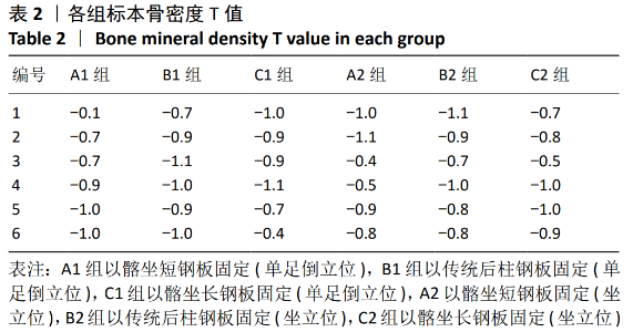
[5] 高应超,郭征,付军,等.坐位骨盆的三维有限元分析[J].中国组织工程研究与临床康复,2011,15(22): 3997-4001.
[6] MATTA JM. Operative treatment of acetabular fractures through the ilioinguinal approach: a 10-year perspective.J Orthop Trauma.2006; 20(1 Suppl):S20-29.
[7] 郭东鸿.五种内固定方式固定髋臼后柱骨折的三维有限元对比研究[D].广州:南方医科大学,2017.
[8] ATTIAS N, LINDSEY RW, STARR AJ, et al. The use of a virtual three-dimensional model to evaluate the intraosseous space available for percutaneous screw fixation of acetabular fractures.J Bone Joint Surg Br. 2005;87(11):1520-1523.
[9] 刘涛,项大业,何少波,等.经坐骨结节至髋臼后柱置钉安全区域的CT测量研究[J].浙江创伤外科,2012,17(6):726-728.
[10] 杨晓东,刘涵,谷城,等.髂坐钢板经腹直肌外侧入路治疗涉及方形区的髋臼骨折[J].中国矫形外科杂志,2019,27(20):1836-1840.
[11] 代元元,章莹,夏远军,等.长板与拉力螺钉固定治疗髋臼后柱骨折的建模及稳定性比较[J].中国临床解剖学杂,2016,34(2):214-219.
[12] KHAJAVI K, LEE AT, LINDSEY DP, et al. Single column locking plate fixation is inadequate in two column acetabular fractures. A biomechanical analysis. J Orthop Surg Res.2010;5:30.
[13] PETER RE. Open reduction and internal fixation of osteoporotic acetabular fractures through the ilioinguinal approach: use of buttress plates to control medial displacement of the quadrilateral surface.Injury. 2015;46 Suppl 1:S2-7.
[14] 高波华,王钢,卢超,等.基于CT三维重建的髋臼方形区骨折线的初步研究[J].中华创伤骨科杂志,2014,16(4):305-310.
[15] ARCHDEACON MT. Comparison of the ilioinguinal approach and the anterior intrapelvic approaches for open reduction and internal fixation of the acetabulum.J Orthop Trauma.2015;29 Suppl 2:S6-S9.
[16] SHAZAR N, ESHED I, ACKSHOTA N, et al. Comparison of acetabular fracture reduction quality by the ilioinguinal or the anterior intrapelvic(modified Rives—Stoppa)surgical approaches.J Orthop Trauma. 2014;28(6):313-319.
[17] 薛飞,贾文超,冯卫,等.改良Stoppa人路内髂坐钢板固定治疗涉及后柱的髋臼骨折[J].中华创伤骨科杂志,2019,21(6):478-483.
[18] 欧艺,陈戈,陈仲,等.斜行髂坐钢板前路固定低位髋臼后柱骨折[J].中华创伤骨科杂志,2018,20(5):382-388.
[19] 陈开放,杨帆,郭晓东,等.髋臼四方区组合钢板治疗老年髋臼骨折的有效性[J].中华创伤杂志,2018,34(4):323-330. |