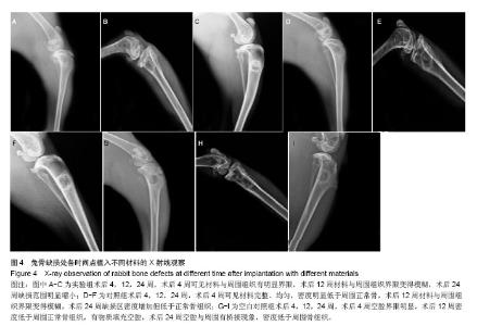
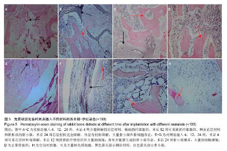
| [1]袁冰,韦卓.骨缺损修复的研究进展[J].生物骨科材料与临床研究, 2014,11(3):38-41.
[2]方志伟,李舒,樊征夫,等.异种骨和人工骨修复骨肿瘤性骨缺损[J].中国组织工程研究,2014,18(16):2468-2473.
[3]王小红,马建彪,王亦农,等.骨修复材料的研究进展[J].生物医学学杂志,2001,18(4):647-652.
[4]孙文晓,张海巷,韦卓,等.骨修复材料的研究应用现状与展望[J].生物骨科材料与临床研究,2009,6(3):35-40.
[5]刘博,贾全章.复合材料修复骨缺损的研究进展[J].吉林医学, 2013, 34(10):1919-1921.
[6]Sagar N,Pandey AK,Gurbani D,et al.In-Vivo Efficacy of Compliant 3D Nano-Composite in Critical-Size Bone DefectRepair: a Six Month Preclinical Study in Rabbit.PLoS One.2013;8(10):e77578.
[7]杨毅,毕鑫,李多玉,等.人工骨材料修复骨缺损:多种复合后的生物学与力学特征[J].中国组织工程研究,2014,18(16): 2582-2587.
[8]肖建德,熊建义,欧阳侃,等.纳米陶瓷人工骨修复骨缺损的实验研究[J].中华创伤骨科杂志,2006,8(11):1048-1052.
[9]王浩,张里程,石涛,等.胶原-羟基磷灰石-硫酸软骨素-骨形态发生蛋白骨修复材料的性质评估[J].北京大学学报:医学版,2011, 43(5):730-734.
[10]邢自宝,刘永刚,苏佳灿.骨缺损修复研究进展[J].临床医学工程, 2010,17(2):145-147.
[11]张建新,张展望,常峰.组织工程化人工骨修复骨缺损的实验研究[J].中国矫形外科杂志,2009,17(16):1258-1261.
[12]刘艳辉,谢利娜.淫羊藿在骨膜附加活性材料nHAC/PLA修复颌骨缺损中的影响[J].广州中医药大学学报,2014,31(5):791-794.
[13]赵红霞.载药壳聚糖/羟基磷灰石复合支架在骨组织工程中的研究进展[J].功能材料,2014,13(45):1306-1312.
[14]吴涛,徐俊昌,南开辉,等.淫羊藿苷促进羊骨髓间充质干细胞的增殖和成骨分化[J].中国组织工程研究与临床康复,2009,13(19): 3725-3729.
[15]贾亮亮,袁丁,王洪武,等.淫羊藿苷药理作用的研究进展[J].现代生物医学进展,2010,10(20):3976-3979.
[16]于波,杨久山,刘岩,等.淫羊藿苷对人成骨细胞的作用[J].中医正骨,2006,18(6):17-19.
[17]李恩中.组织工程研究的新策略-组织诱导性生物材料[J].生物医学工程学杂志,2009, 26(3):461-464.
[18]Rajangam T,An SS.Fibrinogen and fibrin based micro and nano scaffolds incorporated with drugs, proteins, cells and genes for therapeutic biomedical applications.Int J Nanomedicine.2013;8:3641-3662.
[19]朱美忠,李晓斌,陈滔.纳米羟基磷灰石/聚酰胺66复合生物活性人工骨在肢体骨缺损应用87例[J].创伤外科杂志,2014,16(1): 29-31.
[20]王大平,熊建义,朱伟民,等.纳米羟基磷灰石人工骨在骨缺损修复中的临床应用研究[J].中国临床解剖学杂志,2014,32(2): 670-673.
[21]Mok SK,Chen WF,Lai WP,et al.Icariin protects against bone loss induced by oestrogen deficiency and activates oestrogen receptor-dependent osteoblastic functions inUMR 106 cells. Br J Pharmacol.2010;159(4):939-949.
[22]Li XF, Xu H, Zhao YJ,et al.Icariin Augments Bone Formation and Reverses the Phen-otypes of Osteoprotegerin-Deficient Mice through the Activation of Wnt/β-Catenin-BMP Signaling. Evid Based Complement Alternat Med. 2013;2013:652317.
[23]殷晓雪,陈仲强,党耕町,等.淫羊藿苷对人成骨细胞增殖与分化的影响[J].中国中药杂志,2005, 30(4):289-291.
[24]郭海玲,赵咏芳,王翔,等.淫羊藿苷对人成骨细胞增殖及OPG蛋白表达的实验研究[J].中国骨伤,2011,24(7):585-588.
[25]Xue L,Jiao L,Wang Y,et al.Effects and interaction of icariin, curculigoside, and berberine in er-xian decoction, a traditional chinese medicinal formula, on osteoclastic bone resorption. Evid Based Complement Alternat Med. 2012;2012:490843. |