Chinese Journal of Tissue Engineering Research
Previous Articles Next Articles
Nano decalcified bone matrix promotes bone fusion
Huang Kai1, Chen Xiong-sheng2, Jia Lian-shun2, Zhu Wei2, Fang Lei2
- 1 Zhabei Branch of Shanghai Changzheng Hospital, Shanghai 200070, China
2 Shanghai Changzheng Hospital, Shanghai 200043, China
-
Received:2012-11-16Revised:2012-12-04Online:2013-05-21Published:2013-05-21 -
Contact:Chen Xiong-sheng, Master’s supervisor, Chief physician, Shanghai Changzheng Hospital, Shanghai 200043, China chenxiongsheng@vip.sohu.com -
About author:Huang Kai☆, M.D., Attending physician, Zhabei Branch of Shanghai Changzheng Hospital, Shanghai 200070, China 13817784210@163.com -
Supported by:the Shanghai Science and Technology Committee, No. 054119641*, 0852nm03100
CLC Number:
Cite this article
Huang Kai, Chen Xiong-sheng, Jia Lian-shun, Zhu Wei, Fang Lei. Nano decalcified bone matrix promotes bone fusion[J]. Chinese Journal of Tissue Engineering Research, doi: 10.3969/j.issn.2095-4344.2013.21.009.
share this article
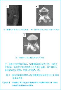
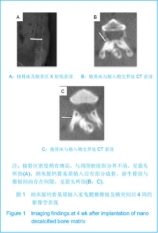
2.1 实验动物数量分析 36只兔均进入结果分析。 2.2 各组术后不同时间点影像学观察结果 纳米脱钙骨基质组、脱钙骨基质组及自体骨各组均取得骨性融合。纳米脱钙骨基质组融合速度、新生骨质质地、密度高均与自体骨组接近;脱钙骨基质组融合速度、质量均弱于纳米脱钙骨基质及自体骨组。 纳米脱钙骨基质组:术后4周,X射线片及CT扫描结果见图1。植骨床及植骨区:植骨区密度稍有增高,与周围软组织分界不清,见图1A。植骨床与植入物交界处:纳米脱钙骨基质植入后有部分成骨,新生骨块与椎板间尚存在间隙,见图1B,C。"

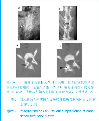
术后8周,X射线片及CT扫描结果见图2。植骨床及植骨区:植骨区密度较周围软组织稍有增高,见图2A,B;植骨床与植入物交界处:植骨床与植入材料间间隙仍存在,见图2C,D。"
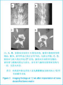
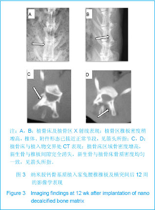
术后12周,X射线片及CT扫描结果见图3。植骨床及植骨区:植骨区椎板密度稍增高,椎体、附件形态已接近正常节段,见图3A,B;植骨床与植入物交界处:植骨床区域骨密度增高,新生骨与椎板间隙完全消失,新生骨与植骨床骨质密度均匀一致,见图3C,D。"
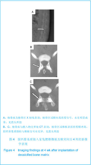
脱钙骨基质组:术后4周,X射线片及CT扫描结果见图4。植骨床及植骨区:植骨区域稍有高密度信号,未见明显成骨,见图4A;植骨床与植入物交界处:植骨区域椎板表面密度稍增高,脱钙骨基质颗粒与椎板信号有差异,见图4B,C。"
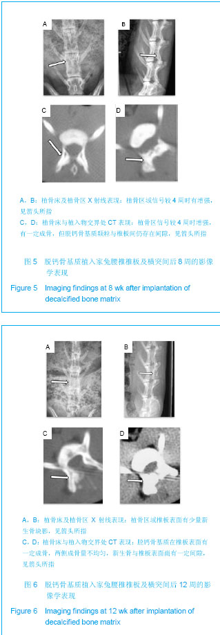
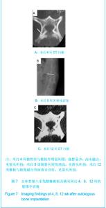
术后8周,X射线片及CT扫描结果见图5。植骨床及植骨区:植骨区域信号较4周时有增强,见图5A,B;植骨床与植入物交界处:植骨区信号较4周时增强,有一定成骨,但脱钙骨基质颗粒与椎板间仍存在间隙,见图5C,D。 术后12周,X射线片及CT扫描结果见图6。植骨床及植骨区:植骨区域椎板表面有少量新生骨块影,见图6A,B;植骨床与植入物交界处:脱钙骨基质在椎板表面有一定成骨,两侧成骨量不均匀,新生骨与椎板表面尚有一定间隙,见图6C,D。"
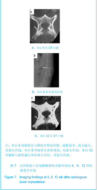
自体骨组:术后4周CT扫描见植骨区高信号影,植骨块与椎板有明显间隙,成骨量少,尚未融合,见图7A。X射线可见植骨区高密度影。术后8周,X射线见植骨区密度增高,见图7B,CT扫描见植骨区有较多成骨,植骨块与椎板间隙基本消失。术后12周,CT扫描可见椎板与植骨融合界面愈合良好,见图7C,X射线可见植骨区均有新生骨块影,融合骨块质地均匀。"
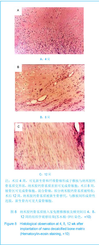
2.3 各组组织学观察结果 纳米脱钙骨基质组:术后4周,可见新生骨和纤维骨痂形成于椎板与纳米脱钙骨基质交界部,纳米脱钙骨基质表面可见成骨细胞;术后8周,植骨区可见成骨细胞、混合骨痂,部分纳米脱钙骨基质被吸收;术后12周,纳米脱钙骨基质被新生骨替代,与椎板间形成骨性连接,新生骨内可见大量骨细胞,见图8。"
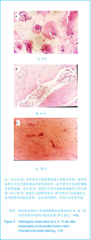
脱钙骨基质组:术后4周,新生骨及纤维骨痂形成于植骨交界部,脱钙骨基质内可见纤维骨痂及纤维结缔组织,新生骨内可见成骨细胞及软骨细胞;术后8周,植骨区可见少量成骨细胞及不均匀成骨;术后12周,植骨区达到骨性愈合,新生骨内可见质地均匀、排列较整齐的板层样骨,也有类骨物质,中间可见血管形成,见图9。"
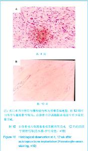
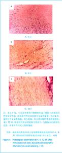
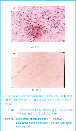
自体骨组:术后4周时自体骨与椎板结合部为纤维骨痂连接,术后8周时纤维骨痂部分成骨,到12周时自体骨与椎板骨性融合。自体骨在远离椎板连接部位有少量死骨形成,图10。"
| [1] Dinopoulos H,Dimitriou R, Giannoudis PV. Bone graft substitutes: What are the options? Surgeon.2012; 10(4): 230-239.[2] Bhatt RA,Rozental TD.Bone Graft Substitutes.Hand Clin. 2012;28(4): 457-468.[3] Enrique Guerado, Carl Hans Fuerstenberg. What bone graft substitutes should we use in post-traumatic spinal fusion? Injury.2011;42 Suppl 2: S64-S71.[4] Seebach C,Schultheiss J,Wilhelm K,et al.Comparison of six bone-graft substitutes regarding to cell seeding efficiency, metabolism and growth behaviour of human mesenchymal stem cells (MSC) in vitro. Injury.2010;41(7): 731-738.[5] Nenad I,Petar N,Zorica A,et al. Biphasic calcium phosphate coated with poly d,1 lactide co glycolide hiomaterial as a hone substitute. Eur Ceram Soc.2007;27: 1589-1594.[6] De Long WG Jr,Einhorn TA,Koval K,et al. Bone grafts and bone graft substitutes in orthopaedic trauma surgery.A critical analysis .J Bone Joint Surg Am. 2007;89(3):649-658.[7] Hsu W,Fuchs D.A Meta-Analysis of Fusion Rates from Bone Graft Substitutes in a Rodent Posterolateral Spine Arthrodesis Model.Spine J.2011;11(10 Suppl): S59-S60.[8] Athanasiou VT,Papachristou DJ,Panagopoulos A,et al.Histological comparison of autograft, allograft- DBM, xenograft,and synthetic grafts in a trabecular bone defect:an experimental study in rabbits.Med Sci Monit.2010;16(1): 24-31.[9] Powell M,Griffin M,Tai S. Bottom-up risk regulation? How nano-technology risk knowledge gaps challenge federal and state environmental agencies. Environ Manage.2008;42(3): 426-443.[10] Hak DJ.The use of osteoconductive bone graft substitutes in orthopaedic trauma.J Am Acad Orthop Surg.2007;15(9): 525-536.[11] Janicki P,Schmidmaier G.What should be the characteristics of the ideal bone graft substitute? Combining scaffolds with growth factors and/or stem cells. Injury.2011;42 Suppl 2: S77-S81.[12] Blokhuis TJ,Arts JJ.Bioactive and osteoinductive bone graft substitutes: Definitions, facts and myths.Injury.2011;42 Suppl 2: S26-S29.[13] Jurczyk MU,Jurczyk K,Miklaszewski A,et al.Nanostructured titanium-45S5 Bioglass scaffold composites for medical applications.Mater Design. 2011;32(10):4882-4889.[14] Caldorera-Moore M,Peppas NA.Micro- and nanotechnologies for intelligent and responsive biomaterial-based medical systems.Adv Drug Del Rev.2009;61(15):1391-1401.[15] Rodgers WB, Gerber EJ. A DBM, BMA, Local Bone Graft Composite in Multi-Level PLIF: Fusion Rates. Spine J. 2010; 10(9 Suppl): S26.[16] Li XQ,Liu TQ,Song KD.Dalian Ligong Daxue Xuebao. 2005; 45(5):653-657.李香琴,刘天庆, 宋克东.成骨细胞在纳米材料表面上粘附特性[J].大连理工大学学报,2005,45(5):653-657.[17] Qian Y,Zhang ZF,Shen ZL,et al.Zhongguo Jiaoxing Waike Zazhi. 2005;13(12):911-914.钱鋆,张兆锋,沈尊理,等.冻干脱钙骨表面纳米结构对细胞行为的影响[J].中国矫形外科杂志,2005,13(12):911-914.[18] Yoshikawa H,Myoui A.Bone tissue engineering with porous hydroxyapatite ceramics.Artif Organs.2005;8(3): 131-136.[19] Bohner M.Resorbable biomaterials as bone graft substitutes. Mater Today.2010;13(1-2): 24-30.[20] Zimmermann G,Moghaddam A.Allograft bone matrix versus synthetic bone graft substitutes. Injury.2011;42 Suppl 2: S16-S21.[21] Geurts J,Chris Arts JJ,Walenkamp GH.Bone graft substitutes in active or suspected infection. Contra-indicated or not? Injury.2011;42 Suppl 2:S82-S86.[22] Drosos GI,Babourda E,Magnissalis EA,et al. Mechanical characterization of bone graft substitute ceramic cements. Injury.2012;43(3): 266-271.[23] Chitsazi MT,Shirmohammadi A,Faramarzie M,et al.A clinical comparison of nano-crystalline hydroxyapatite (Ostim) and autogenous bone graft in the treatment of periodontal intrabony defects. Med Oral Patol Oral Cir Bucal. 2011;16(3): E448-E453.[24] Kweon H, Lee KG, Chae CH,etal.Development of Nano-Hydroxyapatite Graft With Silk Fibroin Scaffold as a New Bone Substitute. J Oral Maxillofac Surg.2011; 69(6): 1578-1586.[25] Chai YC,Kerckhofs G,Roberts SJ,et al. Ectopic bone formation by 3D porous calcium phosphate-Ti6Al4V hybrids produced by perfusion electrodeposition. Biomaterials.2012; 33(16):4044-4058.[26] Huang K,Chen XS,Jia LS,et al.Zhongguo Jiaoxing Waike Zazhi. 2009;17(13):1017-1019.黄凯,陈雄生,贾连顺,等. 纳米脱钙骨基质的制备及其性能检测[J].中国矫形外科杂志,2009,17(13):1017-1019.[27] Zhao XM.Dianda Ligong. 2005;27(4):35-36.赵晓明.纳米材料的特性及应用[J].电大理工,2005,27(4):35-36.[28] Qian Y,Shen ZL,Zhang ZF,et al.Zhongguo Xiufu Chongjian Waike Zazhi. 2006;20(5):561-564.钱鋆,沈尊理,张兆锋,等.运用组织工程原理结合纳米技术构建骨组织的实验研究[J].中国修复重建外科杂志,2006,20(5): 561-564.[29] Kailasanathan C,Selvakumar N,Naidu V. Structure and properties of titania reinforced nano-hydroxyapatite/gelatin bio-composites for bone graft materials. Ceran Int.2012;38(1): 571-579.[30] Tang ZB,Cao JK,Wen N,et al.Posterolateral spinal fusion with nano-hydroxyapatite-collagen/PLA composite and autologous adipose-derived mesenchymal stem cells in a rabbit model.J Tissue Eng Regen Med. 2012;6(4):325-336. |
| [1] | Li Li, Ma Li. Immobilization of lactase on magnetic chitosan microspheres and its effect on enzymatic properties [J]. Chinese Journal of Tissue Engineering Research, 2021, 25(4): 576-581. |
| [2] | Zhou Anqi, Tang Yufei, Wu Bingfeng, Xiang Lin. Designing of periosteum tissue engineering: combination of generality and individuality [J]. Chinese Journal of Tissue Engineering Research, 2021, 25(22): 3551-3557. |
| [3] | Huo Hua, Cheng Yuting, Zhou Qian, Qi Yuhan, Wu Chao, Shi Qianhui, Yang Tongjing, Liao Jian, Hong Wei. Effects of drug coating on implant surface on the osseointegration [J]. Chinese Journal of Tissue Engineering Research, 2021, 25(22): 3558-3564. |
| [4] | Lang Limin, He Sheng, Jiang Zengyu, Hu Yiyi, Zhang Zhixing, Liang Minqian. Application progress of conductive composite materials in the field of tissue engineering treatment of myocardial infarction [J]. Chinese Journal of Tissue Engineering Research, 2021, 25(22): 3584-3590. |
| [5] | Wei Qin, Zhang Xue, Ma Lei, Li Zhiqiang, Shou Xi, Duan Mingjun, Wu Shuo, Jia Qiyu, Ma Chuang. Platelet-derived growth factor-BB induces the differentiation of rat bone marrow mesenchymal stem cells into osteoblasts [J]. Chinese Journal of Tissue Engineering Research, 2021, 25(19): 2953-2957. |
| [6] | Guo Zhibin, Wu Chunfang, Liu Zihong, Zhang Yuying, Chi Bojing, Wang Bao, Ma Chao, Zhang Guobin, Tian Faming. Simvastatin stimulates osteogenic differentiation of bone marrow mesenchymal stem cells [J]. Chinese Journal of Tissue Engineering Research, 2021, 25(19): 2963-2968. |
| [7] | Xie Jian, Su Jiansheng. Advantages and characteristics of electrospun aligned nanofibers as scaffolds for tissue engineering [J]. Chinese Journal of Tissue Engineering Research, 2021, 25(16): 2575-2581. |
| [8] | Ji Qi, Yu Zhengwen, Zhang Jian. Problems and trends of technique and clinical application of metallic biomaterials prepared by three-dimensional printing technology [J]. Chinese Journal of Tissue Engineering Research, 2021, 25(16): 2597-2604. |
| [9] | Wu Yukun, Han Jie, Wen Shuaibo. Mechanism of Runx2 gene in fracture healing [J]. Chinese Journal of Tissue Engineering Research, 2021, 25(14): 2274-2279. |
| [10] | Qian Nannan, Zhang Qian, Yang Rui, Ao Jun, Zhang Tao. Mesenchymal stem cells in the treatment of spinal cord injury: cell therapy and combination of new drugs and biomaterials [J]. Chinese Journal of Tissue Engineering Research, 2021, 25(13): 2114-2120. |
| [11] | Jia Wei, Zhang Mandong, Chen Weiyi, Wang Chenyan, Guo Yuan. Effects of femoral prosthetic materials on artificial knee arthroplasty performance [J]. Chinese Journal of Tissue Engineering Research, 2021, 25(10): 1477-1481. |
| [12] | Wang Qian, Li Lu, Shu Jingyuan, Dong Zhiheng, Jin Youshi, Wang Qingshan. Micro-morphology and phase of zirconia-based nano-hydroxyapatite functional gradient biomaterials [J]. Chinese Journal of Tissue Engineering Research, 2021, 25(10): 1517-1521. |
| [13] | Chen Qiang, Zhuo Hongwu, Xia Tian, Ye Zhewei . Toxic effects of different-concentration isoniazid on newborn rat osteoblasts in vitro [J]. Chinese Journal of Tissue Engineering Research, 2020, 24(8): 1162-1167. |
| [14] | Lin Ming, Pan Jinyong, Zhang Huirong. Knockout of NIPBL gene down-regulates the abilities of proliferation and osteogenic differentiation in mouse bone marrow mesenchymal stem cells [J]. Chinese Journal of Tissue Engineering Research, 2020, 24(7): 1002-1008. |
| [15] | Li Jinyu, Yu Xing, Jiang Junjie, Xu Lin, Zhao Xueqian, Sun Qi, Zheng Chenying, Bai Chunxiao, Liu Chuyin, Jia Yusong. Promoting effect of osteopractic total flavone combined with nano-bone materials on proliferation and differentiation of MC3T3-E1 cells [J]. Chinese Journal of Tissue Engineering Research, 2020, 24(7): 1030-1036. |
| Viewed | ||||||
|
Full text |
|
|||||
|
Abstract |
|
|||||