Chinese Journal of Tissue Engineering Research ›› 2019, Vol. 23 ›› Issue (17): 2770-2775.doi: 10.3969/j.issn.2095-4344.1695
Previous Articles Next Articles
Cerebrospinal fluid promotes neuron-like differentiation of mesenchymal stem cells
Ren Chao1, Ji Yongqiang2, Guan Lina3
- 1Department of Neurology, 2Department of Nephrology, 3Department of Neurology Intensive Care Unit, the Affiliated Yantai Yuhuangding Hospital of Qingdao University, Yanta 264000, Shandong Province, China
-
Revised:2019-01-16Online:2019-06-18Published:2019-06-18 -
Contact:Guan Lina, Master, Attending physician, Department of Neurology Intensive Care Unit, the Affiliated Yantai Yuhuangding Hospital of Qingdao University, Yanta 264000, Shandong Province, China -
About author:Ren Chao, Master, Attending physician, Department of Neurology, the Affiliated Yantai Yuhuangding Hospital of Qingdao University, Yanta 264000, Shandong Province, China -
Supported by:the National Natural Science Foundation of China (Youth Science Fund Project), No. 81501185 (to RC); Shandong Key Research and Development Program (Public Welfare Special Project), No. 2017GSF218043 (to RC); Yantai City Key Research and Development Plan, No. 2016WS017 (to GLN)
CLC Number:
Cite this article
Ren Chao1, Ji Yongqiang2, Guan Lina3. Cerebrospinal fluid promotes neuron-like differentiation of mesenchymal stem cells[J]. Chinese Journal of Tissue Engineering Research, 2019, 23(17): 2770-2775.
share this article
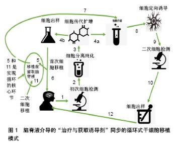
2.1 脑脊液对间充质干细胞的影响 骨髓间充质干细胞具有多向分化和增殖能力,且免疫原性低,易于获得、分离和培养。颜连启等[18]使用自体脑脊液诱导骨髓间充质干细胞,培养四五天即可出现神经样细胞形态,分化为神经元、星形胶质细胞和少突胶质细胞,并表现相应的特征结构和生物学特性。有研究将骨髓间充质干细胞与神经细胞共培养,尝试建立更加类似人体的微环境从而诱导骨髓间充质干细胞的分化,培养1周后发现骨髓间充质干细胞形成突起,细胞逐渐变为星状,星状细胞数目逐渐增并互相形成连接,具有神经样形态结构,免疫荧光显示神经元特异性烯醇化酶阳性。而对照组骨髓间充质干细胞未形成神经样形态结构,免疫荧光显示神经元特异性烯醇化酶呈阴性。 因此,研究者认为神经细胞生长过程中可分泌一些诱导骨髓间充质干细胞分化的物质,其提供的微环境对骨髓间充质干细胞分化为神经元具有促进作用。课题组的研究团队亦在既往研究中发现类似的结果[4,13,19-26],且发现脐源间充质干细胞有类似骨髓间充质干细胞的结果[4,13,19-20,24]。 Farivar等[27]也证明脑脊液可以诱导脐源间充质干细胞分化为神经样细胞,不过使用分化诱导的脑脊液浓度不同、诱导分化获得神经细胞所需的天数略有不同。以上说明间充质干细胞可在脑脊液中生长,并可维持其分化为神经细胞的潜能。沈忆新等[28]认为间充质干细胞在脑脊液中和在一般培养液中具有相似的生长特性,和课题组认为的一样:间充质干细胞迅速生长的同时,悬浮的各种血细胞减少,提示造血干细胞的生长或分化受抑,表明脑脊液培养液环境有助于间充质干细胞的生长而不利于造血干细胞的生长,该培养环境有助于从骨髓/脐血(脐带)中分离、培养间充质干细胞,但要注意脑脊液培养、诱导方案的具体制定,这是因为由于间充质干细胞具有多向分化潜能,在不同的培养液中可分化为不同的组织细胞。 因此不同的培养目的需要有不同的培养成分,但是具体机制目前探讨较少,理论上推导认为:间充质干细胞在脑内或脊髓内可诱导分化为神经细胞,脑内或脊髓内的微环境为其的定向诱导分化提供了必要的条件,具体条件仍不确定。而近期Zhu等[29]研究发现脑脊液是通过胰岛素样生长因子1调节干细胞增殖和迁移的;而Glage等[30]却发现胰高血糖素样肽1可能是其中的重要调控因子。也有学者认为是脑脊液中的脉络丛细胞与间充质干细胞两种细胞之间直接相互作用的结果[31-34],亦或是两种细胞融合的结果,甚至有学者提出脉络丛微生态信号是调节干细胞分化、迁移的重要因素[35-36],并有研究发现PRDM16可能是该信号通路上的重要调控因子[36]。另外,有学者发现肌萎缩侧索硬化患者脑脊液也能促进间充质干细胞分化成为神经元样细胞[37]。 脑脊液对间充质干细胞影响的研究不仅局限于诱导实验,目前已经有将诱导后获得神经细胞用于治疗的相关研究[4,19,21,26,38],且提示一定的疗效。当然,也有临床试验未取得预期效果,如有研究将间充质细胞注入9例阿尔茨海默症患者的海马和楔前叶,观察24个月,患者认知状态未取得任何改善[39]。 2.2 脑脊液对胚胎干细胞的影响 卞林等[40]使用正常成人脑脊液能诱导人胚胎源干细胞分化为神经样细胞,如神经元、星形胶质细胞、少突胶质细胞,但所得不同类型细胞的比例不同,其中以胶质细胞比例较高,认为这可能由于脑脊液中含有更多能使干细胞偏向于胶质细胞方向分化的因子有关。 陈新生等[41]研究中,血性脑脊液比正常脑脊液组神经干细胞分化为神经元的比例高,能获得更多比例的胶质细胞,这可能与脑损伤后脑脊液中促胶质细胞增生的成分增多有一定的关系。尹国才等[42]在研究“人胎脑神经干细胞在发育期脑脊液中的迁移和分化”时发现在胚胎期,处于不同发育阶段的胚胎干细胞分泌的信息物质差异大,且脑屏障发育尚未完成,处于开放状态,因而其对脑脊液的分泌有影响,而不同发育时期的脑脊液所承载的效应成分有差异,并进一步证实胶质细胞的出现可能与特定的脑脊液环境密切相关。和以上研究诱导获得胶质细胞为主不同,徐杰等[43]在其专利中指出“先采用一定浓度的抗坏血酸诱导分化,后采用正常成人脑脊液进一步使胚胎中脑来源的干细胞向多巴胺能神经细胞方向分化,可以获得最高比例的多巴胺能神经元”。 Zappaterra等[44]发现脑脊液的流体压力是促使胚胎干细胞分化、迁移的重要促发因素。而Martín等[45]提出成纤维细胞生长因子2是脑脊液诱导胚胎干细胞分化的重要因子。Parada等[46]通过基因分析证实了脑脊液中的因子对早期胚胎发育过程,尤其是神经细胞的分化、形成的有重要影响。Kiiski等[47]发现健康人脑脊液促进人类胚胎干细胞分化为神经细胞并能促进其形成保留自发活动的神经元网络,但Ma等[48]的研究却认为成人脑脊液不支持胚胎干细胞的神经发生,也有研究表明脑脊液抑制胚胎干细胞分化成神经元但却可促进其分化为胶质细胞[49],这可能和他们使用的胚胎干细胞和脑脊液来源不同所致。 2.3 脑脊液对神经干细胞的影响 李青[50]研究发现脊髓损伤后通过改变脑脊液的成分会对脊髓内源性神经干细胞增殖和分化产生影响。刘国平[51]通过对比研究发现:①神经干细胞在脑外伤血性脑脊液和脑积水清亮脑脊液中均能存活、增殖和分化;②神经干细胞在脑外伤血性脑脊液中贴壁分化的速度比在脑积水清亮脑脊液中要快,贴壁分化比例要高;③脑外伤血性脑脊液和脑积水清亮脑脊液对神经干细胞分化的种类上有差异。 神经干细胞在脑外伤血性脑脊液中倾向于向胶质细胞方向分化,在脑积水清亮脑脊液中倾向于向神经元方向分化。滕弘等[52]发现脑缺血大鼠脑脊液可促进体外培养神经干细胞存活并且促进神经干细胞向神经元和星形胶质细胞分化。Nozaki等[53]研究蛛网膜下腔出血患者的脑脊液发现含血脑脊液是激活并促进内源性神经干细胞增殖、分化的有效刺激物。Haines等[54]发现多发性硬化患者的脑脊液可影响神经干细胞中的少突胶质细胞祖细胞的转录变化。以上说明病态下的脑脊液可能是患者启动自身内源性神经修复的一个重要因素[55-56],但修复中脑脊液促进神经干细胞分化方向有一定差异[57],多以胶质细胞为主[16],少数可见神经元样细胞为主[58]。 如何获得想要的目的细胞用于治疗,目前含药脑脊液[59],尤其是中药脑脊液药理学的创立是目前的诱导内源性干细胞治疗神经系统疾病的研究热点,课题组团队正在开展此方面的探索(EGb761脑脊液药理学方法介导循环式干细胞移植治疗痴呆研究),并提出了脑脊液介导的“治疗与获取诱导剂”同步的循环式干细胞移植模式,见图1,这是将来临床值得推广的、可以“一箭双雕”的个体化干细胞移植治疗优选方案之一。"
| [1] Kuhn HG, Svendsen CN. Origins functions and potential of adult neural stem cells. Bioessays. 1999;21(8):625-630.[2] Vogel G. Europe: dismay, confusion greet human stem cell patent ban. Science. 2011;334(6055):441-442. [3] Freedman MS, Uccelli A. Neurorepair with mesenchymal stem cells: hope or hype? Lancet Neurol. 2012;11(2): 123-125.[4] Ren C, Geng RL, Ge W, et al. An observational study of autologous bone marrow-derived stem cells transplantation in 7 patients with nervous system diseases: a 2 year follow-up. Cell Biochem Biophys. 2014;69(1):179-187. [5] Low CB, Liou YC, Tang BL. Neural differentiation and potential use of stem cells from the human umbilical cord for central nervous system transplantation therapy. J Neurosci Res. 2008;86(8):1670-1679. [6] Kang XQ, Zang WJ, Bao L, et al. Differentiating characterization of human umbilical cord blood-derived mesenchymal stem cells in vitro. Cell Biol Int. 2006;30(7): 569-575.[7] Woodbury D, Schwarz EJ, Prockop DJ, et al. Adult rat and human bone marrow stromal cells differentiate into neurons. J Neurosci Res. 2000;61(4):364-370. [8] Kögler G, Sensken S, Airey JA, et al. A new human somatic stem cell from placental cord blood with intrinsic pluripotent differentiation potential. J Exp Med. 2004;200(2):123-135. [9] Yong-zhou S, Hui-xian C, Zhe L, et al. Effects of brain homogenate on the differentiation of rat bone mcsenchymal stem cells into neuron-like cells following traumatic brain injury[J]. Zhongguo Zuzhi Gongcheng Yanjiu yu Linchuang Kangfu. 2008;12(3):461-464. [10] Lim JY, Park SI, Oh JH, et al. Brain-derived neurotrophic factor stimulates the neural differentiation of human umbilical cord blood-derived mesenchymal stem cells and survival of differamtiated cells through MAPK/ERK and PIK/AKT-dependent signaling pathways. J Neumsci Res. 2008;86(10):2168-2178.[11] 贾延,杨于嘉,周燕,等.黄芩甙诱导大鼠骨髓基质细胞向神经细胞分化的研究[J].中华医学杂志,2002,82(19):1337-1341.[12] Tureyen K, Vemuganti R, Bowen KK, et al. EGF and FGF-2 in fusion increases post-ischemic neural progenitor cell proliferation in the adult rat brain. Neurosurgery. 2005;57(6): 1254-1263. [13] Ge W, Ren C, Duan X, et al. Differentiation of mesenchymal stem cells into neural stem cells using cerebrospinal fluid. Cell Biochem Biophys. 2015;71(1):449-455. [14] Illes S. More than a drainage fluid: the role of CSF in signaling in the brain and other effects on brain tissue. Handbook Clin Neurol. 2017;146:33-46. [15] Rivera FJ, Sierraha WD, Mingnell JJ, et al. Adult hippocampus derived soluble factors induce a neuronal-like phenotype in mesenchymal stem cells. Neurosci Lett. 2006; 406(1-2):49-54.[16] Pandamooz S, Naji M, Alinezhad F, et al. The influence of cerebrospinal fluid on epidermal neural crest stem cells may pave the path for cell-based therapy. Stem Cell Res Ther. 2013;4(4):1-9. [17] Imitola J, Durvasula S. The Cerebrospinal fluid-stem cells interactions as target for regenerative therapy in neurological diseases. Stem Cells Dev. 2015;24(2):145-146. [18] 颜连启,李小磊,孙钰,等.自体脑脊液定向诱导骨髓间充质干细胞分化为神经干细胞[J].江苏医药,2013,39(6):645-647.[19] 任超,张才溢,丁红梅,等.脑脊液诱导的间充质干细胞治疗神经变性疾病的初步研究[J].中国急救复苏与灾害医学杂志,2015, 10(7):608-611.[20] 叶英,万美荣,戴如飞,等.脑脊液诱导人间充质干细胞定向分化为神经干细胞神经前体细胞的研究[J].国际麻醉学与复苏杂志, 2009,30(5):394-398.[21] 叶英,谢熙,刘筱,等.脑脊液诱导的骨髓源性神经样细胞移植的安全性研究[J].中国急救复苏与灾害医学杂志,2015,10(6): 521-525.[22] 冯婷婷,叶英,许铁.脑脊液诱导的大鼠BMSC-Ns移植对脊髓损伤大鼠神经营养因子分泌的影响[J].中国急救复苏与灾害医学杂志,2015,10(7):612-615.[23] Ye Y, Zeng YM, Wan MR, et al. Induction of human bone marrow mesenchymal stem cells differentiation into neural-like cells using cerebrospinal fluid. Cell Biochem Biophys. 2011;59(3):179-184.[24] Ren C, Liu X, Wan M, et al.A comparative study on inducing non-homologous mesenchymal stem cells to differentiate into neural stem cells using non-homologous cerebrospinal fluid. J Biomed Eng. 2013;30(6):1290-1297[25] 陈娟,谢熙,彭怡然,等.人脑脊液诱导大鼠骨髓间充质干细胞定向分化为神经样细胞研究[J].徐州医学院学报,2016,36(4): 254-257.[26] Ye Y, Peng Y, Hu S, et al. In vitro differentiation of bone marrow mesenchymal stem cells into neuron-like cells by cerebrospinal fluid improves motor function of middle cerebral artery occlusion rats. Front Neurol. 2016;7(2):183. [27] Farivar S, Mohamadzade Z, Shiari R, et al. Neural differentiation of human umbilical cord mesenchymal stem cells by cerebrospinal fluid. Iran J Child Neurol. 2015;9(1): 87-93.[28] 沈忆新,王鹏,石恩东.脑脊液体外培养骨髓间充质干细胞[J].中国组织工程研究,2011,15(36):6802-6806.[29] Zhu M, Feng Y, Dangelmajer S, et al. Human cerebrospinal fluid regulates proliferation and migration of stem cells through insulin-like growth factor 1. Stem Cells Dev. 2015; 24(2):160-171. [30] Glage S, Klinge PM, Miller MC, et al. Therapeutic concentrations of glucagon-like peptide-1 in cerebrospinal fluid following cell-based delivery into the cerebral ventricles of cats. Fluids Barriers CNS. 2011;8(1):1-7. [31] 杨海云,顾锐,王文军,等.移植骨髓间充质干细胞后的脑脊液促进神经干细胞的分化[J].中国实验诊断学,2009,13(3):300-302.[32] Wu S, Suzuki Y, Ejiri Y, et al. Bone marrow stromal cells enhance differentiation of cocultured neurosphere cells and promote regeneration of injured spinal cord. J Neurosci Res. 2003;72(3):343-351.[33] Roballo KC, Gonçalves NJ, Pieri NC, et al. Regulation of neural stem cells by choroid plexus cells population. Neurosci Lett. 2016;626:35-41. [34] Moore SA. The spinal ependymal layer in health and disease. Vet Pathol. 2016;53(4):746-753. [35] Silva-Vargas V, Maldonado-Soto A, Mizrak D, et al. Age-dependent niche signals from the choroid plexus regulate adult neural stem cells. Cell Stem Cell. 2016;19(5): 643-652. [36] Kazanis I, Ffrench-Constant C. The number of stem cells in the subependymal zone of the adult rodent brain is correlated with the number of ependymal cells and not with the volume of the niche. Stem Cells Dev. 2012;21(7):1090-1096. [37] 王云甫,杨超,孙延鹏,等.肌萎缩侧索硬化患者脑脊液对间充质干细胞增殖分化的影响[J].卒中与神经疾病,2012,19(6):328-331.[38] Kim DH, Lee D, Chang EH, et al. GDF-15 secreted from human umbilical cord blood mesenchymal stem cells delivered through the cerebrospinal fluid promotes hippocampal neurogenesis and synaptic activity in an Alzheimer's disease model. Stem Cells Dev. 2015;24(20): 2378-2390. [39] Choi DH, Kim JH, Kim SM, et al., Therapeutic potential of induced neural stem cells for parkinson’s disease. Int J Mol Sci. 2017;18(1):224.[40] 卞林,惠国桢,陆华,等.正常成人脑脊液诱导人胚来源的神经干细胞分化的实验研究[J].实用临床医药杂志,2003,7(3):202-204.[41] 陈新生,尹国才,郑爱芳,等.血性脑脊液对人胎脑神经干细胞影响的体外研究[J].武汉大学学报(医学版),2011,32(4):480-483.[42] 尹国才,陈新生,郑爱芳,等.人胎脑神经干细胞在发育期脑脊液中的迁移和分化[J].中国组织工程研究,2010,14(1):24-27.[43] 徐杰,房文峰,朱爱华.人胚中脑来源的神经干细胞体外定向分化为多巴胺能神经元方法: CN103045538A[P].2013.[44] Zappaterra MW, Lamantia AS, Walsh CA, et al. Isolation of cerebrospinal fluid from rodent embryos for use with dissected cerebral cortical explants. J Vis Exp. 2013;(73): e50333. [45] Martin C, Bueno D, Alonso MI, et al. FGF2 plays a key role in embryonic cerebrospinal fluid trophic properties over chick embryo neuroepithelial stem cells. Dev Biol. 2006;297(2): 402-416. [46] Parada C, Martin C, Alonso MI, et al. Embryonic cerebrospinal fluid collaborates with the isthmic organizer to regulate mesencephalic gene expression. J Neurosci Res. 2005;82(3):333-345. [47] Kiiski H, Äänismaa R, Tenhunen J, et al. Healthy human CSF promotes glial differentiation of hESC-derived neural cells while retaining spontaneous activity in existing neuronal networks. Biol Open. 2013;2(6):605-612.[48] Ma Y, Liu M, He B. Adult cerebrospinal fluid does not support neurogenesis from fetal rat neural stem cells. Neurol Sci. 2013;34(5):735-739. [49] Buddensiek J, Dressel A, Kowalski M, et al. Adult cerebrospinal fluid inhibits neurogenesis but facilitates gliogenesis from fetal rat neural stem cells. J Neurosci Res. 2009;87(14):3054-3066. [50] 李青.脑脊液对内源性神经干细胞增殖分化影响的实验研究[D].天津:天津医科大学,2012.[51] 刘国平.脑脊液对神经干细胞增殖分化影响的实验研究[D].湖南:中南大学,2007.[52] 滕弘,高丹宇,朱晓峰.脑缺血大鼠脑脊液对神经干细胞存活及分化的影响[J].黑龙江医药科学,2003,26(5):10-12.[53] Nozaki K, Boccalini P, Moskowitz M A. Expression of c-fos-like immunoreactivity in brainstem after meningeal irritation by blood in the subarachnoid space. Neuroscience. 1992;49(3):669-680. [54] Haines JD, Vidaurre OG, Zhang F, et al. Multiple sclerosis patient-derived CSF induces transcriptional changes in proliferating oligodendrocyte progenitors. Mult Scler. 2015; 21(13):1655-1669. [55] Peirouvi T, Yekani F, Azarnia M, et al. High neuronal/astroglial differentiation plasticity of adult rat hippocampal neural stem/progenitor cells in response to the effects of embryonic and adult cerebrospinal fluids. Iran J Vet Res. 2015;16(1): 83-89. [56] Delgado AC, Ferrón SR, Vicente D, et al. Endothelial NT-3 delivered by vasculature and CSF promotes quiescence of subependymal neural stem cells through nitric oxide induction. Neuron. 2014;83(3):572-585. [57] Cristofanilli M, Cymring B, Lu A, et al. Cerebrospinal fluid derived from progressive multiple sclerosis patients promotes neuronal and oligodendroglial differentiation of human neural precursor cells in vitro. Neuroscience. 2013;250(8):614-621. [58] Buddensiek J, Dressel A, Kowalski M, et al. Cerebrospinal fluid promotes survival and astroglial differentiation of adult human neural progenitor cells but inhibits proliferation and neuronal differentiation. BMC Neurosci. 2010;11(1):48. [59] 王凯,张琳琳,宋宛珊,等.从JAK2/STAT3信号通路探讨益肾化浊方含药脑脊液对神经干细胞增殖与分化的影响[J].中华中医药杂志,2016,31(5):1879-1882.[60] Lehtinen MK, Zappaterra MW, Chen X, et al. The cerebrospinal fluid provides a proliferative niche for neural progenitor cells. Neuron. 2011;69(5):893-905. [61] Alonso MI, Gato A.Cerebrospinal fluid and neural stem cell niche control.Neural Regen Res. 2018;13(9):1546-1547.[62] Zappaterra MW, Lehtinen MK. The cerebrospinal fluid: regulator of neurogenesis, behavior, and beyond. Cell Mol Life Sci. 2012;69(17):2863-2878. [63] Kim HJ, Seo SW, Chang JW, et al. Stereotactic brain injection of human umbilical cord blood mesenchymal stem cells in patients with Alzheimer’s disease dementia: a phase 1 clinical trial. Alzheimers Dement (N Y). 2015;1(2):95-102. [64] Einsiedel EF, Adamson H. Stem cell tourism and future stem cell tourists: policy and ethical implications. Dev World Bioeth. 2012;12(1):35-44. [65] Grochowski C, Radzikowska E, Maciejewski R, Neural stem cell therapy-Brief review. Clin Neurol Neurosurg. 2018;173: 8-14. |
| [1] | Pu Rui, Chen Ziyang, Yuan Lingyan. Characteristics and effects of exosomes from different cell sources in cardioprotection [J]. Chinese Journal of Tissue Engineering Research, 2021, 25(在线): 1-. |
| [2] | Lin Qingfan, Xie Yixin, Chen Wanqing, Ye Zhenzhong, Chen Youfang. Human placenta-derived mesenchymal stem cell conditioned medium can upregulate BeWo cell viability and zonula occludens expression under hypoxia [J]. Chinese Journal of Tissue Engineering Research, 2021, 25(在线): 4970-4975. |
| [3] | Zhang Xiumei, Zhai Yunkai, Zhao Jie, Zhao Meng. Research hotspots of organoid models in recent 10 years: a search in domestic and foreign databases [J]. Chinese Journal of Tissue Engineering Research, 2021, 25(8): 1249-1255. |
| [4] | Yuan Mei, Zhang Xinxin, Guo Yisha, Bi Xia. Diagnostic potential of circulating microRNA in vascular cognitive impairment [J]. Chinese Journal of Tissue Engineering Research, 2021, 25(8): 1299-1304. |
| [5] | Hou Jingying, Yu Menglei, Guo Tianzhu, Long Huibao, Wu Hao. Hypoxia preconditioning promotes bone marrow mesenchymal stem cells survival and vascularization through the activation of HIF-1α/MALAT1/VEGFA pathway [J]. Chinese Journal of Tissue Engineering Research, 2021, 25(7): 985-990. |
| [6] | Shi Yangyang, Qin Yingfei, Wu Fuling, He Xiao, Zhang Xuejing. Pretreatment of placental mesenchymal stem cells to prevent bronchiolitis in mice [J]. Chinese Journal of Tissue Engineering Research, 2021, 25(7): 991-995. |
| [7] | Liang Xueqi, Guo Lijiao, Chen Hejie, Wu Jie, Sun Yaqi, Xing Zhikun, Zou Hailiang, Chen Xueling, Wu Xiangwei. Alveolar echinococcosis protoscolices inhibits the differentiation of bone marrow mesenchymal stem cells into fibroblasts [J]. Chinese Journal of Tissue Engineering Research, 2021, 25(7): 996-1001. |
| [8] | Fan Quanbao, Luo Huina, Wang Bingyun, Chen Shengfeng, Cui Lianxu, Jiang Wenkang, Zhao Mingming, Wang Jingjing, Luo Dongzhang, Chen Zhisheng, Bai Yinshan, Liu Canying, Zhang Hui. Biological characteristics of canine adipose-derived mesenchymal stem cells cultured in hypoxia [J]. Chinese Journal of Tissue Engineering Research, 2021, 25(7): 1002-1007. |
| [9] | Geng Yao, Yin Zhiliang, Li Xingping, Xiao Dongqin, Hou Weiguang. Role of hsa-miRNA-223-3p in regulating osteogenic differentiation of human bone marrow mesenchymal stem cells [J]. Chinese Journal of Tissue Engineering Research, 2021, 25(7): 1008-1013. |
| [10] | Lun Zhigang, Jin Jing, Wang Tianyan, Li Aimin. Effect of peroxiredoxin 6 on proliferation and differentiation of bone marrow mesenchymal stem cells into neural lineage in vitro [J]. Chinese Journal of Tissue Engineering Research, 2021, 25(7): 1014-1018. |
| [11] | Zhu Xuefen, Huang Cheng, Ding Jian, Dai Yongping, Liu Yuanbing, Le Lixiang, Wang Liangliang, Yang Jiandong. Mechanism of bone marrow mesenchymal stem cells differentiation into functional neurons induced by glial cell line derived neurotrophic factor [J]. Chinese Journal of Tissue Engineering Research, 2021, 25(7): 1019-1025. |
| [12] | Duan Liyun, Cao Xiaocang. Human placenta mesenchymal stem cells-derived extracellular vesicles regulate collagen deposition in intestinal mucosa of mice with colitis [J]. Chinese Journal of Tissue Engineering Research, 2021, 25(7): 1026-1031. |
| [13] | Pei Lili, Sun Guicai, Wang Di. Salvianolic acid B inhibits oxidative damage of bone marrow mesenchymal stem cells and promotes differentiation into cardiomyocytes [J]. Chinese Journal of Tissue Engineering Research, 2021, 25(7): 1032-1036. |
| [14] | Guan Qian, Luan Zuo, Ye Dou, Yang Yinxiang, Wang Zhaoyan, Wang Qian, Yao Ruiqin. Morphological changes in human oligodendrocyte progenitor cells during passage [J]. Chinese Journal of Tissue Engineering Research, 2021, 25(7): 1045-1049. |
| [15] | Li Cai, Zhao Ting, Tan Ge, Zheng Yulin, Zhang Ruonan, Wu Yan, Tang Junming. Platelet-derived growth factor-BB promotes proliferation, differentiation and migration of skeletal muscle myoblast [J]. Chinese Journal of Tissue Engineering Research, 2021, 25(7): 1050-1055. |
| Viewed | ||||||
|
Full text |
|
|||||
|
Abstract |
|
|||||