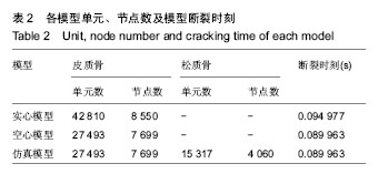
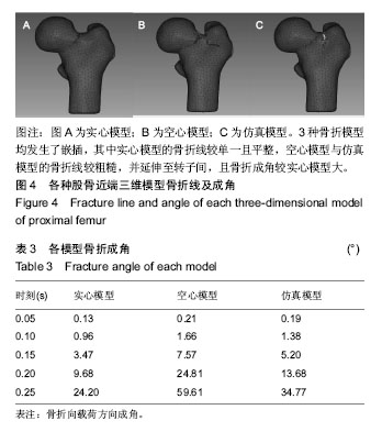
| [1] 郭鱼波,王丽丽,马如风,等. 骨质疏松的中医病因病机分析及其中医药治疗的前景探讨[J]. 世界科学技术-中医药现代化, 2015, 17(4):768-772.[2] Resnick NM, Greenspan SL. 'Senile' osteoporosis reconsidered. JAMA. 1989;261(7):1025-1029.[3] Roberts B J, Thrall E, Muller J A, et al. Comparison of hip fracture risk prediction by femoral aBMD to experimentally measured factor of risk. Bone. 2010;46(3):742-746.[4] Holzer G, von Skrbensky G, Holzer LA, et al. Hip fractures and the contribution of cortical versus trabecular bone to femoral neck strength. J Bone Miner Res. 2009;24(3):468-474.[5] Thomas CDL, Mayhew PM, Power J, et al. Femoral neck trabecular bone: loss with aging and role in preventing fracture. J Bone Miner Res.2009;24(11):1808-1818.[6] Huiskes R. On the modelling of long bones in structural analyses. J Biomech. 1982;15(1):65-69.[7] 李鹏飞,杜根发,林梓凌,等. 基于LS-DYNA模拟老年股骨颈骨折的有限元分析[J]. 中国组织工程研究, 2016,22(44):6606-6611.[8] Zhong RY, Wu XP, Liao EY, et al. Relationship between age-related bone mass of the lumbar spine and skeletal size and their effects on the diagnosis of osteoporosis in women. Chin J Osteoporos. 2012;18(2):99-105.[9] Hayes WC, Piazza SJ, Zysset PK. Biomechanics of fracture risk prediction of the hip and spine by quantitative computed tomography. Radiol Clin North Am.1991;29(1):1-18.[10] 彭李华,陈世荣,唐进,等. 骨质疏松股骨三维有限元模型的建立[J]. 中国组织工程研究与临床康复, 2010,14(9):1545-1548.[11] Morgan EF, Keaveny TM. Dependence of yield strain of human trabecular bone on anatomic site. 2001;34(2001): 569-577.[12] Rieger R, Auregan JC, Hoc T. Micro-finite-element method to assess elastic properties of trabecular bone at micro- and macroscopic level. Morphologie. 2018;102(336):12-20.[13] 李平.不同跌倒姿势与髋部应力变化关系的有限元分析[D]. 广州: 广州中医药大学, 2014.[14] 常文举,丁海. 股骨近端解剖与生物力学研究进展[J]. 医用生物力学, 2016,31(2):188-192.[15] Kopperdahl DL, Aspelund T, Hoffmann PF, et al. Assessment of incident spine and hip fractures in women and men using finite element analysis of CT scans. J Bone Miner Res. 2014; 29(3):570-580.[16] Marco M, Giner E, Larraínzar-Garijo R, et al. Numerical Modelling of Femur Fracture and Experimental Validation Using Bone Simulant. Ann Biomed Eng.2017;45(10): 2395-2408.[17] Ariza O, Gilchrist S, Widmer RP, et al. Comparison of explicit finite element and mechanical simulation of the proximal femur during dynamic drop-tower testing. J Biomech. 2015; 48(2):224-232.[18] 王清远, Pidaparti RM. 骨疲劳损伤研究[J]. 力学进展, 2002, 32(2):285-292.[19] 丁海,朱振安,薛晶,等. 骨质疏松症对松质骨骨小梁应力与微损伤关系的影响[J]. 医用生物力学, 2015,30(1):68-73.[20] Carter DR, Hayes WC. The compressive behavior of bone as a two-phase porous structure. J Bone Joint Surg Am. 1977; 59(7):954-962.[21] Koivumaki JE, Thevenot J, Pulkkinen P, et al. Cortical bone finite element models in the estimation of experimentally measured failure loads in the proximal femur. Bone. 2012; 51(4):737-740.[22] Nawathe S, Nguyen BP, Barzanian N, et al. Cortical and trabecular load sharing in the human femoral neck. J Biomech. 2015;48(5):816-822.[23] Lu Y, Wang L, Hao Y, et al. Analysis of trabecular distribution of the proximal femur in patients with fragility fractures. BMC Musculoskelet Disord. 2013;14:130.[24] Bell KL, Loveridge N, Power J, et al. Regional differences in cortical porosity in the fractured femoral neck. Bone. 1999; 24(1):57-64.[25] Granke M, Makowski A J, Uppuganti S, et al. Prevalent role of porosity and osteonal area over mineralization heterogeneity in the fracture toughness of human cortical bone. J Biomech. 2016;49(13):2748-2755.[26] 梁伟,吴斗,赵恩哲,等. 皮质厚度在骨质疏松性髋部骨折中的应用研究[J]. 中华老年骨科与康复电子杂志, 2018,4(3):184-188.[27] 杜根发. 基于断裂力学探讨骨密度影响老年股骨颈骨折的有限元分析[D]. 广州:广州中医药大学, 2017.[28] Zani L, Erani P, Grassi L, et al. Strain distribution in the proximal Human femur during in vitro simulated sideways fall. J Biomech. 2015;48(10):2130-2143.[29] Nishiyama KK, Gilchrist S, Guy P, et al. Proximal femur bone strength estimated by a computationally fast finite element analysis in a sideways fall configuration. J Biomech. 2013; 46(7):1231-1236.[30] Pashah S, Massenzio M, Jacquelin E. Prediction of structural response for low velocity impact. Int J Impact Eng. 2008;35(2): 119-132.[31] Haider IT, Speirs AD, Frei H. Effect of boundary conditions, impact loading and hydraulic stiffening on femoral fracture strength. J Biomech. 2013;46(13):2115-2121.[32] 孙培栋. 侧方跌倒高度及髋保护器对髋部冲击影响的实验及有限元分析[D].广州:南方医科大学, 2012.[33] Robinovitch SN, Inkster L, Maurer J, et al. Strategies for avoiding hip impact during sideways falls. J Bone Miner Res. 2003;18(7):1267-1273.[34] Szivek JA, Benjamin JB, Anderson PL. An experimental method for the application of lateral muscle loading and its effect on femoral strain distributions. Med Eng Phys. 2000; 22(2):109-116. |