Chinese Journal of Tissue Engineering Research ›› 2019, Vol. 23 ›› Issue (12): 1881-1886.doi: 10.3969/j.issn.2095-4344.1126
Previous Articles Next Articles
Effect of stiffness change of calcified cartilage zone on the stress of osteochondral structure using finite element analysis
Chen Kaining1, Nong Mingshan1, Ye Qing1, Luo Liuning1, Yang Xing1, Chen Cheng2, Wang Fuyou2
- 1Department of Orthopedics, Guangxi General Hospital of Chinese Armed Police Force, Nanning 530003, Guangxi Zhuang Autonomous Region, China; 2Department of Joint Surgery, Southwest Hospital, Army Medical University (the Third Military Medical University), Chongqing 400038, China
-
Online:2019-04-28Published:2019-04-28 -
Contact:Wang Fuyou, MD, Associate professor, Department of Joint Surgery, Southwest Hospital, Army Medical University (the Third Military Medical University), Chongqing 400038, China -
About author:Chen Kaining, MD, Attending physician, Department of Orthopedics, Guangxi General Hospital of Chinese Armed Police Force, Nanning 530003, Guangxi Zhuang Autonomous Region, China -
Supported by:the National Natural Science Foundation of China, No. 81271981 (to WFY); the Natural Science Foundation of Guangxi Zhuang Autonomous Region, China, No. 2015GXNSFAA139168 (to CKN)
CLC Number:
Cite this article
Chen Kaining, Nong Mingshan, Ye Qing, Luo Liuning, Yang Xing, Chen Cheng, Wang Fuyou. Effect of stiffness change of calcified cartilage zone on the stress of osteochondral structure using finite element analysis [J]. Chinese Journal of Tissue Engineering Research, 2019, 23(12): 1881-1886.
share this article
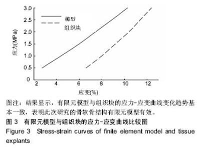
2.1 验证有限元模型的有效性 选取10个正常成年的牛膝髌骨标本,从每个髌骨中垂直关节面钻取1个骨软骨组织块(直径1.0 cm,高度0.5 cm)。使组织块的透明软骨面朝上,放入电子万能材料试验机的压盘上,以200 N/min的速度逐渐施加压缩载荷至3.0 MPa。然后计算出组织块的应力-应变曲线。另一方面,对骨软骨结构有限元模型也逐渐施加压缩载荷至3.0 MPa,计算出模型的应力-应变曲线,并与组织块的曲线进行比较,见图3。结果显示,有限元模型与组织块的应力-应变曲线变化趋势基本一致,表明此次研究的骨软骨结构有限元模型有效。"
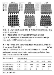
2.2 钙化软骨层的应力分布 在0.5 MPa压缩载荷作用下,钙化软骨层正常、坚硬和柔软模型中钙化软骨层的应力分布见图4,各个载荷(0.5-3.0 MPa)所形成的应力分布基本相同。在正常模型中,钙化软骨层上表面球冠突起受到了中等强度的应力,而下表面指状突起受到的应力较小,最大应力分布在钙化软骨层中间部分指状突起之间的区域,见图4A。在坚硬模型中,钙化软骨层上表面球冠突起受到了较小的应力,而下表面指状突起受到了中等强度的应力,最大应力分布在指状突起之间的区域,与正常模型相似,见图4B。在柔软模型中,钙化软骨层下表面的指状突起受到的应力较小,最大应力分布在指状突起根部的周围区域,与正常和坚硬模型明显不同,见图4C。在0.5-3.0 MPa的载荷作用下,钙化软骨层正常时和变硬后的应力峰值均随载荷的增加而增加,并且变硬后的应力峰值依次比正常时增加26.67%,27.12%,25.84%,27.12%,25.69%及26.59%,平均增加26.51%。而当钙化软骨层变软后,在0.5,1.0 MPa载荷作用下其应力峰值分别比正常时减少53.33%和50.85%,平均减少52.09%,在载荷大于1.0 MPa时,柔软模型发生失效,见表2。"

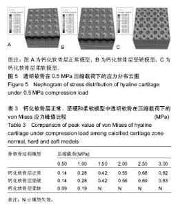
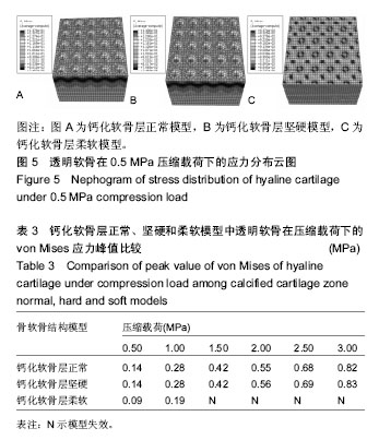
2.3 透明软骨的应力分布 钙化软骨层正常、坚硬和柔软模型中的透明软骨在各个压缩载荷(0.5-3.0 MPa)作用下所形成的应力分布云图基本一致,见图5。在正常和坚硬模型中,透明软骨的大部分区域受到的应力较小,应力主要集中在下表面的球冠凹陷中,尤其底部受到的应力最大,见图5A,B。在柔软模型中,透明软骨的大部分区域受到中等强度的应力,最大应力的区域与正常和坚硬模型相似,见图5C。在0.5-3.0 MPa的压缩载荷作用下,正常和坚硬模型中透明软骨的应力峰值均随载荷的增加而增加,并且两者几乎相同。在0.5,1.0 MPa压缩载荷作用下,柔软模型中透明软骨的应力峰值分别比正常时减少35.71%和32.14%,平均减少33.93%,但在大于1.0 MPa载荷下,模型发生失效,见表3。"
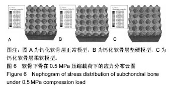
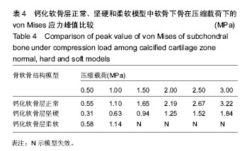
2.4 软骨下骨的应力分布 在钙化软骨层正常、坚硬和柔软模型中,各个压缩载荷(0.5-3.0 MPa)对软骨下骨所形成的应力分布与0.5 MPa压缩载荷下的应力分布差别不大,见图6。在正常和柔软模型中,软骨下骨的最大应力分布在上表面指状凹陷的上部周缘,而凹陷底部受到的应力较小,见图6A,C。在坚硬模型中,软骨下骨的最大应力分布在指状凹陷之间,见图6B。在0.5-3.0 MPa的压缩载荷作用下,正常和坚硬模型中软骨下骨的应力峰值均随载荷的增加而增加,但坚硬模型中软骨下骨的应力峰值依次比正常模型减少43.64%,42.73%,43.03%,42.92%,43.07%及42.86%,平均减少43.04%。柔软模型在0.5,1.0 MPa压缩载荷作用下,其软骨下骨的应力峰值与正常模型相似,在载荷大于1.0 MPa时,模型发生失效,见表4。"


"
| [1] 王富友,杨柳,段小军,等. 人体正常膝关节钙化软骨层组成成分研究[J]. 第三军医大学学报, 2008, 30(8): 687-690.[2] Wang F, Ying Z, Duan X, et al. Histomorphometric analysis of adult articular calcified cartilage zone. J Struct Biol. 2009; 168(3): 359-365.[3] Gupta HS et al. Two different correlations between nanoindentation modulus and mineral content in the bone-cartilage interface. J Struct Biol. 2005;149(2):138-148.[4] Flachsmann R, Broom ND, Hardy AE, et al. Why is the adolescent joint particularly susceptible to osteochondral shear fracture? Clin Orthop Relat Res. 2000;1(381): 212-221.[5] Hargrave-Thomas E, van Sloun F, Dickinson M, et al. Multi-scalar mechanical testing of the calcified cartilage and subchondral bone comparing healthy vs early degenerative states. Osteoarthritis Cartilage. 2015;23(10): 1755-1762.[6] Burr DB. Anatomy and physiology of the mineralized tissues: role in the pathogenesis of osteoarthrosis . Osteoarthritis Cartilage. 2004;12 Suppl A: S20-30.[7] 宋伟,王富友,杨柳. 关节软骨钙化层研究进展[J]. 中国修复重建外科杂志, 2011,25(11):1339-1342.[8] Norrdin RW, Kawcak CE, Capwell BA, et al. Calcified cartilage morphometry and its relation to subchondral bone remodeling in equine arthrosis. Bone. 1999;24(2):109-114.[9] Zizak I, Roschger P, Paris O, et al. Characteristics of mineral particles in the human bone/cartilage interface. J Struct Biol. 2003;141(3): 208-217.[10] Mente PL, Lewis JL. Elastic modulus of calcified cartilage is an order of magnitude less than that of subchondral bone . J Orthop Res. 1994; 12(5):637-647.[11] Allan KS, Pilliar RM, Wang J, et al. Formation of biphasic constructs containing cartilage with a calcified zone interface. Tissue Eng. 2007; 13(1):167-177.[12] St-Pierre JP, Gan L, Wang J, et al. The incorporation of a zone of calcified cartilage improves the interfacial shear strength between in vitro-formed cartilage and the underlying substrate. Acta Biomater. 2012;8(4):1603-1615.[13] Campbell SE, Ferguson VL, Hurley DC. Nanomechanical mapping of the osteochondral interface with contact resonance force microscopy and nanoindentation. Acta Biomater. 2012;8(12):4389-4396.[14] Stender ME, Carpenter RD, Regueiro RA, et al. An evolutionary model of osteoarthritis including articular cartilage damage, and bone remodeling in a computational study . J Biomech. 2016;49(14): 3502-3508.[15] Anderson DD, Brown TD, Radin EL. The influence of basal cartilage calcification on dynamic juxtaarticular stress transmission. Clin Orthop. 1993;286: 298-307.[16] Dar FH, Aspden RM. A finite element model of an idealized diarthrodial joint to investigate the effects of variation in the mechanical properties of the tissues. Proc Inst Mech Eng H. 2003;217(5): 341-348.[17] Taylor AM. Metabolic and endocrine diseases, cartilage calcification and arthritis. Curr Opin Rheumatol. 2013; 25(2):198-203.[18] Goldring SR, Goldring MB. Changes in the osteochondral unit during osteoarthritis: structure, function and cartilage-bone crosstalk . Nat Rev Rheumatol. 2016;12(11): 632-644.[19] Cheng HW, Luk KD, Cheung KM, et al. In vitro generation of an osteochondral interface from mesenchymal stem cell-collagen microspheres . Biomaterials. 2011;32(6): 1526-1535.[20] Lee WD, Hurtig MB, Pilliar RM, et al. Engineering of hyaline cartilage with a calcified zone using bone marrow stromal cells. Osteoarthritis Cartilage. 2015; 23(8): 1307-1315.[21] 王富友,杨柳,段小军,等. 正常膝关节软骨钙化层形态结构研究[J]. 中国修复重建外科杂志, 2008,22(5): 524-527.[22] Simha NK, Jin H, Hall ML, et al. Effect of indenter size on elastic modulus of cartilage measured by indentation. J Biomech Eng. 2007; 129(5): 767-775.[23] Knecht S, Vanwanseele B, Stussi E. A review on the mechanical quality of articular cartilage-implications for the diagnosis of osteoarthritis . Clin Biomech (Bristol, Avon). 2006;21(10): 999-1012.[24] Byers PD, Brown RA. Cell columns in articular cartilage physes questioned: a review. Osteoarthritis Cartilage. 2006; 14(1): 3-12.[25] Frisbie DD, CrossM W, McIlwraith C W. A comparative study of articular cartilage thickness in the stifle of animal species used in human preclinical studies compared to articular cartilage thickness in the human knee. Vet Comp Orthop Traumatol. 2006;19(3): 142-146.[26] Daubs BM, Markel MD, Manley PA. Histomorphometric analysis of articular cartilage, zone of calcifed cartilage, and subchondral bone plate in femoral heads from clinically normal dogs and dogs with moderate or severe osteoarthritis. Am J Vet Res. 2006;67(10): 1719-1724.[27] Ferguson VL, Bushby AJ, Boyde A. Nanomechanical properties and mineral concentration in articular calcified cartilage and subchondral bone. J Anat. 2003;203(2): 191-202.[28] Malekipour F, Oetomo D, Lee PV. Equine subchondral bone failure threshold under impact compression applied through articular cartilage . J Biomech. 2016;49(10): 2053-2059.[29] Turley SM, Thambyah A, Riggs CM, et al. Microstructural changes in cartilage and bone related to repetitive overloading in an equine athlete model. J Anat. 2014; 224(6): 647-658.[30] 宋伟,杨柳,王富友. 膝关节原发性骨关节炎软骨和软骨下骨病理改变的定量研究[J]. 中国修复重建外科杂志, 2011, 25(12): 1434-1439.[31] Burr DB, Radin EL. Microfractures and microcracks in subchondral bone: are they relevant to osteoarthrosis? Rheum Dis Clin North Am. 2003; 29(4): 675-685. |
| [1] | Xu Feng, Kang Hui, Wei Tanjun, Xi Jintao. Biomechanical analysis of different fixation methods of pedicle screws for thoracolumbar fracture [J]. Chinese Journal of Tissue Engineering Research, 2021, 25(9): 1313-1317. |
| [2] | Zhang Tongtong, Wang Zhonghua, Wen Jie, Song Yuxin, Liu Lin. Application of three-dimensional printing model in surgical resection and reconstruction of cervical tumor [J]. Chinese Journal of Tissue Engineering Research, 2021, 25(9): 1335-1339. |
| [3] | Chen Xinmin, Li Wenbiao, Xiong Kaikai, Xiong Xiaoyan, Zheng Liqin, Li Musheng, Zheng Yongze, Lin Ziling. Type A3.3 femoral intertrochanteric fracture with augmented proximal femoral nail anti-rotation in the elderly: finite element analysis of the optimal amount of bone cement [J]. Chinese Journal of Tissue Engineering Research, 2021, 25(9): 1404-1409. |
| [4] | Zhou Jihui, Li Xinzhi, Zhou You, Huang Wei, Chen Wenyao. Multiple problems in the selection of implants for patellar fracture [J]. Chinese Journal of Tissue Engineering Research, 2021, 25(9): 1440-1445. |
| [5] | Wu Xun, Meng Juanhong, Zhang Jianyun, Wang Liang. Concentrated growth factors in the repair of a full-thickness condylar cartilage defect in a rabbit [J]. Chinese Journal of Tissue Engineering Research, 2021, 25(8): 1166-1171. |
| [6] | Li Jiacheng, Liang Xuezhen, Liu Jinbao, Xu Bo, Li Gang. Differential mRNA expression profile and competitive endogenous RNA regulatory network in osteoarthritis [J]. Chinese Journal of Tissue Engineering Research, 2021, 25(8): 1212-1217. |
| [7] | Geng Qiudong, Ge Haiya, Wang Heming, Li Nan. Role and mechanism of Guilu Erxianjiao in treatment of osteoarthritis based on network pharmacology [J]. Chinese Journal of Tissue Engineering Research, 2021, 25(8): 1229-1236. |
| [8] | Zeng Yanhua, Hao Yanlei. In vitro culture and purification of Schwann cells: a systematic review [J]. Chinese Journal of Tissue Engineering Research, 2021, 25(7): 1135-1141. |
| [9] | Xu Yulin, Shen Shi, Zhuo Naiqiang, Yang Huilin, Yang Chao, Li Yang, Zhao Heng, Zhao Lu. Biomechanical comparison of three different plate fixation methods for acetabular posterior column fractures in standing and sitting positions [J]. Chinese Journal of Tissue Engineering Research, 2021, 25(6): 826-830. |
| [10] | Cai Qunbin, Zou Xia, Hu Jiantao, Chen Xinmin, Zheng Liqin, Huang Peizhen, Lin Ziling, Jiang Ziwei. Relationship between tip-apex distance and stability of intertrochanteric femoral fractures with proximal femoral anti-rotation nail: a finite element analysis [J]. Chinese Journal of Tissue Engineering Research, 2021, 25(6): 831-836. |
| [11] | Song Chengjie, Chang Hengrui, Shi Mingxin, Meng Xianzhong. Research progress in biomechanical stability of lateral lumbar interbody fusion [J]. Chinese Journal of Tissue Engineering Research, 2021, 25(6): 923-928. |
| [12] | Liu Zhao, Xu Xilin, Shen Yiwei, Zhang Xiaofeng, Lü Hang, Zhao Jun, Wang Zhengchun, Liu Xuzhuo, Wang Haitao. Guiding role and prospect of staging and classification combined collapse prediction method for osteonecrosis of femoral head [J]. Chinese Journal of Tissue Engineering Research, 2021, 25(6): 929-934. |
| [13] | He Xiangzhong, Chen Haiyun, Liu Jun, Lü Yang, Pan Jianke, Yang Wenbin, He Jingwen, Huang Junhan. Platelet-rich plasma combined with microfracture versus microfracture in the treatment of knee cartilage lesions: a meta-analysis [J]. Chinese Journal of Tissue Engineering Research, 2021, 25(6): 964-969. |
| [14] | Liu Xin, Yan Feihua, Hong Kunhao. Delaying cartilage degeneration by regulating the expression of aquaporins in rats with knee osteoarthritis [J]. Chinese Journal of Tissue Engineering Research, 2021, 25(5): 668-673. |
| [15] | Xie Chongxin, Zhang Lei. Comparison of knee degeneration after anterior cruciate ligament reconstruction with or without remnant preservation [J]. Chinese Journal of Tissue Engineering Research, 2021, 25(5): 735-740. |
| Viewed | ||||||
|
Full text |
|
|||||
|
Abstract |
|
|||||