Chinese Journal of Tissue Engineering Research ›› 2018, Vol. 22 ›› Issue (30): 4757-4762.doi: 10.3969/j.issn.2095-4344.0969
Effects of domestic porous tantalum on biological behavior and function of rabbit osteoblasts
Wang Qian1, Teng Xue-feng1, Gan Hong-quan2, Zhang Hui3, Cui Yi-shuang4, Chen Jing-jing4, Li Qi-jia1, Wang Zhi-qiang2
- 1Medical Experimental Center, North China University of Science and Technology, Tangshan 063210, Hebei Province, China; 2Department of Orthopedics, Affiliated Hospital of North China University of Science and Technology, Tangshan 063000, Hebei Province, China; 3First Department of Joint, Second Hospital of Tangshan, Tangshan 063000, Hebei Province, China; 4Graduate School, North China University of Science and Technology, Tangshan 063009, Hebei Province, China
-
Received:2018-07-10Online:2018-10-28Published:2018-10-28 -
Contact:Wang Zhi-qiang, Professor, Doctoral supervisor, Department of Orthopedics, Affiliated Hospital of North China University of Science and Technology, Tangshan 063000, Hebei Province, China -
About author:Wang Qian, MD, Associate professor, Medical Experimental Center, North China University of Science and Technology, Tangshan 063210, Hebei Province, China -
Supported by:the Supporting Program for Science and Technology Research of the Ministry of Science and Technology of China, No. 2012BAE06B03; the Supporting Program for Science and Technology Research in Hebei Province, No. 16277776D; the Medical Science Research Project of Hebei Province, No. 20160225; the Doctoral Research Startup Fund of the North China University of Technology, No. 28606299
CLC Number:
Cite this article
Wang Qian, Teng Xue-feng, Gan Hong-quan, Zhang Hui, Cui Yi-shuang, Chen Jing-jing, Li Qi-jia, Wang Zhi-qiang. Effects of domestic porous tantalum on biological behavior and function of rabbit osteoblasts[J]. Chinese Journal of Tissue Engineering Research, 2018, 22(30): 4757-4762.
share this article
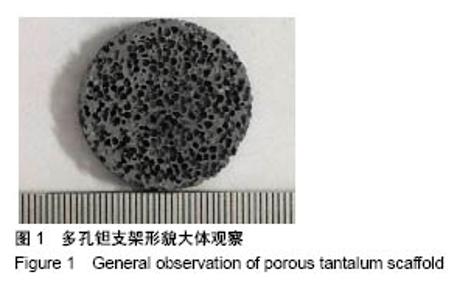
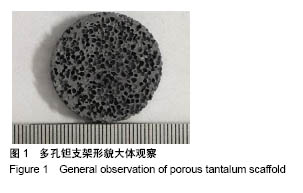
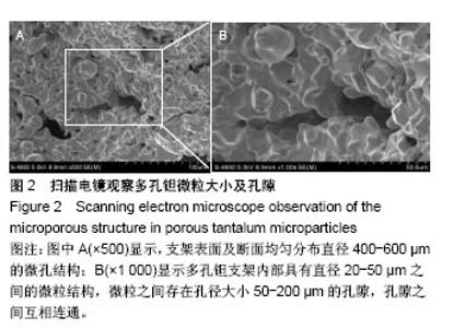
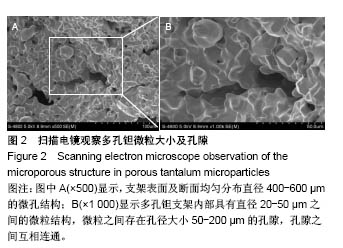
2.1 多孔钽材料观察 大体观察多孔钽支架材料外观呈灰色,表面光洁,材料表面及断面内可见均匀分布着蜂窝状孔隙(图1)。 扫描电镜500倍镜下观察可见,支架表面及断面均匀分布直径400-600 µm的微孔结构;1 000倍镜下可见,多孔钽支架内部具有直径20-50 μm之间的微粒结构,微粒之间存在孔径大小50-200 µm的孔隙,孔隙之间互相连通(图2)。"
"
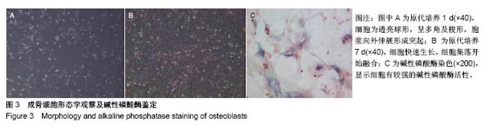
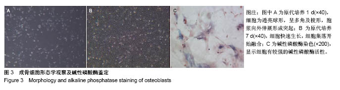
2.2 成骨细胞的形态特征观察及鉴定 倒置相差显微镜观察原代细胞接种后为透亮球形,呈多角及梭形,胞浆向外伸展形成突起;第3天细胞体积增大,数量增加,相互连接,形成分散集落;第4-7天细胞快速生长,细胞集落开始融合(图3A,B);待细胞生长汇合至80%-90%时进行传代培养,传代后细胞多呈长梭形、三角形,体积变大,生长速度加快,5-7 d长满瓶底;第3代细胞形态呈长梭形,趋于单一化。碱性磷酸酶染色呈阳性细胞胞浆内可见蓝染颗粒,显示细胞有较强的碱性磷酸酶活性及钙化能力(图3C)。"
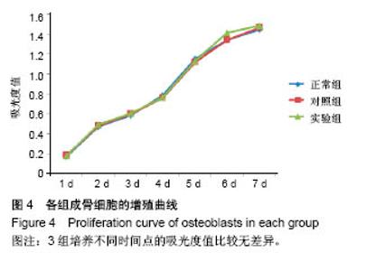
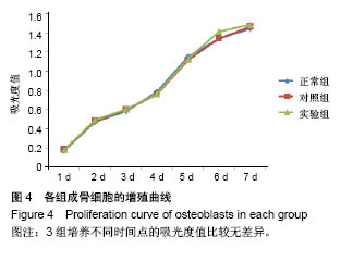
2.3 CCK-8法检测成骨细胞增殖 采用CCK-8法测定各组成骨细胞1-7 d的吸光度值,根据所测得的吸光度值绘制成骨细胞增殖曲线图(图4),经方差分析显示,3组培养不同时间点的吸光度值比较无差异。"

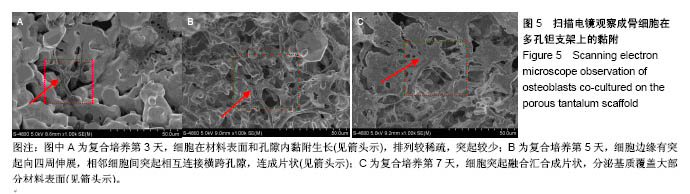
2.4 扫描电镜观察成骨细胞在多孔钽支架上的黏附生长 多孔钽支架材料与成骨细胞复合培养后,随时间延长,细胞在材料表面黏附生长,并逐渐覆盖材料表面。复合培养第3天,细胞在材料表面和孔隙内黏附生长,排列较稀疏,突起较少,随培养时间延长,细胞逐渐伸展、拉长并伸出伪足;第5天,细胞边缘有突起向四周伸展,相邻细胞间突起相互连接横跨孔隙,连成片状;第7天,细胞突起融合汇合成片状,分泌基质覆盖大部分材料表面(图5)。"

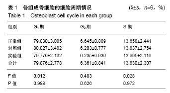
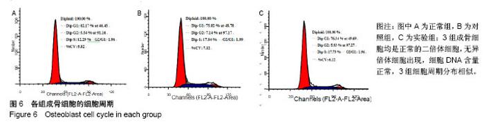
2.5 流式细胞仪检测各组成骨细胞的细胞周期 采用流式细胞仪对细胞周期和倍体进行检测并分析,结果显示3组成骨细胞均是正常的二倍体细胞,无异倍体细胞出现,细胞DNA含量正常,3组细胞周期分布相似;3组细胞均是G1期比例较高,多数细胞处于静止期,提示多孔钽支架没有细胞毒性,不会引起细胞DNA含量变化。对3组间G1、G2、S期细胞比例进行单因素方差分析,结果显示差异无显著性意义(P > 0.05),见表1,图6。"
"
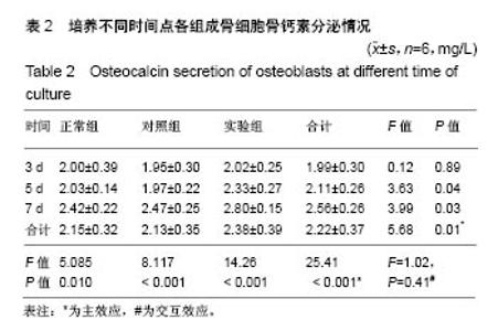
2.6 Elisa法测定各组成骨细胞骨钙素分泌 随着培养时间延长,各组成骨细胞骨钙素分泌量逐渐增多。实验组培养第5,7天的骨钙素分泌量多于其余两组(P < 0.05);对照组与正常组培养不同时间点的骨钙素分泌量无差异,见表2。"

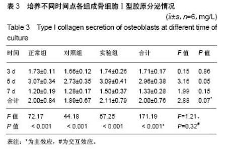
2.7 Elisa法测定各组成骨细胞Ⅰ型胶原分泌 随培养时间的延长,各组成骨细胞Ⅰ型胶原分泌量初期逐渐增多,在第5天达高峰,第7天分泌下降;实验组Ⅰ型胶原分泌量均较其他两组略有升高,但3组间比较差异无显著性意义 (P > 0.05),见表3。"
| [1] 杨化娟,杨柯,张炳春.医用不锈钢的发展及展望[J].材料导报, 2005,19(6): 56-59.[2] Bermudez MD, Carrion FJ, Lopez R, et al. Erosion corrosion of stainless steels, titanium, tantalum and zirconium. Wear. 2005;258(1-4):693-700. [3] Zhang M, Wang GL, Zhang HF, et al. Repair of segmental long bone defect in a rabbit radius nonunion model: comparison of cylindrical porous titanium and hydroxyapatite scaffolds. Artif Organs. 2014;38(6): 493-502. [4] 于思荣.生物医学钛合金的研究现状及发展趋势[J].材料科学与工程, 2000, 18(2):131-134.[5] Bertrand E, Gloriant T, Gordin DM, et al. Synthesis and characterization of a new superelastic Ti-25Ta-25Nb biomedical alloy. J Mech Behav Biomed. 2010;3:559-564.[6] 赵晋,闫振宇,张立智.骨性关节炎软骨和软骨下骨之间信号通路[J].中华骨质疏松和骨矿盐疾病杂志,2016,9(2):193-198.[7] 单鹏程,曹永平.软骨下骨在骨关节炎发病机制中的作用[J].中国矫形外科杂志,2009,17(23):1792-1794.[8] Hanzlik JA, Day JS, Clare M, et al. Is There A Difference in Bone Ingrowth in Modular Versus Monoblock Porous Tantalum Tibial Trays. J Arthroplasty. 2015;30(6):1073-1078. [9] Li X, Lin Z, Duan Y, et al. Repair of large segmental bone defects in rabbits using BMP and FGF composite xenogeneic bone. Genet Mol Res. 2015;14(2):6395-6400. [10] 贺瑞,张文志,尚希福,等.钽金属椎间融合器在腰椎间盘突出症减压术后复发融合术中的应用[J].颈腰痛杂志, 2013,34(5):387-390.[11] 刘永庆,李琪佳,王志强.多孔金属骨科内植物改良策略的研究进展[J].医学综述,2016,22 (20):4021-4024.[12] Wang H, Li Q, Wang Q, et al. Enhanced repair of segmental bone defects in rabbit radius by porous tantalum scaffolds modified with the RGD peptide. J Mater Sci Mater Med. 2017;28:50. [13] 张大鹏,刘永庆,王志强.医用多孔钽的特性及其表面改性的研究进展[J].中国煤炭工业医学杂志,2017,20(2):236-239.[14] 于晓明,谭丽丽,杨柯.医用金属表面的钽涂层制备及其临床应用趋势[J].中国骨科临床与基础研究杂志,2013,5(2):115-119.[15] 赖振权,崔逸爽,陈超,等.新型骨植入材料多孔钽-骨结合界面成骨以及相关成骨因子的表达及意义[J].中国组织工程研究, 2017,21(18):2789-2795.[16] Sagomonyants KB, Hakim-Zargar M, Jhaveri A, et al. Porous tantalum stimulates the proliferation and osteogenesis of osteoblasts from elderly female patients. J Orthop Res. 2011;29(4):609-616. [17] Bobyn JD, Stackpool GJ, Hacking SA, et al. Characteristics of bone ingrowth and interface mechanics of a new porous tantalum biomatcrial. J Bone Joint Surg Br. 1999;81(5):907-914. [18] Wang Q, Zhang H, Li Q, et al. Biocompatibility and osteogenic properties of porous tantalum. Exp Ther Med. 2015;9(3):780-786. [19] 王辉,王茜,张辉,等.带蒂筋膜瓣包裹国产多孔钽修复兔桡骨节段性骨缺损实验研究[J].中国修复重建外科杂志, 2017,31(10):1200-1207.[20] 李琪佳,王茜,甘洪全,等.多孔钽材料细胞毒性、生物相容性及体内成骨性研究[J].中华骨科杂志,2014,34(9):954-961.[21] Findlay DM, Welldon K, Atkins GJ, et al. The proliferation and phenotypic expression of human osteoblasts on tantalum metal. Biomaterials. 2004; 25(12):2215-2227. [22] ANSI/AAMI. ISO 10993-5. Biological evaluation of medical devices Part 5:Tests for cytotoxicity: in vitro methods[S]. Arlington, VA.[23] ANSI /AAMI. ISO 10993-12. Biological evaluation of medical devices Part 12: Sample preparation and reference materials[S]. Arlington, VA.[24] 武垚森,池永龙.小梁金属在骨科中的应用[J].中国骨科杂志, 2007,27(12): 939-941.[25] 董伟,刘洪臣.钽及多孔钽表面改性技术在组织工程学及口腔医学的研究进展[J].中华老年口腔医学杂志,2017,15(2):113-116.[26] 欧阳建安,王大平.多孔钽骨科临床应用概况[J].国际骨科杂志, 2011,32(5): 314-315.[27] 武垚森,池永龙.小梁金属(多孔钽)在骨科的应用现状[J].中华骨科杂志,2007, 27(12):939-941.[28] 初殿伟,王玮,张华亮,等.骨软骨组织工程支架的研究现状及发展趋势[J].国际生物医学工程杂志,2009,32(2):105-116.[29] Guldberg RE, Duvall CL, Peister A, et al. 3D imaging of tissue integration with porous biomaterials. Biomaterials. 2008;29(28):3757-3761. [30] 张彦博,李瑞延,刘贯聪,等.多孔钽的理化/生物特性及其骨整合能力研究进展[J].生物骨科材料与临床研究,2017,14(5):7-11.[31] Cardaropoli F, Alfieri V, Caiazzo F, et al. Manufacturing of porous biomaterials for dental implant applications through selective laser melting. Adv Mater Res. 2012;6(535-537):1222-1229. [32] Wan Y, Wang Y, Liu Z, et al. Adhesion and proliferation of OCT-1osteoblast- like cells on micro-and nano-scale topography structured poly(L-lactide). Biomaterials. 2005;26(21):4453-4459. [33] Mustafa K, Wroblewski J, Hultenby K, et al. Effect of titanium surface blasted with Ti02 particles on the initial attachment of cells derived from human mandibular bone. Clin Oral Implants Res. 2000;11(2):116-128. [34] Marelli B, Ghezzi CE, Barralet JE, et al. Three-dimensional mineralization of dense nanofibrillar collagen-bioglass hybrid scaffolds. Biomacromolecules. 2010;11(6):1470-1479. [35] 张辉,李亮,王茜,等.骨形成蛋白-7对多孔钽-软骨细胞复合物分泌功能以及COL-II、AGG和Sox9基因表达的影响[J].北京大学学报(医学版), 2015, 47(1): 216-222.[36] 王茜,张辉,耿丽鑫,等.国产多孔钽复合MG63细胞培养Col-1、OC和OPN蛋白表达及意义[J].中国矫形外科杂志, 2015,23(5):441-449.[37] 史伟,甘洪全,王茜,等.不同程度低氧培养对多孔钽-人成骨细胞复合物细胞形态及COL-1、OC蛋白表达的影响[J].中国矫形外科杂志, 2017,25(8): 729-736.[38] Pagani F, Francucci CM, Moro L. Markers of bone turnover: biochemical and clinical perspectives. J Endocrinol Invest. 2005;28(Suppl 10):8-13. [39] Lin Z, Solomon KL, Zhang X, et al. In vitro Evaluation of Natural Marine Sponge Collagen as a Scaffold for Bone Tissue Engineering. Int J Biol Sci. 2011;7(7):968-977. [40] Hauschka PV, Lian JB, Cole DEC, et al. Osteocalcin and matrix GLA protein: vitamin K-dependent proteins in bone. Physiol Rev. 1989;69(3): 990-1047. [41] 丁思阳,夏露,陈宁,等.微弧氧化纯钛表面对成骨细胞蛋白合成功能的影响[J].口腔医学研究,2012,28(2):125-128. |
| [1] | Zhang Tongtong, Wang Zhonghua, Wen Jie, Song Yuxin, Liu Lin. Application of three-dimensional printing model in surgical resection and reconstruction of cervical tumor [J]. Chinese Journal of Tissue Engineering Research, 2021, 25(9): 1335-1339. |
| [2] | Li Cai, Zhao Ting, Tan Ge, Zheng Yulin, Zhang Ruonan, Wu Yan, Tang Junming. Platelet-derived growth factor-BB promotes proliferation, differentiation and migration of skeletal muscle myoblast [J]. Chinese Journal of Tissue Engineering Research, 2021, 25(7): 1050-1055. |
| [3] | Liu Cong, Liu Su. Molecular mechanism of miR-17-5p regulation of hypoxia inducible factor-1α mediated adipocyte differentiation and angiogenesis [J]. Chinese Journal of Tissue Engineering Research, 2021, 25(7): 1069-1074. |
| [4] | Zeng Yanhua, Hao Yanlei. In vitro culture and purification of Schwann cells: a systematic review [J]. Chinese Journal of Tissue Engineering Research, 2021, 25(7): 1135-1141. |
| [5] | Ma Zetao, Zeng Hui, Wang Deli, Weng Jian, Feng Song. MicroRNA-138-5p regulates chondrocyte proliferation and autophagy [J]. Chinese Journal of Tissue Engineering Research, 2021, 25(5): 674-678. |
| [6] | Xu Dongzi, Zhang Ting, Ouyang Zhaolian. The global competitive situation of cardiac tissue engineering based on patent analysis [J]. Chinese Journal of Tissue Engineering Research, 2021, 25(5): 807-812. |
| [7] | Wang Yujiao, Liu Dan, Sun Song, Sun Yong. Biphasic calcium phosphate loaded with advanced platelet rich fibrin can promote the activity of rabbit bone marrow mesenchymal stem cells [J]. Chinese Journal of Tissue Engineering Research, 2021, 25(4): 504-509. |
| [8] | Zhou Jihui, Yao Meng, Wang Yansong, Li Xinzhi, Zhou You, Huang Wei, Chen Wenyao. Influence of novel nanoscaffolds on biological behaviors of neural stem cells and the related gene expression [J]. Chinese Journal of Tissue Engineering Research, 2021, 25(4): 532-536. |
| [9] | Ma Zhijie, Li Jingyu, Cao Fang, Liu Rong, Zhao Dewei. Influencing factors and biological property of novel biomedical materials: porous silicon carbide coated with bioactive tantalum [J]. Chinese Journal of Tissue Engineering Research, 2021, 25(4): 558-563. |
| [10] | Wu Zijian, Hu Zhaoduan, Xie Youqiong, Wang Feng, Li Jia, Li Bocun, Cai Guowei, Peng Rui. Three-dimensional printing technology and bone tissue engineering research: literature metrology and visual analysis of research hotspots [J]. Chinese Journal of Tissue Engineering Research, 2021, 25(4): 564-569. |
| [11] | Shi Xiaoxiu, Mao Shilong, Liu Yang, Ma Xingshuang, Luo Yanfeng. Comparison of tantalum and titanium (alloy) as orthopedic materials: physical and chemical indexes, antibacterial and osteogenic ability [J]. Chinese Journal of Tissue Engineering Research, 2021, 25(4): 593-599. |
| [12] | Chang Wenliao, Zhao Jie, Sun Xiaoliang, Wang Kun, Wu Guofeng, Zhou Jian, Li Shuxiang, Sun Han. Material selection, theoretical design and biomimetic function of artificial periosteum [J]. Chinese Journal of Tissue Engineering Research, 2021, 25(4): 600-606. |
| [13] | Liu Fei, Cui Yutao, Liu He. Advantages and problems of local antibiotic delivery system in the treatment of osteomyelitis [J]. Chinese Journal of Tissue Engineering Research, 2021, 25(4): 614-620. |
| [14] | Li Xiaozhuang, Duan Hao, Wang Weizhou, Tang Zhihong, Wang Yanghao, He Fei. Application of bone tissue engineering materials in the treatment of bone defect diseases in vivo [J]. Chinese Journal of Tissue Engineering Research, 2021, 25(4): 626-631. |
| [15] | Zhang Zhenkun, Li Zhe, Li Ya, Wang Yingying, Wang Yaping, Zhou Xinkui, Ma Shanshan, Guan Fangxia. Application of alginate based hydrogels/dressings in wound healing: sustained, dynamic and sequential release [J]. Chinese Journal of Tissue Engineering Research, 2021, 25(4): 638-643. |
| Viewed | ||||||
|
Full text |
|
|||||
|
Abstract |
|
|||||