Chinese Journal of Tissue Engineering Research ›› 2018, Vol. 22 ›› Issue (36): 5852-5857.doi: 10.3969/j.issn.2095-4344.0590
Previous Articles Next Articles
Roles and application values of P75 neurotrophin receptor in bone tissue engineering
Shen Mengjie, Yang Kun, Liu Qi
- (Department of Periodontology, Stomatological Hospital Affiliated to Zunyi Medical University, Zunyi 563003, Guizhou Province, China)
-
Received:2018-08-07Online:2018-12-28Published:2018-12-28 -
Contact:Liu Qi, MD, Professor, Department of Periodontology, Stomatological Hospital Affiliated to Zunyi Medical University, Zunyi 563003, Guizhou Province, China -
About author:Shen Mengjie, Master candidate, Department of Periodontology, Stomatological Hospital Affiliated to Zunyi Medical University, Zunyi 563003, Guizhou Province, China -
Supported by:the National Natural Science Foundation of China, No. 81760199
CLC Number:
Cite this article
Shen Mengjie, Yang Kun, Liu Qi. Roles and application values of P75 neurotrophin receptor in bone tissue engineering [J]. Chinese Journal of Tissue Engineering Research, 2018, 22(36): 5852-5857.
share this article
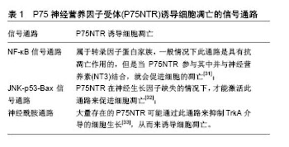
2.1 P75NTR生物学特性 神经生长因子不仅对神经损伤的再生修复具有重要作用,而且对细胞的增殖、分化具有促进作用[8]。其具有两种受体:即具有高亲和力的酪氨酸激酶受体A(TrkA)和低亲和力的P75神经营养因子受体(P75NTR)。P75NTR是相对分子质量为 75 000的Ⅰ型膜蛋白,它的结构主要包括胞外结构域、单跨膜结构域和胞内结构域[9]。胞外结构域主要与肿瘤坏死因子受体超家族结合,负责受体构象和配体结合;跨膜结构域在受体二聚化、配体结合引起的构象变化和信号转导中起着重要作用[10];而胞内结构域就相对比较保守,且较短,但是它有3种不同功能的结构域:①类似与肿瘤坏死因子受体相关因子结合的近膜区;②有死亡结构域;③位于C末端的蛋白结合结构域[11-12]。另外P75NTR属于肿瘤坏死因子受体,同时又是所有神经营养因子的共同受体,包括神经生长因子、神经营养素3、新生营养素等[13]。P75NTR在生物活性上具有多样性,它可以通过激活多种下游信号来调节细胞增殖、凋亡、迁移等多种细胞反应[6]。P75NTR可以单独起作用,也可以与TrkA一起协同作用。这种协同作用不仅可以促进细胞存活,而且对组织的再生修复具有非常重要的作用[14]。 2.2 P75NTR促进骨组织修复 近年来,人们发现许多信号分子在骨组织再生修复过程中有所表达,而且起到了积极的作用。随着对细胞和分子生物学技术的研究不断深入,关于骨组织再生修复过程中的信号因子调节系统引起了学者们的广泛关注,其中神经生长因子开始进入了人们的视线,P75NTR在促进骨组织再生修复方面的应用也引起了很多研究者的兴趣。有报道显示P75NTR不仅可以直接在骨组织再生修复过程中起作用,而且还可以调控干细胞,使其干细胞特性更强、更稳定,从而可以通过体外筛选出优质的干细胞而间接的促进骨组织的再生修复。下面就P75NTR在骨组织工程中的应用进行阐述。 2.2.1 体外研究 目前,关于P75NTR在骨组织修复方面上的应用,在基础研究中关于其作为干细胞表面标记物筛选出的P75NTR阳性的干细胞具有更优良的干细胞特性。Wen等[15]报道了使用P75NTR筛选外胚间充质干细胞,发现筛选出的P75NTR阳性的胚间充质干细胞,在细胞传代过程中呈均匀一致的成纤维细胞形态,细胞表型、增殖能力稳定。而且体外也证实P75NTR阳性的胚间充质干细胞具有分化为多种细胞类型的能力且稳定存在。同样Zhao等[16]也证明了P75NTR阳性的胚间充质干细胞具有更稳定的增殖分化能力和干细胞生物学特性。另外Rada等[17]报道了用抗P75NTR抗体分离的脂肪干细胞具有很高的成骨分化潜能。很多研究已经证明了P75NTR不仅可以作为干细胞表面的标记物,而且筛选出来的P75NTR阳性的干细胞具有更强的稳定性和干细胞特性。目前虽然还没有研究报道应用筛选出的P75NTR阳性的干细胞修复骨损伤,但体外研究证实筛选出的P75NTR阳性的干细胞具有更强的成骨分化能力,这为筛选出理想的种子细胞应用到骨组织工程奠定了坚实的理论基础。 很多的文献也报道了关于P75NTR直接作用于间充质干细胞和成骨细胞,并对其关于成骨分化做了研究,但是对于P75NTR是通过什么机制起作用的目前还在探索中。Ju等[18]报道了不同胚龄的SD大鼠外胚间充质干细胞在矿化过程中P75NTR的表达量,通过流式分析发现P75NTR在12.5,15.5和18.5 d的胚间充质干细胞中均有表达,且随着时间延长P75NTR在胚间充质干细胞中阳性表达率越高,通过ARS染色和碱性磷酸酶活性检测,结果也显示在第18.5天胚间充质干细胞的成骨能力更强。同样Yang等[19]也报道了关于P75NTR对SD大鼠胚间充质干细胞成骨能力的影响,通过免疫组织化学法检测P75NTR在牙发育过程中的表达,发现大鼠神经嵴源性胚间充质干细胞的体外矿化过程中P75NTR参与其矿化,并且认为P75NTR可能是通过与Mage-D1结合来增强其的成骨分化。同时Xing等[20]将外胚层间充质干细胞在牙上皮细胞培养基进行诱导,分别在诱导4,8, 12 d后进行成骨基因和成骨蛋白以及茜素红染色检测,结果也发现P75阳性外胚间充质干细胞较P75阴性外胚间充质干细胞具有更强的成骨分化能力,而且高表达牙本质基质蛋白和牙本质涎磷蛋白,并且发现了P75NTR的矿化作用可能与TGF-β/Smad4的信号通路有关。另外Liu等[21]认为脑源性神经营养因子对人骨髓间充质干细胞的神经发生和成骨的形成具有积极的作用,将脑源性神经营养因子与人骨髓间充质干细胞共培养7 d后,发现人骨髓间充质干细胞高表达神经源性标志物P75和S100,通过茜素红染色和碱性磷酸酶活性检测证实了人骨髓间充质干细胞成骨分化的能力得到了增强;同时体内实验也发现脑源性神经营养因子不仅可以直接促进人骨髓间充质干细胞成骨,而且还可以通过增加神经的发生间接促进成骨。而Mikami等[22]却报道了相反的结果:P75NTR过表达于小鼠胚胎间充质干细胞C3 H10T1/2中时,反而抑制了干细胞的成骨矿化能力,出现这种相反结论的原因可能是P75NTR在不同种类的细胞系体外矿化过程中有不同的调控机制以及体外实验所处微环境的不同而导致的。 对于P75NTR调控细胞增殖作用,认为可能与神经生长因子的存在有关系,当神经生长因子大量存在的时候能够启动TrkA存活通路,关闭P75NTR诱导的死亡通路[23];另外P75NTR的存在能够增强神经生长因子与TrkA结合,从而可以促进细胞的生长。Akiyama等[24]将外源性P75NTR转染到MG63细胞和MC3T3-E1细胞,培养6 d后,流式细胞分析P75NTR对细胞增殖的影响,发现转染了P75NTR的MC3T3-E1细胞的增殖速度比未转染的细胞快,另外关于碱性磷酸酶活性检测,在诱导3 d后,发现转染P75NTR后的MG63细胞和MC3T3-E1细胞与对照组相比碱性磷酸酶染色均表现出强阳性,茜素红染色也得到了相似的效果。RT-PCR检测发现转染P75NTR 12 d后均高表达成骨相关基因(Runx2、OSX、BSP和OC)。另外实验还证明了P75NTR增强细胞的成骨分化可能与P75NTR结合肌钙蛋白受体激酶(Trk)基因TrkA、TrkB、TrkC和Nogo受体(NGR)有关。Mikami等[25]近期的研究也得出相类似的结论。通过对正常骨中P75NTR表达的变化以及P75NTR促进骨组织再生修复通路机制的研究,可以为骨骼疾病的治疗提供新的方案和奠定理论基础。 2.2.2 体内研究 目前,由于P75NTR修复骨组织的机制原理还不是很清楚,关于应用P75NTR来修复骨缺损的实验还比较少,但是也有相关文献报道了P75NTR在骨组织的定位以及修复过程中表达的变化。Chartier 等[26]报道了P75在骨组织中有显著的表达,通过组织学检测,证实C57BL/6J小鼠股骨中P75几乎完全是由位于神经生长因子+血管附近的神经纤维表达,而P75在股骨中表达的部位是关节软骨深层区域。在骨髓和骨膜中,P75存在于具有感觉(CGRP)和交感神经(TH)神经纤维形态的神经纤维和类似TrkA神经纤维中,实验中作者对股骨进行脱钙处理,发现未脱钙的股骨相对于脱钙后的股骨高表达P75,并且对于不同鼠龄所表达P75的量也是不同的,这种老化及损伤后P75表达量的变化可以为以后治疗骨组织疾病提供新的思路。另外王莹莹等[27]利用Micro-CT对野生型小鼠和敲除了P75NTR的小鼠进行左侧股骨扫描,并对股骨的松质骨的骨量、骨小梁的数量、骨小梁的厚度、骨表面积等进行评估,结果发现:敲除了P75NTR的小鼠股骨的相关骨量相较于野生型小鼠显著下降;通过注射钙黄绿素来检测矿化沉积速率,结果也同样表明了敲除P75NTR的小鼠股骨矿化沉积速率(MRA)明显低于野生型;通过RT-PCR、Western Blot对小鼠股骨的总RNA、蛋白质以及相关基因进行检测,得到了与上述相同的结果。Yu等[28]报道了骨折后P75的表达情况,实验中制作了纯种大白兔的右下颌骨折模型,分别在术后1,3,5,7,14,28 d后取材,通过苏木精-伊红染色和免疫组织化学检测,结果表明在实验组骨折处神经生长因子受体P75的阳性率随着时间的延长逐渐增加,而对照组仅发现少量P75表达,推测P75可能通过与神经生长因子的结合来增强成骨细胞的活性,从而在骨折愈合中起到积极的修复作用。张伟等[29]也做了相类似的报道。另外有学者发现在骨折处注射神经生长因子,可以加速骨折愈合,且神经生长因子表达于骨折愈合后成骨样细胞中,这表明神经生长因子及其配体可能与促进骨组织再生修复有关[30]。 2.3 P75NTR抑制骨组织修复 P75NTR在生物活性上表现为多样性,它不仅可以调控细胞的生长、组织的再生,还可以通过激活多种下游信号参与细胞凋亡、迁移和细胞周期阻滞等多种细胞反应[5]。关于P75NTR抑制骨组织修复的机制原理还不是很清楚,但是有学者已经发现了3条关于P75NTR诱导细胞凋亡的信号通路,见表1。"

| [1] Sun H, Zhe Q, Ying G, et al. In vitro and in vivo effects of rat kidney vascular endothelial cells on osteogenesis of rat bone marrow mesenchymal stem cells growing on polylactide-glycoli acid (PLGA) scaffolds. Biomed Eng Online. 2007;6:41. [2] 韩倩,张志宏,刘红红,等. 犬下颌骨骨缺损自我修复能力的电镜观察[J]. 安徽医药,2012,16 (3):326-328. [3] 刘伟,赵光锋,喻任. 转移生长因子-β1、碱性成纤维细胞生长因子、骨形态发生蛋白-2在大鼠骨质疏松骨折愈合骨痂中的表达[J].中华急诊医学杂志,2003,12 (8):530-532. [4] Levimontalcini R. The nerve growth factor 35 years later. Science. 1987;237(4819):1154-1162. [5] 魏传银,王丽梅,陈雪红,等.神经营养素受体p75^NTR介导的信号转导[J].细胞与分子免疫学杂志,2005,21(2):258-60.[6] 李家勇,王铭,彭称飞,et al.P75NTR在兔骨折不愈合局部组织中的表达及意义[J].中国骨质疏松杂志,2017,23(4):437-40.[7] 吴小莹,杨斌.脑源性神经营养因子与骨/牙组织发育代谢:促成或抑制细胞的增殖与分化?[J].中国组织工程研究. 2015,19(2): 283-288.[8] 钟湘平,廖勇仕.TrkA、p75NTR在神经系统中的研究进展[J].社区医学杂志,2013,11(19):14-17.[9] XL H, KC G. Structure of nerve growth factor complexed with the shared neurotrophin receptor p75. Science. 2004;304 (5672):870-875. [10] Vilar M, Charalampopoulos I, Kenchappa RS, et al. Activation of the p75 neurotrophin receptor through conformational rearrangement of disulphide-linked receptor dimers. Neuron. 2009;62(1):72-83. [11] Coulson EJ, May LM, Sykes AM, et al. The role of the p75 neurotrophin receptor in cholinergic dysfunction in Alzheimer's disease. Neuroscientist. 2009;15(4):317-323. [12] 白氡,黄秉仁.神经营养因子低亲和力受体p75NTR信号通路的研究进展[J].中华危重病急救医学,2001,13(7):439-441.[13] Tomellini E, Lagadec C, Polakowska R, et al. Role of p75 neurotrophin receptor in stem cell biology: more than just a marker. Cell Mol Life Sci. 2014;71(13):2467-2481. [14] Blöchl A, Blöchl R. A cell‐biological model of p75NTR signaling. J Neurochem. 2007, 102(2):289-305. [15] Wen X, Liu L, Deng M, et al. Characterization of p75(+) ectomesenchymal stem cells from rat embryonic facial process tissue. Biochem Biophys Res Commun. 2012;427(1): 5-10. [16] Zhao M, Wen X, Li G, et al. [The capacity and biological characteristic of the p75 neurotrophin receptor-positive ectomesenchymal stem cell in vitro]. Zhonghua Kou Qiang Yi Xue Za Zhi. 2015;50(2):103-109. [17] Rada T, Reis RL, Gomes ME. Distinct stem cells subpopulations isolated from human adipose tissue exhibit different chondrogenic and osteogenic differentiation potential. Stem Cell Rev. 2011;7(1):64-76. [18] Ju YX, Wen XJ, Yang K, et al. [Expression of p75 neurotrophin receptor during the mineralization of ectomesenchymal stem cells in vitro]. Zhonghua Kou Qiang Yi Xue Za Zhi. 2016;51(7):426-431. [19] Yang K, Wang Y, Ju Y, et al. p75 neurotrophin receptor regulates differential mineralization of rat ectomesenchymal stem cells. Cell Proliferation. 2016; 50(1):e12290.[20] Xing Y, Nie X, Chen G, et al. Comparison of P75 NTR-positive and -negative etcomesenchymal stem cell odontogenic differentiation through epithelial-mesenchymal interaction. Cell Prolif. 2016;49(2):185-194. [21] Liu Q, Lei L, Yu T, et al. Effect of brain-derived neurotrophic factor on the neurogenesis and osteogenesis in bone engineering. Tissue Engineering Part A. 2018;10(5):462. [22] Mikami Y, Ishii Y, Watanabe N, et al. CD271/p75(NTR) inhibits the differentiation of mesenchymal stem cells into osteogenic, adipogenic, chondrogenic, and myogenic lineages. Stem Cells Dev. 2011;20(5):901-913. [23] Majdan M, Walsh G, Aloyz R, et al. TrkA mediates developmental sympathetic neuron survival in vivo by silencing an ongoing p75NTR-mediated death signal. Journal of Cell Biology. 2001;155(7):1275-1286. [24] Akiyama Y, Mikami Y, Watanabe E, et al. The P75 neurotrophin receptor regulates proliferation of the human MG63 osteoblast cell line. Differentiation. 2014; 87(3-4): 111-118. [25] Mikami Y, Suzuki S, Ishii Y, et al. The p75 neurotrophin receptor regulates MC3T3-E1 osteoblastic differentiation. Differentiation. 2012;84(5):392-399. [26] Chartier SR, Mitchell SAT, Majuta LA, et al. Immunohistochemical localization of nerve growth factor, tropomyosin receptor kinase A, and p75 in the bone and articular cartilage of the mouse femur. Mol Pain. 2017;13: 1744806917745465.. [27] 王莹莹,储庆,杨琨,等. p75NTR敲除对小鼠股骨矿化发育的抑制作用[J]. 第三军医大学学报,2017,39 (12):1245-1250. [28] 于立明,张伟,陈坤,等. 神经生长因子及其受体在下颌骨骨折愈合中的表达及意义[J]. 实用口腔医学杂志,2011,27(4):460-464. [29] 张伟,于立明,吴亚东,等.神经生长因子受体(p75)在下颌骨骨折愈合中的表达及意义的研究[J].中国实验诊断学, 2006,23(7): 255-257.[30] Wang L, Zhou S, Liu B, et al. Locally applied nerve growth factor enhances bone consolidation in a rabbit model of mandibular distraction osteogenesis. J Orthop Res. 2006; 24(12):2238-2245. [31] 王铭,彭称飞,段江涛,等.P75NTR介导细胞凋亡在骨折愈合过程中的研究进展[J].世界最新医学信息文摘,2018,18(6):81-83.[32] Shen J, Chen X, Li H, et al. p75 neurotrophin receptor and its novel interaction partner, NIX, are involved in neuronal apoptosis after intracerebral hemorrhage. Cell Tissue Res. 2017;368(1):13-27. [33] Ahn BY, Saldanhagama RFG, Rahn JJ, et al. Glioma invasion mediated by the p75 neurotrophin receptor (p75NTR/CD271) requires regulated interaction with PDLIM1. Oncogene. 2016; 35(11):1411-1422. [34] Kang IK, Coimbra LS, Chen T, et al. Diabetes reduces mesenchymal stem cells in fracture healing through a TNFα-mediated mechanism. Diabetologia. 2015;58(3): 633-642. [35] Karnes JM, Daffner SD, Watkins CM. Multiple roles of tumor necrosis factor-alpha in fracture healing. Bone. 2015;78: 87-93. [36] 刘凯,乔剑波,杨德文,等.骨折患者血清C-反应蛋白、纤维蛋白原表达变化及其意义[J]. 包头医学院学报, 2016,32 (12):21-22.[37] Barcelona PF, Sitaras N, Galan A, et al. p75NTR and Its Ligand ProNGF Activate Paracrine Mechanisms Etiological to the Vascular, Inflammatory, and Neurodegenerative Pathologies of Diabetic Retinopathy. J Neurosci. 2016;36(34): 8826-8841. [38] Schwabe P, Simon P, Kronbach Z, et al. A pilot study investigating the histology and growth factor content of human non-union tissue. Int Orthop. 2014;38(12):2623-2629. [39] Koga T, Lee SY, Niikura T, et al. Effect of low-intensity pulsed ultrasound on bone morphogenetic protein 7-induced osteogenic differentiation of human nonunion tissue-derived cells in vitro. J Ultrasound Med. 2013;32(6):915-922. [40] Sachs B, Passino M, Sikorski S, et al. ID: 248 p75 Neurotrophin Receptor Regulates Fibrinolysis via a cAMP/PKA Pathway. Journal of Thrombosis & Haemostasis. 2006, 4(s1):142. [41] Maes C, Kobayashi T, Selig MK, et al. Osteoblast Precursors, but Not Mature Osteoblasts, Move into Developing and Fractured Bones along with Invading Blood Vessels. Developmental Cell. 2010, 19(2):329-344. [42] Caporali A, Meloni M, Nailor A, et al. p75NTR-dependent activation of NF-κB regulates microRNA-503 transcription and pericyte–endothelial crosstalk in diabetes after limb ischaemia. Nature Communications. 2015;6:8024. [43] Shanab AY, Mysona BA, Matragoon S, et al. Silencing p75NTR prevents proNGF-induced endothelial cell death and development of acellular capillaries in rat retina. Mol Ther Methods Clin Dev. 2015;2:15013. [44] Luukko K, Moshnyakov M, Sainio K, et al. Expression of neurotrophin receptors during rat tooth development is developmentally regulated, independent of innervation, and suggests functions in the regulation of morphogenesis and innervation. Dev Dyn. 1996; 206(1):87-99. [45] Mitsiadis TA, Pagella P. Expression of Nerve Growth Factor (NGF), TrkA, and p75NTR in Developing Human Fetal Teeth. Front Physiol. 2016;7:338, [46] 张风河,魏奉才,杨丕山,等.对小鼠面神经损伤后脑内神经元再生的影响[J].口腔颌面外科杂志,2007,17(2):139-142. [47] Oakley B, Witt M. Building sensory receptors on the tongue. J Neurocytol. 2004;33(6):631-646. |
| [1] | Zhang Tongtong, Wang Zhonghua, Wen Jie, Song Yuxin, Liu Lin. Application of three-dimensional printing model in surgical resection and reconstruction of cervical tumor [J]. Chinese Journal of Tissue Engineering Research, 2021, 25(9): 1335-1339. |
| [2] | Wang Feng, Zhou Liyu, Saijilafu, Qi Shibin, Ma Yanxia, Wei Shanwen. CaMKII-Smad1 promotes axonal regeneration of peripheral nerves [J]. Chinese Journal of Tissue Engineering Research, 2021, 25(7): 1064-1068. |
| [3] | Wan Ran, Shi Xu, Liu Jingsong, Wang Yansong. Research progress in the treatment of spinal cord injury with mesenchymal stem cell secretome [J]. Chinese Journal of Tissue Engineering Research, 2021, 25(7): 1088-1095. |
| [4] | Zeng Yanhua, Hao Yanlei. In vitro culture and purification of Schwann cells: a systematic review [J]. Chinese Journal of Tissue Engineering Research, 2021, 25(7): 1135-1141. |
| [5] | Deng Zhenhan, Huang Yong, Xiao Lulu, Chen Yulin, Zhu Weimin, Lu Wei, Wang Daping. Role and application of bone morphogenetic proteins in articular cartilage regeneration [J]. Chinese Journal of Tissue Engineering Research, 2021, 25(5): 798-806. |
| [6] | Xu Dongzi, Zhang Ting, Ouyang Zhaolian. The global competitive situation of cardiac tissue engineering based on patent analysis [J]. Chinese Journal of Tissue Engineering Research, 2021, 25(5): 807-812. |
| [7] | Wang Yujiao, Liu Dan, Sun Song, Sun Yong. Biphasic calcium phosphate loaded with advanced platelet rich fibrin can promote the activity of rabbit bone marrow mesenchymal stem cells [J]. Chinese Journal of Tissue Engineering Research, 2021, 25(4): 504-509. |
| [8] | Liu Jiangfeng. Nano-hydroxyapatite/polyamide 66 composite filling combined with locking plate in the treatment of fibrous dysplasia of femoral bone [J]. Chinese Journal of Tissue Engineering Research, 2021, 25(4): 542-547. |
| [9] | Wu Zijian, Hu Zhaoduan, Xie Youqiong, Wang Feng, Li Jia, Li Bocun, Cai Guowei, Peng Rui. Three-dimensional printing technology and bone tissue engineering research: literature metrology and visual analysis of research hotspots [J]. Chinese Journal of Tissue Engineering Research, 2021, 25(4): 564-569. |
| [10] | Chang Wenliao, Zhao Jie, Sun Xiaoliang, Wang Kun, Wu Guofeng, Zhou Jian, Li Shuxiang, Sun Han. Material selection, theoretical design and biomimetic function of artificial periosteum [J]. Chinese Journal of Tissue Engineering Research, 2021, 25(4): 600-606. |
| [11] | Liu Fei, Cui Yutao, Liu He. Advantages and problems of local antibiotic delivery system in the treatment of osteomyelitis [J]. Chinese Journal of Tissue Engineering Research, 2021, 25(4): 614-620. |
| [12] | Li Xiaozhuang, Duan Hao, Wang Weizhou, Tang Zhihong, Wang Yanghao, He Fei. Application of bone tissue engineering materials in the treatment of bone defect diseases in vivo [J]. Chinese Journal of Tissue Engineering Research, 2021, 25(4): 626-631. |
| [13] | Zhang Zhenkun, Li Zhe, Li Ya, Wang Yingying, Wang Yaping, Zhou Xinkui, Ma Shanshan, Guan Fangxia. Application of alginate based hydrogels/dressings in wound healing: sustained, dynamic and sequential release [J]. Chinese Journal of Tissue Engineering Research, 2021, 25(4): 638-643. |
| [14] | Chen Jiana, Qiu Yanling, Nie Minhai, Liu Xuqian. Tissue engineering scaffolds in repairing oral and maxillofacial soft tissue defects [J]. Chinese Journal of Tissue Engineering Research, 2021, 25(4): 644-650. |
| [15] | Xing Hao, Zhang Yonghong, Wang Dong. Advantages and disadvantages of repairing large-segment bone defect [J]. Chinese Journal of Tissue Engineering Research, 2021, 25(3): 426-430. |
| Viewed | ||||||
|
Full text |
|
|||||
|
Abstract |
|
|||||