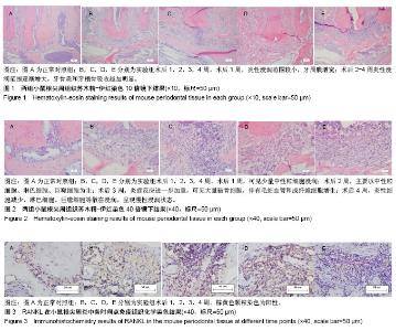
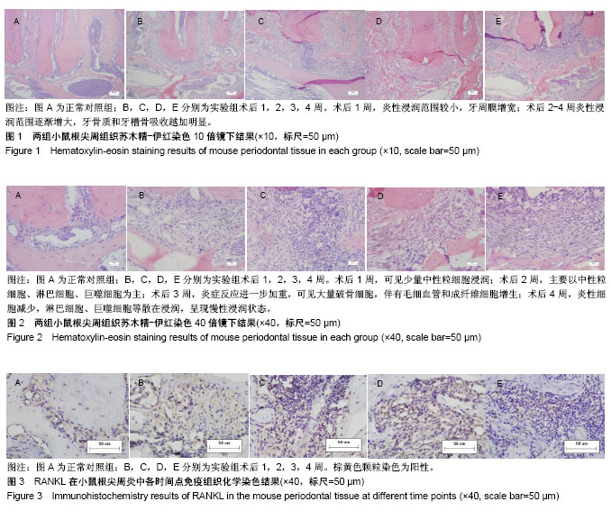
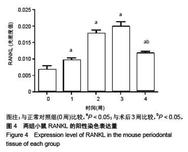
| [1] Márton IJ,Kiss C.Protective and destructive immune reactions in apical periodontitis. Oral Microbiol Immunol. 2000;15(3):139-150.[2] Atao?lu T,Ungör M,Serpek B,et al.Interleukin-1beta and tumour necrosis factor-alpha levels in periapical exudates. Int Endod J. 2002; 35(2):181-185.[3] Nair PN.Pathogenesis of apical periodontitis and the causes of endodonticfa IL ures. Crit Rev Oral Biol Med.2004;15(6):348-381.[4] Reichert S,Machulla HK,Klapproth J,et al.Interferon-gamma and interleukin-12 gene polymorphisms and their relation to aggre-ssive and chronic periodontitis and key periodontalpathogens. J Periodontol. 2008;79(8):1434-1443.[5] Pizzo G,Guiglia R,Lo Russo L,et al. Dentistry and internal medicine: from the focal infection theory to the periodontal medicine concept. Eur J Intern Med.2010;21(6):496-502. [6] Zoellner H.Dental infection and vascular disease.Semin Thromb Hemost. 2011;37(3):181-192. [7] 何飞,吴勇,周艳.Notch信号促进核因子κB受体活化因子配体诱导的破骨细胞分化的体外研究[J].华西口腔医学杂志, 2015,1(33): 25-28.[8] 蒋鹏,宋科官.破骨细胞及其分化调节机制的研究进展[J]中国骨与关节杂志,2017,3(6):223-227.[9] 蔡辉,张蓓蓓.核因子-κB受体活化因子配体抑制剂狄诺塞麦抗骨质疏松的临床研究进展[J]临床内科杂志, 2016,10(33):714-716.[10] 敏杰.骨破坏因RANK-RANKL-骨保护素 在口腔领域研究进展 [J]内蒙古医学杂志,2014,6(46):690-692.[11] Yasuda H, Shima N, Nakagawa N,et al. Osteoclast differentiation factor is a ligandfor osteoprotegerin/ osteoclastogenesis inhibitory factor and is identical toTRANCE/RANKL. Proc Natl Acad Sci U S A. 1998 Mar 31;95(7):3597-3602. [12] Boyce BF, Xing L.Functions of RANKL/RANK/OPG in bone modeling andremodeling, Arch Biochem Biophys. 2008;473(2): 139-146.[13] Rossol M,Heine H,Meusch U,et al.LPS-induced cytokine production in humanmonocytes and macrophages. Crit Rev Immunol. 2011;31(5): 379-446.[14] He W,Qu T,Yu Q,et al.LPS induces IL-8 expression through TLR4, MyD88, NFkappaand MAPK pathways in human dental pulp stem cells. Int Endod J. 2013;46(2):128-136.[15] 胡菊花,李倩,王艳青,等. 白细胞介素-21和核因子κB受体活化因子配体在人根尖囊肿和肉芽肿中的表达及临床意义[J]. 华西口腔医学杂志, 2015, 3(33):244-248.[16] 李永崾,朱鹏娜,王冬香,等.骨保护素/核因子KB受体活化因子配体在甲状旁腺激素调控下颌骨牵张成骨中的表达效应[J].华西口腔医学杂志, 2016, 3(34):234-238.[17] 王华贝.OPG/RANKL/RANK轴在骨相关疾病中的研究现状[J].中国临床研究,2012,1(25):80-81.[18] 严斌,何沛恒,罗震,等.OPG与RANKL在骨折愈合期的表达及其意义[J].实用医学杂志,2016,9(32):1464-1467.[19] 任嫒姝,付钢,邱雨,等.缺氧对人牙周膜细胞OPG和RANKL基因表达的影响 [J] .重庆医学,2015,35(44):4955-4957.[20] 史新连,胡碧波,任曼曼,等.低氧调控大鼠骨髓间充质干细胞OPG/ RANKL mRNA的表达[J].上海口腔医学,2017,3(26):258-262.[21] 赵凌杰,邱艳,商玮,等.姜黄素对佐剂性关节炎大鼠血清骨保护素和核因子κB受体活化因子配体蛋白表达及骨密度的影响[J].环球中医药, 2015,3(8): 316-319.[22] 李卓凯,冯唯嘉,王成龙,等.双氢青蒿素抑制核因子κB受体激动剂配体诱导RAW264.7细胞分化破骨细胞的研究[J].中华解剖与临床杂志, 2017, 2(22):148-152.[23] 吴侗,孙晓麟,杜燕,等.小白菊内酯对核因子κB受体活化因子配体诱导的RAW264.7细胞向破骨细胞分化的影响[J].中华风湿病学杂志, 2015, 7(19):468-472.[24] 娄苇苇,杜晓楠,袁慧慧,等.靶向于肿瘤坏死因子和RANKL的人源化抗体对DBA/1小鼠单关节炎的保护作用[J].微生物学免疫学进展, 2017,2(45): 8-16.[25] 黄诗言,饶南荃,徐舒豪,等.调控牙萌出的细胞及分子机制研究进展[J].华西口腔医学杂志,2016,3(34):317-321.[26] 谢冰洁,冯捷,韩向龙.破骨细胞生物学特征的研究与进展[J].中国组织工程研究,2017,21(11):1770-1775. [27] Fouad AF, Acosta AW. Periapical lesion progression andcytokine expression in an LPS hyporesponsive model. Int Endod J. 2001; 34(7):506-513.[28] Oseko F,Yamamoto T,Akamatsu Y,et al.IL-17 is involved in bone resorption in mouse periapical lesions. Microbiol Immunol. 2009; 53(5):287-294.[29] Vaira S,Alhawagri M,Anwisye I,et al. RelA/p65 promotes osteoclast differentiation by blocking a RANKL-induced apoptotic JNK pathway in mice. J Clin Invest. 2008;118(6):2088-2097.[30] Novack DV.Role of NF-kappaB in the skeleton.Cell Res. 2011; 21(1): 169-182.[31] Boyce BF,Xing L.Functions of RANKL/RANK/OPG in bone modeling and remodeling. Arch Biochem Biophys. 2008;473(2): 139-146.[32] 李伟娟,谢保平,石丽颖,等.从ERα/RANK 通路探讨淫羊藿苷抑制破骨细胞分化作用[J].中国实验方剂学杂志,2017,23(7):121-126.[33] Takeno A, Yamamoto M, Notsu M, et al.Administration of anti-receptor activator of nuclear factor-kappa B ligand (RANKL) antibody for the treatment of osteoporosis was associated with amelioration of hepatitis in a female patient with growth hormone deficiency: a case report. BMC Endocr Disord. 2016;16(1):66.[34] Chen C, Cheng P, Xie H,et al.MiR-503 Regulates osteoclastogenesis via targeting RANK.J Bone Miner Res.2014;29( 2) :338-347. [35] 丁欣欣,李淑慧,葛翘诚,等.转化生长因子β1对牙周膜干细胞OPG/ RANKL基因表达的影响[J].中华实用诊断与治疗杂志,2017,31(12): 1156-1159.[36] Carneiro E,Parolin AB,Wichnieski C,et al.Expression levels of the receptor activator of NF-kB ligand and osteoprotegerin and the number of gram-negative bacteria in symptomatic and asymptomatic periapical lesions. Arch Oral Biol. 2017;73: 166-171.[37] Hanif JK,Adrian WSH,Roberto S,et al.The Syk-NFAT-IL-2Pathway in Dendritic Cells Is Required for Optimal Sterile Immunity Elicited by Alum Adjuvants.J Immunol. 2017;198(1):196-204 [38] Rhoda B,Krisna M,Jiming L,et al.NFATc1 releases BCL6 dependent repression of CCR2 agonist expression in peritoneal macrophages from Saccharomyces cerevisiae infected mice.Eur J Immunol.2016;46: 634-646. [39] 李忠浩,丁宁,杨全增,等.破骨细胞中NFATc1相关调节研究进展[J].中国骨质疏松杂志,2017,23(5):695-700. |