Chinese Journal of Tissue Engineering Research ›› 2021, Vol. 25 ›› Issue (23): 3654-3659.doi: 10.12307/2021.035
Previous Articles Next Articles
Serine/threonine protein kinases can promote bone destruction in mouse models of chronic periapical periodontitis
Yang Caihui1, Liu Qicheng1, Dong Ming1, Wang Lina1, Zuo Meina2, Lu Ying1, Niu Weidong1
- 1School of Stomatology, Dalian Medical University, Dalian 116044, Liaoning Province, China; 2Stomatological Hospital, School of Stomatology, Dalian Medical University, Dalian 116000, Liaoning Province, China
-
Received:2020-05-19Revised:2020-05-20Accepted:2020-06-29Online:2021-08-18Published:2021-01-26 -
Contact:Niu Weidong, MD, Professor, Doctoral supervisor, School of Stomatology, Dalian Medical University, Dalian 116044, Liaoning Province, China -
About author:Yang Caihui, Master candidate, School of Stomatology, Dalian Medical University, Dalian 116044, Liaoning Province, China Liu Qicheng, Master, Associate professor, School of Stomatology, Dalian Medical University, Dalian 116044, Liaoning Province, China Yang Caihui and Liu Qicheng contributed equally to this work. -
Supported by:the National Natural Science Foundation of China, No. 81700962 (to LY)
CLC Number:
Cite this article
Yang Caihui, Liu Qicheng, Dong Ming, Wang Lina, Zuo Meina, Lu Ying, Niu Weidong. Serine/threonine protein kinases can promote bone destruction in mouse models of chronic periapical periodontitis[J]. Chinese Journal of Tissue Engineering Research, 2021, 25(23): 3654-3659.
share this article
Add to citation manager EndNote|Reference Manager|ProCite|BibTeX|RefWorks
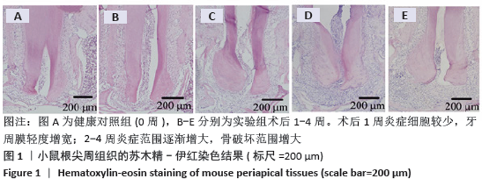
2.1 实验动物数量分析 实验选用小鼠25只,分为5组,实验过程无脱失,全部进入结果分析。 2.2 小鼠根尖组织的苏木精-伊红染色结果 苏木精-伊红染色结果显示,健康对照组小鼠根尖周组织中牙周膜纤维没有明显病变,牙周膜未见增宽,仅可见极少量炎性细胞;术后1周的根尖周组织中可见到小范围的炎症细胞的浸润,属于炎症的早期,牙周膜轻度增宽;术后2周,炎症范围增大进入炎症急性期,大量巨噬细胞、中性粒细胞、淋巴细胞聚集根尖周围,破骨细胞较活跃,此时出现了牙槽骨的吸收;术后3周炎症范围进一步增大,根尖周围主要是淋巴细胞,可见成纤维细胞和毛细血管,亦出现了大量的破骨细胞;4周时骨破坏范围最大,见图1。 "

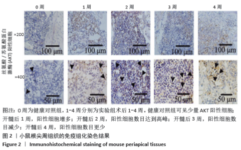
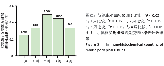
2.3 AKT在小鼠慢性根尖周炎中的表达 免疫组织化学染色结果表明,在根尖周炎的发展过程中均可见到AKT的阳性细胞。AKT阳性细胞多为圆形、椭圆形或分叶状,表达在深褐色的胞质中,且主要表达在中性粒细胞和破骨细胞中。健康对照组见到少量阳性细胞;开髓术后1周,阳性细胞数目增多;开髓术后2周,阳性细胞数目达到高峰;3周时,数目减少;术后4周,阳性细胞数目更少,见图2。各实验组AKT阳性细胞数均大于健康对照组,AKT统计学结果为1周与4周之间差异无显著性意义(P > 0.05),其余各组间相比差异均有显著性意义(P < 0.05),见图3。 "

"
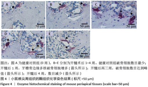
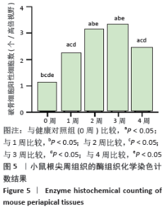
2.4 酶组织化学染色结果 酶组织化学染色结果显示,健康对照组小鼠根尖周围破骨细胞数目最少;术后1周,由于炎症的浸润,牙槽骨被吸收,牙槽骨边缘多核破骨细胞增多;开髓后二三周,破骨细胞数目达到峰值,更加活跃;术后4周,数目减少,骨吸收范围继续增大,见图4。2周与3周之间破骨细胞阳性细胞数相比差异无显著性意义(P > 0.05),其余各组之间相比差异均有显著性意义(P < 0.05),见图5。"

"

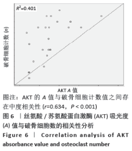
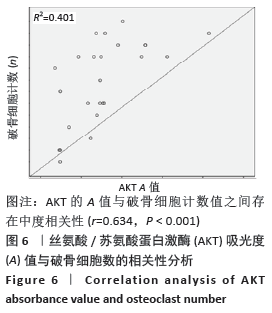
2.5 AKT A值与破骨细胞数的相关性 Pearson相关分析结果显示,AKT A值与破骨细胞计数值之间存在中度相关性(r=0.634,P < 0.001),见图6。"
| [1] MAKKAWI H, HOCH S, BURNS E, et al. Porphyromonasgingivalis stimulates TLR2-PI3K signaling to escape immune clearance and induce bone resorption independently of MyD88. Front Cell Infect Microbiol. 2017;7:359. [2] ZHOU Y, CHEN M, RICUPERO CL, et al. Profiling of Stem/Progenitor Cell Regulatory Genes of the Synovial Joint by Genome-Wide RNA-Seq Analysis. Biomed Res Int. 2018;2018:9327487. [3] PAN ST, QIN Y, ZHOU ZW, et al. Plumbagin induces G2/M arrest, apoptosis, and autophagy via p38 MAPK-and PI3K/Akt/mTOR-mediated pathways in human tongue squamous cell carcinoma cells. Drug Design Dev Ther. 2015;9:1601. [4] YAMANAKA Y, CLOHISY JC, ITO H, et al. Blockade of JNK and NFAT pathways attenuates orthopedic particle-stimulated osteoclastogenesis of human osteoclast precursors and murine calvarialosteolysis. J Orthop Res. 2012;31(1):67-72. [5] KIM TH, CHOI SJ, LEE YH, et al. Combined therapeutic application of mTOR inhibitor and vitamin D(3) for inflammatory bone destruction of rheumatoid arthritis. Med Hypoth. 2012;79(6):757-760. [6] ZHONG JT, YU J, WANG HJ, et al. Effects of endoplasmic reticulum stress on the autophagy, apoptosis, and chemotherapy resistance of human breast cancer cells by regulating the PI3K/AKT/mTOR signaling pathway. Tumour Boil. 2017;39(5):1010428317697562. [7] KAWAMURA N, KUGIMIYA F, OSHIMA Y, et al. Akt1 in osteoblasts and osteoclasts controls bone remodeling. PloS One. 2007;2(10):e1058. [8] SHI ZM, HAN YW, HAN XH, et al. Upstream regulators and downstream effectors of NF-κB in Alzheimer’s disease. J Neurol Sci. 2016;366: 127-134. [9] HOESEL B, SCHMID JA. The complexity of NF-κB signaling in inflammation and cancer. Mol Cancer. 2013;(12):86. [10] ZHANG H, SHAN Y, WU Y, et al. Berberine suppresses LPS-induced inflammation through modulating Sirt1/NF-κB signaling pathway in RAW264.7 cells. Int Immunopharmacol. 2017;52:93-100. [11] lin c, shao y, zeng c, et al. Blocking PI3K/AKT signaling inhibits bone sclerosis in subchondral bone and attenuates post‐traumatic osteoarthritis. J Cell Physiol. 2018;233(8):6135-6147. [12] CHEN L, HUANG M, TAN J, et al. PI3K/AKT Pathway Involvement in the Osteogenic Effects of Osteoclast Culture Supernatants on Preosteoblast Cells. Tissue EngPart A. 2013;19(19-20):2226-2232. [13] SUGATANI T, HRUSKA KA. Akt1/Akt2 and mammalian target of rapamycin/Bim play critical roles in osteoclast differentiation and survival, respectively, whereas Akt is dispensable for cell survival in isolated osteoclast precursors. J Biol Chem. 2005;280(5):3583-3589. [14] BOYCE BF. Advances in osteoclast biology reveal potential new drug targets and new roles for osteoclasts. J Bone Miner Res. 2013; 28(4):711-722. [15] JOHNSON SM, GULHATI P, RAMPY BA, et al. Novel expression patterns of PI3K/Akt/mTOR signaling pathway components in colorectal cancer. J Am Coll Surg. 2010;210(5):767-776. [16] YEON JT, KIM KJ, SON YJ, et al. Idelalisib inhibits osteoclast differentiation and pre-osteoclast migration by blocking the PI3Kδ-Akt-c-Fos/NFATc1 signaling cascade. Arch Pharm Res. 2019;42(2):712-721. [17] XIAO L, ZHU L, YANG S, et al. Different correlation of sphingosine-1-phosphate receptor 1 with receptor activator of nuclear factor kappa B ligand and regulatory T cells in rat periapical lesions. J Endod. 2015;41(4):479-486. [18] Wang L, Zhang H, Dong M, et al.Role of the Btk-PLCγ2 Signaling Pathway in the Bone Destruction of Apical Periodontitis. Mediators Inflamm. 2019;2019:8767529. [19] MOON JB, KIM JH, KIM K, et al. Akt induces osteoclast differentiation through regulating the GSK3β/NFATc1 signaling cascade. J Immunol. 2012;188(1):163-169. [20] KE K, SUL OJ, CHOI EK, et al. Reactive oxygen species induce the association of SHP-1 with c-Src and the oxidation of both to enhance osteoclast survival. Am J Physiol Endocrinol Metab. 2014;307(1): E61-E70. [21] CEJKA D, HAYER S, NIEDERREITER B, et al. Mammalian target of rapamycin signaling is crucial for joint destruction in experimental arthritis and is activated in osteoclasts from patients with rheumatoid arthritis. Arthritis Rheum. 2010;62(8):2294-2302. [22] 徐杨,傅文贞,何进卫,等. AKT1基因嵌合性体细胞突变导致Proteus综合征临床研究[J].中华内科杂志,2019,58(7):508-513. [23] KAWAMURA N, KUGIMIYA F, OSHIMA Y, et al. Akt1 in osteoblasts and osteoclasts controls bone remodeling. Plos One. 2007;2(10):1058. [24] ULICI V, HOENSELAAR KD, AGOSTON H, et al. The role of Akt1 in terminal stages of endochondral bone formation: angiogenesis and ossification. Bone. 2009;45(6):1133-1145. [25] PENG X, XU PZ, CHEN ML, et al. Dwarfism, impaired skin development, skeletal muscle atrophy, delayed bone development, and impeded adipogenesis in mice lacking Akt1 and Akt2. Genes Dev. 2003;17(11): 1352-1365. [26] BRAZIL DP, YANG ZZ, HEMMINGS BA. Advances in protein kinase B signalling: AKTion on multiple fronts. Trends Biochem Sci. 2004;29(5): 233-242. [27] MANNING BD, CANTLEY LC. AKT/PKB signaling: navigating downstream. Cell. 2007;129(7):1261-1274. [28] LIU X, BRUXVOORT KJ, ZYLSTRA CR, et al. Lifelong accumulation of bone in mice lacking Pten in osteoblasts. Proc Natl Acad Sci U S A . 2007;104(7):2259-2264. [29] VISWANATH K, BODIGA S, BALOGUN V, et al. Cardioprotective effect of zinc requires ErbB2 and Akt during hypoxia/reoxygenation. Biometals. 2011;24(1):171-180. [30] ORTIZ PA, HONG NJ, GARVIN JL. Luminal flow induces eNOS activation and translocation in the rat thick ascending limb. Ajp Renal Physiol. 2004;287(2):F274-F280. [31] CHEN JX, LAWRENCE ML, CUNNINGHAM G, et al. HSP90 and Akt modulate Ang-1-induced angiogenesis via NO in coronary artery endothelium. J Appl Physiol. 2004;96(2):612-620. [32] 汪林芳,袁喆晨,苏芬芬,等. IRF3在绿脓杆菌感染时抑制中性粒细胞粘附的机制研究[J]. 杭州师范大学学报(自然科学版), 2019, 18(5):516-520. [33] MAZAKI Y, NISHIMURA Y, SABE H. GBF1 bears a novel phosphatidylinositol-phosphate binding module, BP3K, to link PI3Kγ activity with Arf1 activation involved in GPCR-mediated neutrophil chemotaxis and superoxide production. Mol Biol Cell. 2012;23(13): 2457-2467. [34] STARK AK, SRISKANTHARAJAH S, HESSEL EM, et al. PI3K inhibitors in inflammation, autoimmunity and cancer. Curr Opin Pharmacol. 2015; 23:82-91. [35] DONG M, HU N, HUA Y, et al. Chronic Akt activation attenuated lipopolysaccharide-induced cardiac dysfunction via Akt/GSK3β-dependent inhibition of apoptosis and ER stress. Biochim Biophys Acta. 2013;1832(6):848-863. [36] TAKAKI A, SATOSHI O, MIYUKI T, et al. Enhanced AKT Phosphorylation of Circulating B Cells in Patients with Activated PI3KδSyndrome. Front Immunol. 2018;9:568. [37] CHEN PJ, KO IL, LEE CL, et al. Targeting allosteric site of AKT by 5,7-dimethoxy-1,4-phenanthrenequinone suppresses neutrophilic inflammation. Ebiomedicine. 2019;40:528-540. [38] LIAN Z, HAN J, HUANG L, et al. A005, a novel inhibitor of phosphatidylinositol 3-kinase/mammalian target of rapamycin, prevents osteosarcoma-induced osteolysis. Carcinogenesis. 2019;40(2): E1-E13. |
| [1] | Li Jing, Xie Jianshan, Cui Huilin, Cao Ximei, Yang Yanping, Li Hairong. Expression and localization of diacylglycerol kinase zeta and protein kinase C beta II in mouse back skin with different coat colors [J]. Chinese Journal of Tissue Engineering Research, 2021, 25(8): 1196-1200. |
| [2] | Zhang Mi, Wu Saixuan, Dong Ming, Lu Ying, Niu Weidong. Expression of interleukin-24 in a mouse model of periapical periodontitis [J]. Chinese Journal of Tissue Engineering Research, 2021, 25(5): 679-684. |
| [3] | Yang Junhui, Luo Jinli, Yuan Xiaoping. Effects of human growth hormone on proliferation and osteogenic differentiation of human periodontal ligament stem cells [J]. Chinese Journal of Tissue Engineering Research, 2021, 25(25): 3956-3961. |
| [4] | Sun Jianwei, Yang Xinming, Zhang Ying. Effect of montelukast combined with bone marrow mesenchymal stem cell transplantation on spinal cord injury in rat models [J]. Chinese Journal of Tissue Engineering Research, 2021, 25(25): 3962-3969. |
| [5] | Fan Junchao, Chen Yong, Song Junjie. Sevoflurance combined with xenon pretreatment protects against spinal cord ischemia-reperfusion injury in a rat model [J]. Chinese Journal of Tissue Engineering Research, 2021, 25(23): 3660-3665. |
| [6] | Yan Peng, Ma Yufei, Cui Jingfu, Hao Shaofei, Liu Jinhui, Guan Chunlei, Wang Xiaoran, Yang Xiaoyu. Mechanism of anodic block electrical stimulation of sacral nerve root to reconstruct bladder function [J]. Chinese Journal of Tissue Engineering Research, 2021, 25(23): 3684-3689. |
| [7] | Huo Hua, Cheng Yuting, Zhou Qian, Qi Yuhan, Wu Chao, Shi Qianhui, Yang Tongjing, Liao Jian, Hong Wei. Effects of drug coating on implant surface on the osseointegration [J]. Chinese Journal of Tissue Engineering Research, 2021, 25(22): 3558-3564. |
| [8] | Jiang Shengyuan, Li Dan, Jiang Jianhao, Shang-you Yang, Yang Shuye. Biological response of Co2+ to preosteoblasts during aseptic loosening of the prosthesis [J]. Chinese Journal of Tissue Engineering Research, 2021, 25(21): 3292-3299. |
| [9] | Chen Xinling, Wang Shenglan. Cell autophagy, pathway, regulation and its multiple correlations with pulmonary hypertension [J]. Chinese Journal of Tissue Engineering Research, 2021, 25(2): 311-316. |
| [10] | Liu Xing, Wei Xiaohan, Deng Jie, Li Zhongming . Preparing a blunt contusion model of rabbit skeletal muscle under different blow strengths [J]. Chinese Journal of Tissue Engineering Research, 2021, 25(2): 196-200. |
| [11] | Wu Yukun, Han Jie, Wen Shuaibo. Mechanism of Runx2 gene in fracture healing [J]. Chinese Journal of Tissue Engineering Research, 2021, 25(14): 2274-2279. |
| [12] | Liu Yan, Gao Xiang, Zhao Xiaoxia, Chen Qingyu, Gao Junwu. Effects of conditioned medium of human periodontal ligament stem cells on proliferation and osteogenic differentiation of inflammatory tissue-derived human periodontal ligament stem cells [J]. Chinese Journal of Tissue Engineering Research, 2021, 25(13): 2005-2010. |
| [13] | Long Qian, Guan Xiaoyan, Wang Qian, Hu Huan, Liu Jianguo. Transcriptome sequencing technology and its application in oral diseases, dental implants and regeneration [J]. Chinese Journal of Tissue Engineering Research, 2021, 25(11): 1791-1798. |
| [14] | Zhou Liyu, Ma Yanxia, Qi Shibin, Saijilafu, Wei Shanwen, Ni Li. Regulatory effects of ADT-OH on the proliferation of neural precursor cells [J]. Chinese Journal of Tissue Engineering Research, 2021, 25(1): 96-100. |
| [15] | Xu Nuo, Cao Zhen, Li Xiaojie, Shi Chun. MicroRNA-21 regulates proliferation and differentiation of osteoclasts in periodontitis [J]. Chinese Journal of Tissue Engineering Research, 2020, 24(8): 1225-1230. |
| Viewed | ||||||
|
Full text |
|
|||||
|
Abstract |
|
|||||