Chinese Journal of Tissue Engineering Research ›› 2018, Vol. 22 ›› Issue (6): 952-957.doi: 10.3969/j.issn.2095-4344.0074
Previous Articles Next Articles
Silk fibroin/chitosan scaffolds for corneal tissue engineering: theoretical development and applications
- Department of Ophthalmology, Lanzhou University Second Hospital, Lanzhou 730030, Gansu Province, China
-
Received:2017-12-09Online:2018-02-28Published:2018-02-28 -
Contact:Zhang Wen-fang, M.D., Chief physician, Department of Ophthalmology, Lanzhou University Second Hospital, Lanzhou 730030, Gansu Province, China -
About author:Wang Jia-qi, Studying for master’s degree, Physician, Department of Ophthalmology, Lanzhou University Second Hospital, Lanzhou 730030, Gansu Province, China -
Supported by:Major Disease Research Program of Gansu Province, No. 201405
CLC Number:
Cite this article
Wang Jia-qi, Zhang Wen-fang . Silk fibroin/chitosan scaffolds for corneal tissue engineering: theoretical development and applications[J]. Chinese Journal of Tissue Engineering Research, 2018, 22(6): 952-957.
share this article
Add to citation manager EndNote|Reference Manager|ProCite|BibTeX|RefWorks

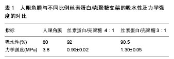
2.1 丝素蛋白/壳聚糖复合支架制备方法 目前该复合支架的制备主要有两种方法:一种是冷冻干燥法,另一种是乙醇浸泡法。 2.1.1 冷冻干燥法[16] 将家蚕丝于无水碳酸钠中煮沸脱胶并烘干拉松,在75 ℃时置入三元溶液中,搅拌制备纤维溶液,随后经透析得到纯化的丝素蛋白溶液。将其配置为2%的浓度后加入2%的壳聚糖混合而成。利用超声脱去混合液的气泡,置于-80 ℃冷冻12 h,待冻制品完成后,干燥24 h。最后将样品于甲醇中处理,使其产生β-折叠,此时,材料具备疏水性并且韧性增加。用去离子水反复洗涤并再次冷冻干燥,最终制得复合支架。 2.1.2 乙醇浸泡法[17] 将蚕丝在高温碳酸氢钠液中脱胶,并于真空箱中烘干,随后将制备出的脱胶丝于溴化锂水溶液中溶丝过滤。3 d内不间断的去水离子透析去除溴化锂后离心保存。将制备好的再生蚕丝溶液置于烧杯中低速搅拌,同时逐渐加入壳聚糖醋酸水溶液,继续低速搅拌而成共混液后于-20 ℃冷冻24 h。随后转移置于-5 ℃的乙醇溶液里长时浸泡,待到共混支架结构成熟时,多次洗涤去除乙醇并用碳酸氢钠中和掉支架里的醋酸,即得到高孔隙的复合支架. 2.2 丝素蛋白壳聚糖复合材料理化性质 丝素蛋白与壳聚糖互为改性,Hirano等[18]通过电镜扫描指出,混入了壳聚糖的丝素蛋白支架的孔隙结构比单纯的丝素蛋白的孔隙结构更加均匀有序,这是因为壳聚糖抑制了丝素蛋白的片层结构。Park等[19]指出,丝素蛋白壳聚糖复合支架的内部为三维多孔结构。Kweon等[20]的研究表明,壳聚糖以合理的比例加入丝素蛋白后,其理化性质得到增强。 2.2.1 孔径大小 柳磊等[21]实验显示,除了纯丝素蛋白因为溶蚀率高而无法计算其孔径大小外,无论壳聚糖和丝素蛋白以何种比例进行配比,其孔径率均高于90%,合适的孔径大小是细胞黏附的结构基础。当壳聚糖含量比重大时,复合材料的孔径会变小,连通性差,而丝素蛋白含量比重增大时,其孔径增大但却向着片层结构发展[22]。因此当二者以60%丝素蛋白和40%壳聚糖比例混合制备时,该复合支架的连通性较好且孔径大小合理,为(210±23.71) μm。 2.2.2 吸水性 张霞只等[16]的实验指出,当壳聚糖与丝素蛋白的在混合材料中的质量比例为1∶2时,该材料的吸水率最高,约为37%,而随着丝素蛋白比例的增加,吸水率降至30%。作为角膜组织工程的材料,其渗透性能受到平衡含水量的影响[22]。植入角膜后,材料前表面接触到泪液,后表面与前房房水相同,低吸水性导致外液中的阳离子因为较高的渗透压向材料转移从而形成自身沉淀,导致支架的脆性增加。 2.2.3 力学强度 作为角膜修复的材料,应当具有一定的力学强度来对抗眼压。丝素蛋白的力学性能要优于壳聚糖,当壳聚糖成比例地加入丝素蛋白中,复合材料的力学强度也较纯丝素蛋白下降[23]。Ruan等[24]的实验表明,纯丝素蛋白的压缩模量为(7.51±0.19) kPa,当壳聚糖与丝素蛋白的混合比例为1∶8时,压缩模量降低到(6.36±0.69) kPa,而当二者比例为1∶2时,其压缩模量为(4.70±0.14) kPa。正常眼压的上限为2.80 kPa,因此,二者成比例制备时,随着壳聚糖相对质量的增加,其力学强度也将相应的下降(表1)。 2.2.4 生物降解速率 良好的生物支架,其降解速率应与角膜细胞的生长速率吻合。Kang等[25]的研究表明,当复合支架的配成比为40%丝素蛋白和60%壳聚糖时,在最初的15 d里,该复合材料迅速失去了14.43%的原始重量,随后降解速率开始减慢,在第64天里时,材料仅仅失去了18.25%的重量,其降解速率方程拟合曲线与人体组织的新生率相匹配。Baran等[26]指出,最开始降解速率快的原因可能是因为来自壳聚糖的降解反应。同时,在材料降解的两个月的时间段内,其降解液的pH值始终波动在7.42-7.62之间,与人体微环境的类似[27]。 2.2.5 合理的制备比 综合各实验结果,在统筹考虑复合支架的吸水率,力学强度,降解率等方面,壳聚糖与丝素蛋白的配成比随着壳聚糖的增加,其力学性能下降,吸水率升高,但考虑到生物降解的合理曲线,二者的构成比为1∶1至1∶1.5时,各项指标满意[25]。 2.3 丝素蛋白壳聚糖复合材料的生物相容性 支架材料与人体组织的生物相容性是衡量其实际效用的关键,目前通常采用接种成纤维细胞株或者间充质干细胞在支架材料上观察其细胞毒性、增殖活力以及黏附作用,再通过电镜扫描观察细胞株的生长情况来评价支架的生物相容性[28]。 2.3.1 细胞毒性 Sha等[29]利用细胞毒性相对增殖率对支架材料的细胞毒性进行分级。叶鹏等[30]利用丝素蛋白壳聚糖构建的生物支架于兔骨髓间充质干细胞进行体外共培养,分别置于25%,50%以及100%的支架浸提液中,观察24,48以及72 h,经测定相对增殖率值均大于100,根据细胞毒性反应分级标准,其结果均为合格。 2.3.2 增殖活力 佘荣峰[31]通过提取第3代经诱导后的间充质干细胞接种于丝素蛋白壳聚糖支架中,培养箱中培养并加入MTT,孵化4 h滴入盐酸异丙醇,通过ELISA测定吸光度并采用扫描电镜观察,随着时间的推移,细胞在支架上的数量逐渐增加,分裂增殖速度正常。其机制可能与天然生物材料可以促进细胞增殖有关[32-33]。 2.3.3 黏附作用 刘宁华[34]通过制备脂肪干细胞植入丝素蛋白壳聚糖支架中,培养3 d后取出通过扫描电镜观察,结果显示脂肪干细胞形态呈现长梭形,表面光滑,足突伸出与丝素蛋白壳聚糖支架相互黏附,作用紧密,而细胞彼此之间也通过伪足相联。 2.3.4 植入角膜的生物相容性 Thuaksuban等[35]指出,不同来源的细胞和同一支架的相容性不同,在角膜组织工程中,植入是否成功在于保持角膜细胞的高度分化性和良好的胶原产生能力。关于这点,Lawrence等[36]提出,良好的角膜移植吻合口,应当具有相对平滑的细胞外基质,而大部分的支架材料,很难使角膜细胞保持表面静止。Gil等[37]的研究表明,将支架的结构制备为单孔或者多孔的膜状多层组织,其移植结果令人满意。Bray等[38]的研究表明,利用丝素蛋白和壳聚糖混合制备的支架,不仅有助于多孔结构的重建,而且会提供一个类似角膜细胞外基质的微环境。 2.4 丝素蛋白壳聚糖支架在角膜组织工程的应用进展 Guan等[39]研究表明,壳聚糖的含量影响着角膜细胞的附着能力,当丝素蛋白和壳聚糖的比例为3∶1的时候,此时支架细胞液中的角膜细胞的黏附力最强,呈现球形三维结构,培养第7天时,角膜细胞对数生长,然而在第14天时,部分细胞生长停滞,推测其原因,可能是因为壳聚糖提供的过于宽敞的结构,导致了细胞处于静止状态。在实验期内,角膜细胞里未检出CD4/CD8的表达,提示复合支架与角膜不会发生排斥反应。后期其课题组利用15只新西兰兔构建出了角膜缺损模型,并将兔角膜上皮细胞和基质细胞种植在丝素蛋白/壳聚糖支架中,制备出体外共培养的材料,将其植入缺损模型中。结果显示,在一开始,构建的角膜并不像兔眼本身的角膜那样透明,苏木精-伊红染色显示,细胞并未皱缩,细胞排列也较为规整;在培养第7天时,细胞呈现树突样的形态,并成功在支架孔隙并形成了网状纤维样结构;在培养14 d后细胞还呈现出叠加的双层片状结构;随后利用Western Blot定量分析的结果显示,丝素蛋白壳聚糖细胞培养物高表达角蛋白。随访期间内,所有实验兔眼的植入部位未感染,置于裂隙灯下也未发现角膜出现新生血管。丝素蛋白壳聚糖支架在4个月之间完全降解,角膜透明度也逐渐恢复,结果表明重建层的角膜结构与天然角膜相似,角膜上皮的K3/12基因高表达,证明支架裙边结构和角膜组织没有发生免疫反应[40]。 Kim等[41]在对丝素蛋白壳聚糖支架进行制备的时候发现,将细胞接种在壳聚糖质量占5%的混合支架后,通过分光光度计测定,在该比例下,支架材料的透明度最佳;并指出,虽然壳聚糖质量占5%的混合支架具有良好的透明度,但是其角膜内皮细胞数量为(236±13)细胞/mm2,显著低于壳聚糖质量占1%的对照组(327±42 细胞/mm2),这一结果表明,壳聚糖的含量影响着角膜内皮的细胞密度;另外,移植支架产生了类似细胞外基质般可进行钠钾阳离子交换的功能,通过对NaK标记物的对比,在壳聚糖质量占3%的对照组中高表达。大量研究指出角膜内皮细胞密度对角膜的增殖至关重要,当数量减少以及分布不均匀时,其功能开始丧失[42-44]。Yee等[45]指出NaK标记物的表达与水分子的渗透效率以及控制水肿并维持角膜透明有关,并发现维持壳聚糖在3%的水平时,角膜更为透明。 Hazra等[46]将从大鼠角膜上获得的角膜外植体在丝素蛋白壳聚糖支架上进行培养,在第4天时,观察到支架中的角膜上皮细胞开始分裂,并附着于支架上向远端迁徙,随后逐渐覆盖模型的缺损部位。Paiva等[47]通过免疫组化测定,发现角膜细胞膜上高表达ABCG2蛋白,表明支架支持角膜缘干细胞的生长。在进行兔眼的动物实验环节时,与Hazra等实验不同之处在于,其未将兔眼角膜细胞培养后接种在支架上再进行移植,而是直接将丝素蛋白壳聚糖支架直接植入构建的损伤模型中。在裂隙灯下观察,植入后兔眼耐受良好,透明度良好,虹膜清晰可见,两个月内支架材料未发生降解。 Wang等[49]的实验证明了丝素蛋白壳聚糖支架在角膜生物相容性方面的肯定性价值。但是他的实验里发现了一例新生血管的形成,不过在第30天的时候消失。证明丝素蛋白壳聚糖支架的植入也是可以短暂的引起炎症反应的,这与前述几位作者的实验结果不尽相同,其炎症刺激机制尚需更加深入的研究去阐明。同时,其也特别指出,该类支架会影响角膜基质的渗透能力,并引起角膜上皮变薄。 "
| [1]潘志强,梁庆丰.重视角膜移植手术的供体材料问题[J].中华眼科杂志,2016,52(9):641-643.[2]Fagerholm P, Lagali NS, Merrett K, et al. A biosynthetic alternative to human donor tissue for inducing corneal regeneration: 24-month follow-up of a phase 1 clinical study. Sci Transl Med. 2010;2(46):46-61.[3]Zhou Y, Wu Z, Ge J, et al. Development and characterization of acellular porcine corneal matrix using sodium dodecylsulfate. Cornea, 2011, 30(1):73-82.[4]Ghezzi C, Rnjakkovacina J, Kaplan DL. Corneal tissue engineering: recent advances and future perspectives. Tissue Eng Part B Rev.2015;91(8):307-319.[5]Altman AM, Yan Y, Matthias N, et al. IFATS collection: human adipose-derived stem cells seeded on a silk fibroin-chitosan scaffold enhance wound repair in a murine soft tissue injury model. Stem Cells. 2009;27(1):250-258.[6]Luangbudnark W, Viyoch J, Laupattarakasem W, et al. Properties and biocompatibility of chitosan and silk fibroin blend films for application in skin tissue engineering. ScientificWorldJournal.2012;2012(2012):697201.[7]Gupta V, Mun GH, Choi B, et al. Repair and reconstruction of a resected tumor defect using a composite of tissue flap–nanotherapeutic–silk fibroin and chitosan scaffold. Ann Biomed Eng. 2011;39(9):2374-2387.[8]李大为,何进,何凤利,等.丝素蛋白/壳聚糖复合材料在组织工程中应用的研究进展[J].中国生物工程杂志,2017,37(10):111-117.[9]Freddi G, Tsukada M, Beretta S. Structure and physical properties of silk fibroin/polyacrylamide blend films. J Appl Polym Sci. 2015; 71(10):1563-1571.[10]张灿伟.体外构建组织工程角膜的研究进展[J].中华实验眼科杂志,2017,35(2):170-174.[11]Logithkumar R, Keshavnarayan A, Dhivya S, et al. A review of chitosan and its derivatives in bone tissue engineering. Carbohydr Polym.2016;151:172.[12]Ahmed TA, Aljaeid BM. Preparation, characterization, and potential application of chitosan, chitosan derivatives, and chitosan metal nanoparticles in pharmaceutical drug delivery. Drug Des Devel Ther. 2016;10:483–507.[13]Mohammed MA, Syeda JTM, Wasan KM, et al. An overview of chitosan nanoparticles and its application in Non-Parenteral Drug Delivery. Pharmaceutics. 2017; 9(4): 53.[14]Hazra S, Nandi S, Naskar D, et al. non-mulberry silk fibroin biomaterial for corneal regeneration. Sci Rep. 2016; 6: 21840.[15]Bhardwaj N, Kundu SC. Chondrogenic differentiation of rat MSCs on porous scaffolds of silk fibroin/chitosan blends. Biomaterials.2012;33(10):2848-2857.[16]张霞只,司徒方民,彭鹏,等.加入壳聚糖改善丝素蛋白结晶性:力学稳定强度更好的三维支架材料[J].中国组织工程研究,2015, 20(12):1858-1863.[17]董新钰.再生丝素蛋白/壳聚糖三维多孔复合材料的制备[D].复旦大学,2013.[18]Hirano S, Noishiki Y, Kinugawa J, et al. Chitin and chitosan for use as a novel biomedical material. Adva in Bio Poly. 1987: 285-297.[19]Park SJ, Lee KY, Ha WS, et al. Structural changes and their effect on mechanical properties of silk fibroin/chitosan blends. J Appl Polym Sci.2015; 74(11):2571-2575.[20]Kweon HY, Park YH. Structural and conformational changes of regenerated Antheraea pernyi silk fibroin films treated with methanol solution. J Appl Polym Sci. 2015;73(14):2887-2894.[21]柳磊,曾曙光,任力,等.丝素蛋白/壳聚糖三维多孔支架的构建及结构表征[J].中国组织工程研究, 2012,16(12):2197-2202.[22]Gobin AS, Froude VE, Mathur AB. Structural and mechanical characteristics of silk fibroin and chitosan blend scaffolds for tissue regeneration. J Biomed Mater Res A.2010; 74(3):465-473.[23]Ran J, Hu J, Sun G, et al. A novel chitosan-tussah silk fibroin/nano-hydroxyapatite composite bone scaffold platform with tunable mechanical strength in a wide range. Int J Biol Macromol. 2016; 93:87.[24]Ruan Y, Lin H, Yao J, et al. Preparation of 3D fibroin/chitosan blend porous scaffold for tissue engineering via a simplified method. Macromol Biosci. 2011;11(3):419-426.[25]Kang Y, Yao Y, Yin G, et al. A study on the in vitro degradation properties of poly(L-lactic acid)/beta-tricalcuim phosphate (PLLA/beta-TCP) scaffold under dynamic loading. Med Eng Phys. 2009;31(5):589-594.[26]Baran ET, Mano JF, Reis RL. Enzymatic degradation behavior and cytocompatibility of silk fibroin-starch-chitosan conjugate membranes. Mater Sci Eng C Mater Biol Appl. 2012; 32(6): 1314-1322.[27]Chen X, Li W, Zhong W, et al. pH sensitivity and ion sensitivity of hydrogels based on complex‐forming chitosan/silk fibroin interpenetrating polymer network. Journal of Applied Polymer Science, 2015, 65(11):2257-2262.[28]Kim J, Kim YR, Kim Y, et al. Graphene-incorporated chitosan substrata for adhesion and differentiation of human mesenchymal stem cells. J Mater Chem B. 2013;1(7):933-938.[29]Sha JM, Tao YQ, Yan ZY, et al. Cytotoxicity evaluation of hydroxyapatite on human umbilical cord vein endothelial cells for mechanical heart valve prosthesis applications. Thorac Cardiovasc Surg. 2009; 57(2):74-78.[30]叶鹏,田仁元,黄文良,等.丝素/壳聚糖/纳米羟基磷灰石构建的骨组织工程支架[J].中国组织工程研究,2013,17(29):5269-5274.[31]佘荣峰.丝素蛋白/壳聚糖支架复合BMSCs修复兔关节软骨缺损的研究[D].遵义医学院,2012.[32]Qi R, Shen M, Cao X, et al. Exploring the dark side of MTT viability assay of cells cultured onto electrospun PLGA-based composite nanofibrous scaffolding materials. Analyst.2011; 136(14):2897.[33]Allahyari Z, Haghighipour N, Moztarzadeh F, et al. Optimization of electrical stimulation parameters for MG-63 cell proliferation on chitosan/functionalized multiwalled carbon nanotube films. Rsc Advances. 2016;6(111):1-13.[34]刘宁华.脂肪干细胞与丝蛋白-壳聚糖材料生物相容性的初步研究[D].复旦大学,2013.[35]Thuaksuban N, Nuntanaranont T, Pattanachot W, et al. Biodegradable polycaprolactone-chitosan three-dimensional scaffolds fabricated by melt stretching and multilayer deposition for bone tissue engineering: assessment of the physical properties and cellular response. Biomed Mater.2011; 6(1):015009.[36]Lawrence BD, Marchant J K, Pindrus MA, et al. Silk film biomaterials for corneal tissue engineering. Biomaterials. 2009;30(7):1299-1308.[37]Gil ES, Sang HP, Marchant J, et al. Response of Human Corneal Fibroblasts on Silk Film Surface Patterns. Macromol Biosci.2010;10(6):664.[38]Bray LJ, George KA, Hutmacher DW, et al. A dual-layer silk fibroin scaffold for reconstructing the human corneal limbus. Biomaterials.2012;33(13):3529-3538.[39]Guan L, Tian P, Ge H, et al. Chitosan-functionalized silk fibroin 3D scaffold for keratocyte culture. J Mol Histol.2013; 44(5):609-618.[40]Guan L, Ge H, Tang X, et al. Use of a silk fibroin-chitosan scaffold to construct a tissue-engineered corneal stroma. Cells Tissues Organs.2013;198(3):190.[41]Kim DK, Bo RS, Khang G. Nature-derived aloe vera gel blended silk fibroin film scaffolds for cornea endothelial cell regeneration and transplantation. Nat Rev Mater. 2016; 8(24):15160.[42]Wang Y, Kim HJ, Vunjaknovakovic G, et al. Stem cell-based tissue engineering with silk biomaterials. Biomaterials. 2006;27(36):6064-6082.[43]Antonini V, Torrengo S, Marocchi L, et al. Combinatorial plasma polymerization approach to produce thin films for testing cell proliferation. Colloids Surf B Biointerfaces. 2014; 113(1):320.[44]Folkman J, Moscona A. Role of cell shape in growth control. Nature.1978;273(5661):345.[45]Yee RW, Geroski DH, Matsuda M, et al. Correlation of corneal endothelial pump site density, barrier function, and morphology in wound repair. Invest Ophthalmol Vis Sci.1985; 26(9):1191.[46]Hazra S, Nandi S, Naskar D, et al. Non-mulberry silk fibroin biomaterial for corneal regeneration. Sci Rep.2016;6:21840. [47]Paiva CS, Chen Z, Corrales RM, et al. ABCG2 Transporter Identifies a Population of Clonogenic Human Limbal Epithelial Cells. Stem Cells. 2005;23(1):63–73.[48]Wang L, Ma R, Du G, et al. Biocompatibility of helicoidal multilamellar arginine-glycine-aspartic acid-functionalized silk biomaterials in a rabbit corneal model. J Biomed Mater Res B Appl Biomater.2015;103(1):204-211.[49]Shalumon KT, Lai GJ, Chen CH, et al. Modulation of bone-specific tissue regeneration by incorporating bone morphogenetic protein and controlling the shell thickness of filk fibroin/chitosan/nanohydroxyapatite core-shell Nanofibrous Membranes. Nat Rev Mater.2015; 7(38):21170.[50]陈忻,陈纯馨,袁毅桦,等.2-羟丙基三甲基氯化铵壳聚糖的制备及其性能研究[J].合成化学, 2007,15(3):368-371.[51]Xing J F, Zheng ML, Duan XM. Two-photon polymerization microfabrication of hydrogels: an advanced 3D printing technology for tissue engineering and drug delivery. Chem Soc Rev. 2015;44(15):5031.[52]Fedotov AY, Egorov AA, Zobkov YV, et al. 3D printing of mineral-polymer structures based on calcium phosphate and polysaccharides for tissue engineering. Materials (Basel). 2016;7(2):240-243. |
| [1] | Zhang Tongtong, Wang Zhonghua, Wen Jie, Song Yuxin, Liu Lin. Application of three-dimensional printing model in surgical resection and reconstruction of cervical tumor [J]. Chinese Journal of Tissue Engineering Research, 2021, 25(9): 1335-1339. |
| [2] | Zeng Yanhua, Hao Yanlei. In vitro culture and purification of Schwann cells: a systematic review [J]. Chinese Journal of Tissue Engineering Research, 2021, 25(7): 1135-1141. |
| [3] | Xu Dongzi, Zhang Ting, Ouyang Zhaolian. The global competitive situation of cardiac tissue engineering based on patent analysis [J]. Chinese Journal of Tissue Engineering Research, 2021, 25(5): 807-812. |
| [4] | Wu Zijian, Hu Zhaoduan, Xie Youqiong, Wang Feng, Li Jia, Li Bocun, Cai Guowei, Peng Rui. Three-dimensional printing technology and bone tissue engineering research: literature metrology and visual analysis of research hotspots [J]. Chinese Journal of Tissue Engineering Research, 2021, 25(4): 564-569. |
| [5] | Li Li, Ma Li. Immobilization of lactase on magnetic chitosan microspheres and its effect on enzymatic properties [J]. Chinese Journal of Tissue Engineering Research, 2021, 25(4): 576-581. |
| [6] | Chang Wenliao, Zhao Jie, Sun Xiaoliang, Wang Kun, Wu Guofeng, Zhou Jian, Li Shuxiang, Sun Han. Material selection, theoretical design and biomimetic function of artificial periosteum [J]. Chinese Journal of Tissue Engineering Research, 2021, 25(4): 600-606. |
| [7] | Liu Fei, Cui Yutao, Liu He. Advantages and problems of local antibiotic delivery system in the treatment of osteomyelitis [J]. Chinese Journal of Tissue Engineering Research, 2021, 25(4): 614-620. |
| [8] | Li Xiaozhuang, Duan Hao, Wang Weizhou, Tang Zhihong, Wang Yanghao, He Fei. Application of bone tissue engineering materials in the treatment of bone defect diseases in vivo [J]. Chinese Journal of Tissue Engineering Research, 2021, 25(4): 626-631. |
| [9] | Zhang Zhenkun, Li Zhe, Li Ya, Wang Yingying, Wang Yaping, Zhou Xinkui, Ma Shanshan, Guan Fangxia. Application of alginate based hydrogels/dressings in wound healing: sustained, dynamic and sequential release [J]. Chinese Journal of Tissue Engineering Research, 2021, 25(4): 638-643. |
| [10] | Chen Jiana, Qiu Yanling, Nie Minhai, Liu Xuqian. Tissue engineering scaffolds in repairing oral and maxillofacial soft tissue defects [J]. Chinese Journal of Tissue Engineering Research, 2021, 25(4): 644-650. |
| [11] | Xing Hao, Zhang Yonghong, Wang Dong. Advantages and disadvantages of repairing large-segment bone defect [J]. Chinese Journal of Tissue Engineering Research, 2021, 25(3): 426-430. |
| [12] | Chen Siqi, Xian Debin, Xu Rongsheng, Qin Zhongjie, Zhang Lei, Xia Delin. Effects of bone marrow mesenchymal stem cells and human umbilical vein endothelial cells combined with hydroxyapatite-tricalcium phosphate scaffolds on early angiogenesis in skull defect repair in rats [J]. Chinese Journal of Tissue Engineering Research, 2021, 25(22): 3458-3465. |
| [13] | Wang Hao, Chen Mingxue, Li Junkang, Luo Xujiang, Peng Liqing, Li Huo, Huang Bo, Tian Guangzhao, Liu Shuyun, Sui Xiang, Huang Jingxiang, Guo Quanyi, Lu Xiaobo. Decellularized porcine skin matrix for tissue-engineered meniscus scaffold [J]. Chinese Journal of Tissue Engineering Research, 2021, 25(22): 3473-3478. |
| [14] | Mo Jianling, He Shaoru, Feng Bowen, Jian Minqiao, Zhang Xiaohui, Liu Caisheng, Liang Yijing, Liu Yumei, Chen Liang, Zhou Haiyu, Liu Yanhui. Forming prevascularized cell sheets and the expression of angiogenesis-related factors [J]. Chinese Journal of Tissue Engineering Research, 2021, 25(22): 3479-3486. |
| [15] | Li Xinping, Cui Qiuju, Zeng Shuguang, Ran Gaoying, Zhang Zhaoqiang, Liu Xianwen, Fang Wei, Xu Shuaimei. Effect of modification of β-tricalcium phosphate/chitosan hydrogel on growth and mineralization of dental pulp stem cells [J]. Chinese Journal of Tissue Engineering Research, 2021, 25(22): 3493-3499. |
| Viewed | ||||||
|
Full text |
|
|||||
|
Abstract |
|
|||||