Chinese Journal of Tissue Engineering Research ›› 2018, Vol. 22 ›› Issue (6): 958-963.doi: 10.3969/j.issn.2095-4344.0075
Previous Articles Next Articles
Actuality and challenge of biomaterials in annulus fibrosus repair
- 1School of Materials Science and Engineering, Nanchang University, Nanchang 330000, Jiangxi Province, China; 2Department of Orthopedics, First Affiliated Hospital of Nanchang University, Nanchang 330000, Jiangxi Province, China
-
Received:2017-09-06Online:2018-02-28Published:2018-02-28 -
Contact:Cheng Bao-chang, Professor, School of Materials Science and Engineering, Nanchang University, Nanchang 330000, Jiangxi Province, China -
About author:Zhang Lei, Studying for master’s degree, School of Materials Science and Engineering, Nanchang University, Nanchang 330000, Jiangxi Province, China -
Supported by:the National Natural Science Foundation of China, No. 51571107, 81560352
CLC Number:
Cite this article
Zhang Lei, Zhou Song, Cheng Bao-chang. Actuality and challenge of biomaterials in annulus fibrosus repair[J]. Chinese Journal of Tissue Engineering Research, 2018, 22(6): 958-963.
share this article
Add to citation manager EndNote|Reference Manager|ProCite|BibTeX|RefWorks

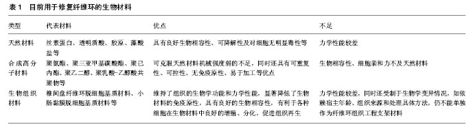
2.1 纤维环的结构特点 纤维环主要由内纤维环和外纤维环两部分构成,内层主要由类软骨细胞及其分泌的Ⅱ型胶原组成,承受椎间盘的压力;外层主要由类成纤维细胞及其分泌的Ⅰ型胶原和纤维蛋白组成,维持纤维环的张力[13-14]。纤维环在椎间盘内受到机械负荷的影响,其结构特点有利于纤维环承受均匀分布的压力,避免椎间盘纤维环因局部压力过大而导致破损[15]。纤维在椎间盘内排列呈同心圆状,胶原纤维在椎体间呈斜行排列,各层纤维环之间的角度约为60°,这些独特的构造使纤维环形成特殊的交叉网状结构,使其具有强大的抗拉伸能力[14-16],能防止髓核向外突出,对维持椎间盘的稳定性起重要作用。 然而,内层纤维环胶原蛋白表达的减少,将降低椎间盘对外界机械负荷的承受能力;而纤维环中蛋白聚糖的损失将影响纤维环细胞的功能,进一步引起椎间盘纤维变性,降低椎间盘的高度,加快椎间盘的退变[17]。纤维环是维持椎间盘正常生理功能的重要结构,椎间盘的退变过程也伴随着纤维环结构和组分的改变,纤维环退变也同样加快了椎间盘的退变进程。因此,构建具有生物学功能的材料以修复损伤的纤维环,对于椎间盘退变的治疗有重要意义[18-19]。此外,由于退变及椎间盘内压力等微环境引起的纤维环组织损伤(如纤维层分离、纤维环断裂等)是引起整个椎间盘退变的重要原因[8]。近年来,组织工程学的不断发展给学者们带来希望,生物材料在组织器官修复再生方面具有突出的优点,通过修复损伤的纤维环以修复退变椎间盘受到了广泛关注。 2.2 生物材料修复纤维环 生物材料在纤维环组织工程中扮演着重要作用,主要通过促进细胞外基质的产生和组织再生,以恢复纤维环的力学功能和结构的完整 性[20],因此,作为纤维环修复的材料应具备一些基本要求[15,19-22]:①低免疫原性:材料在人体内不会产生明显的免疫排斥反应;②生物相容性:将生物材料植入椎间盘后,对外源性细胞及体内组织无毒害作用;③适当的孔隙:便于种子细胞在其内部生长并分泌细胞外基质;④生物可降解性:在体内可降解,并且降解速率与组织的再生速率相匹配;⑤良好的力学性能:在椎间盘的压力下,能保持材料的稳定性。目前,用于纤维环修复的生物材料种类繁多,根据材料本身的来源可分为:一是天然材料,包括丝素蛋白、透明质酸、胶原、藻酸盐等;二是合成高分子材料,包括聚氨酯、聚三亚甲基碳酸酯、聚己内酯、聚乙二醇、聚乳酸-乙醇酸共聚物等[19];三是生物组织材料,如来源于椎间盘纤维环的脱细胞基质材料、小肠黏膜脱细胞基质材料等(表1)。 2.2.1 天然材料 由于天然材料具有良好的生物相容性、对细胞无明显毒性等优点,在纤维环组织工程中被广泛利用,目前已报道的天然材料包括丝素蛋白、纤维蛋白、藻酸盐、胶原、透明质酸等。 丝素蛋白作为一种天然材料,具有力学强度高、生物降解速率可控、生物相容性好和结构稳定等特点,其不仅能独立用于组织工程修复纤维环,也可与其他生物材料联用构建生物材料,因此在纤维环组织工程中应用前景广阔。Park等[23]根据纤维环的板层状特点,将丝素蛋白制作成与椎间盘纤维环结构相似的支架材料,将纤维环细胞接种于材料中培养2周,结果显示纤维环细胞能在板层状支架材料中良好生长,并且能较好地分泌细胞外基质。Bhattacharjee等[24]采用蚕丝构建与纤维环结构相似的薄层状纤维支架,通过与硫酸软骨素交联,增强生物材料的力学性能,在修复纤维环时,使其能在椎间盘压力微环境下保持材料的稳定性,此外,将其与软骨细胞共培养后,能显著分泌与纤维环组织相似的细胞基质。这些研究显示,丝素蛋白可能是一种理想的纤维环组织工程支架生物材料。 由于纤维蛋白生物材料良好的生物相容性、有利于细胞合成细胞外基质及蛋白聚糖等特点,被认为是椎间盘组织工程中理想的生物材料[25-26]。Colombini等[26]将纤维蛋白与胶原合成可注射性的纤维蛋白水凝胶,将纤维环细胞种植于凝胶后植入裸鼠皮下,结果显示与植入前相比,纤维环细胞能在材料内长期存在并合成大量的细胞外基质及与天然纤维软骨组织相似的Ⅰ型胶原。 Guillaume等[27]制作了一种形状记忆藻酸盐-胶原复合材料,该材料具有较好的弹性性能,在外界压力下,能快速恢复其原有形状,此外,纤维环细胞在材料中具有良好的生物活性,并且能很好地黏附于材料中并迁移。体内实验显示,间充质干细胞在材料中也能分泌大量细胞外基质,修复缺损的纤维环。Fuller等[28]制作了一种透明质酸寡糖,体外研究显示该材料对椎间盘细胞无毒性作用,并且能下调MMP13、ADAMTS1、ADAMTS4等的表达,这种改变有望清除缺损部位的瘢痕组织,有利于细胞外基质的形成,羊纤维环损伤的体内实验也证实其能促进椎间盘纤维环的修复。 Grunert等[29]制作了一种含有核黄素的高密度胶原凝胶材料,该材料具有较好的刚性和渗透性能,而核黄素能显著提高材料的这种特性,发现纤维原细胞在水凝胶内逐渐形成纤维帽,以修复缺损的纤维环;动物体内研究同样显示,该材料能更好地维持髓核的结构,增强椎间盘内蛋白聚糖的水合作用,保证盘内的渗透性,抑制椎间盘退变的进展。Borde等[30]同样制备了一种可注射型的高密度胶原凝胶材料,注入鼠纤维环缺损处取得了理想的修复效果。 由于京尼平交联剂能显著增强材料的力学性能,被广泛用于椎间盘组织工程中[31]。Schek等[32]将京尼平交联的纤维蛋白凝胶材料用于修复纤维环,结果显示在超负荷应力下,细胞也能在其表面较好地生长,但是,纤维环细胞在京尼平交联纤维蛋白凝胶材料中生长缓慢,这提示该材料可能更适用于修复较小纤维环缺口,或者用于修复大纤维环缺口的附加手段。Long等[33]采用纤维蛋白-京尼平水凝胶作为修复牛纤维环缺损模型的黏合剂,获得了较理想的结果,此外,其还能恢复椎间盘的生物力学强度,降低椎间盘突出风险。 2.2.2 合成高分子材料 合成高分子材料具有较好的可塑性,可克服天然材料力学性能差的不足,同时还具有可控性好、无免疫原性且易于加工等优点。 Wismer等[34]采用静电纺丝技术构建有方向性和无方向性的聚氨酯静电纺丝支架,结果显示,有方向性静电纺丝支架生物材料能更好地促进细胞黏附,维持细胞表型,促进胶原、蛋白多糖等细胞外基质的沉积,此外,其良好的力学强度使其在椎间盘压力微环境下可保持材料的稳定性,有望成为修复纤维环理想的生物材料。Liu等[35]同样证实了有序静电纺丝生物材料能为纤维环来源干细胞分化为纤维环细胞提供良好的微环境,还能更多地分泌细胞外基质。 Turner等[36]通过静电纺丝技术制备了聚氨酯纳米纤维静电纺丝支架,将其与纤维环细胞共培养,结果显示纤维环细胞能在聚氨酯纳米纤维静电纺丝支架中较好地增殖及合成胶原,而外界拉力能抑制纤维环细胞的细胞学行为,这种抑制作用与材料本身的弹性模量有关。Iu等[37]将内层纤维环细胞和外层纤维环细胞分别接种于聚氨酯支架生物材料内,通过测定并对比多功能蛋白聚糖和COL1A1等基因的表达水平,发现内层纤维环细胞高表达 COL2A1、聚集蛋白聚糖、多功能蛋白聚糖基因,而COL1A1基因低表达,此外,与外层纤维环细胞相比,内层纤维环细胞能合成大量的胶原和蛋白聚糖,该研究认为聚氨酯支架生物材料可维持内层纤维环细胞的表型,该研究可能对未来纤维环组织工程中选择合适的种子细胞修复纤维环具有重要意义。 Zhu等[38]制作了一种6聚氨酯静电纺丝支架材料,将纤维环来源的干细胞种植于材料内,结果显示,纤维环干细胞能很好地贴附于生物材料上,并且分泌大量细胞外基质,随着时间的推移,细胞增殖也越来越多。此外,该研究还显示,在不同弹性模量的聚氨酯静电纺丝生物材料中接种纤维环干细胞后,细胞的基质组成、基因表达和细胞力学方面受到该生物材料的弹性模量调控,并有着向纤维环结构所特有的细胞类型分化的趋势,这为椎间盘纤维环修复提供了新的研究思路与方向。他们还发现,随着该材料刚度的增加,骨髓间充质干细胞在聚氨酯静电纺丝材料中Ⅰ型胶原的表达也增加,而Ⅱ型胶原和蛋白聚糖则呈现相反的趋势[39]。 Pirvu等[40]同样制备了一种聚三亚甲基碳酸酯支架生物材料,该材料表面给予聚氨酯包被,将其移植入破损的纤维环中,结果显示在一定的压力下,其在椎间盘内能保持材料的稳定性,阻止髓核突出,恢复并维持椎间盘的高度,体外也证实其对间充质干细胞无细胞毒性,具有较好的生物相容性,间充质干细胞在材料中同样能分泌细胞外基质,以进一步修复纤维环。Blanquer等将[41]脂肪干细胞接种于聚三亚甲基碳酸酯生物材料中,再加入转化生长因子β3,证实了脂肪干细胞能在生物材料中更好地向纤维环组织方向分化,并且分泌大量胶原蛋白,显示出用于纤维环修复的巨大潜力。 Van Uden等[42]介绍了聚己内酯用于纤维环组织工程的能力,它模拟椎间盘的生理结构及纤维环组织的各向异性,对纤维环细胞无毒害作用,具有良好的细胞相容性,同时可获得比人椎间盘更好的力学性能。Nerurkar等[43]制备了一种纳米纤维聚己内酯生物材料,能模拟纤维环结构的非线性和各向异性特点,有利于纤维环细胞和干细胞的生长、增殖并分泌细胞外基质,增强材料的力学性能。 Jeong等[44]将透明质酸和聚乙二醇制备成复合水凝胶生物材料,结果显示纤维环细胞能在材料内大量增殖,并且分泌细胞外基质和蛋白聚糖产物。由于聚乙酸合成高分子材料具有良好生物相容性和可降解等特性,引起了广大研究者的兴趣[45-47],Xin等[48]制作了一种聚乳酸-乙醇酸共聚物支架材料,将其植入兔椎间盘纤维环缺损模型中,结果显示该材料能显著修复缺损的纤维环,恢复椎间盘的高度,展现出其修复纤维环的潜力。 从上述研究可看出,合成高分子材料在纤维环修复中具有较广阔的应用前景和空间,但合成高分子材料仍有一些不足,如缺少生物活性、细胞亲和力差等。 2.2.3 生物组织材料 脱细胞基质材料是通过物理和化学等方法去除组织中的细胞成分,保留以细胞外基质为主的成分,不仅维持了组织的生物学功能和力学性能,还能显著降低生物材料的免疫原性,具有良好的生物相容性,有利于各种细胞在生物材料中良好的增殖、分化,促进组织再生,可作为生物组织工程的支架材料[49]。此外,脱细胞基质还可为细胞提供接近其体内生长的微环境。 Xu等[50]分别采用Triton X-100、十二烷基硫酸钠及胰蛋白酶对猪来源纤维环进行去细胞化,制作脱细胞基质材料,结果显示这3种方式制作的脱细胞基质材料对细胞均无毒性作用,纤维环细胞在能在脱细胞材料中生长良好,展示了脱细胞基质材料良好的生物相容性。然而,经十二烷基硫酸钠和胰蛋白酶方法处理后,纤维环原有的同心圆排列结构被破坏,而采用Triton X-100方法制作的脱细胞基质材料能保留主要的细胞外基质及纤维环薄层状结构,其力学强度也明显优于十二烷基硫酸钠和胰蛋白酶处理组。具有良好生物相容性脱细胞基质材料,很有可能成为修复纤维环的理想生物材料。McGuire等[51]将猪来源心包制作成与纤维环结构相似的多层结构脱细胞基质材料,结果显示该材料具有良好的抗拉伸能力,其弹性模量与人纤维环相似,将纤维环细胞种于材料后,细胞能在材料内正常增殖和生长,并且分泌细胞外基质。Le Visage等[52]用猪来源小肠黏膜下层制备了一种脱细胞基质材料,将纤维环细胞接种于材料后,细胞生长良好,细胞外基质分泌增加,蛋白聚糖、胶原、Sox-9 基因显著表达。 但脱细胞基质力学性能较差,同时还受制于生物学变异情况,如依赖宿主年龄、组织来源和处理具体方法,仍不能单独作为纤维环组织工程支架材料,限制了其在纤维环组织工程中的应用,因而需要适宜的材料加强其力学性能。 "
| [1]Yurube T,Hrata H,Kakutani K,et al.Notochordal cell disappearance and modes of apoptotic cell death in a rat tail static compression-induced disc degeneration model.Arthritis Res Ther.2014;16(1):R31.[2]Evans CH,Huard J.Gene therapy approaches to regenerating the musculoskeletal system.Nat Rev Rheumatol. 2015;11(4): 234-242.[3]Fontana G,See E,Pandit A.Current trends in biologics delivery to restore intervertebral disc anabolism.Adv Drug Deliv Rev. 2015;84:146-158.[4]Hughes SP,Freemont AJ,Hukins DW,et al.The pathogenesis of degeneration of the intervertebral disc and emerging therapies in the management of back pain.J Bone Joint Surg Br.2012;94(10):1298-1304.[5]Sharifi S,Bulstra SK,Grijpma DW,et al.Treatment of the degenerated intervertebral disc; closure, repair and regeneration of the annulus fibrosus.J Tissue Eng Regen Med.2015;9(10):1120-1132.[6]Sharifi S,Bulstra SK,Grijpma DW,et al.Treatment of the degenerated intervertebral disc; closure, repair and regeneration of the annulus fibrosus.J Tissue Eng Regen Med. 2014;3(13):53-67.[7]Silva-Correia J,Correia SI,Oliveira JM,et al.Tissue engineering strategies applied in the regeneration of the human intervertebral disk.Biotechnol Adv.2013;31(8):1514-1531.[8]Iatridis JC.Tissue engineering: Function follows form.Nat Mater.2009;8(12):923-927.[9]Xiao L,Ding M,Saadoon O,et al.A novel culture platform for fast proliferation of human annulus fibrosus cells.Cell Tissue Res.2017;367(2):339-350.[10]Moriguchi Y,Alimi M,Khair T,et al.Biological Treatment Approaches for Degenerative Disk Disease: A Literature Review of In Vivo Animal and Clinical Data.Global Spine J. 2016;6(5):497-518.[11]Guterl CC,See EY,Blanquer SB,et al.Challenges and strategies in the repair of ruptured annulus fibrosus.Eur Cell Mater.2013;25:1-21.[12]Zhang SJ,Yang W,Wang C,et al.Autophagy: A double-edged sword in intervertebral disk degeneration. Clin Chim Acta. 2016;457:27-35.[13]Driscoll TP,Nakasone RH,Szczesny SE,et al.Biaxial mechanics and inter-lamellar shearing of stem-cell seeded electrospun angle-ply laminates for annulus fibrosus tissue engineering.J Orthop Res. 2013;31(6):864-870.[14]Li J,Liu C,Guo Q,et al.Regional variations in the cellular, biochemical, and biomechanical characteristics of rabbit annulus fibrosus.PLoS One.2014;9(3):e91799.[15]Yu J,Schollum ML,Wade KR,et al.ISSLS Prize Winner: A Detailed Examination of the Elastic Network Leads to a New Understanding of Annulus Fibrosus Organization.Spine(Phila Pa 1976).2015; 40(15):1149-1157.[16] Pattappa G,Li Z,Peroglio M,et al.Diversity of intervertebral disc cells: phenotype and function. J Anat.2012;221(6):480-496.[17]Vergari C,Mansfield J,Meakin JR,et al.Lamellar and fibre bundle mechanics of the annulus fibrosus in bovine intervertebral disc.Acta Biomater.2016;37:14-20.[18]Hudson KD,Alimi M,Grunert P,et al.Recent advances in biological therapies for disc degeneration: tissue engineering of the annulus fibrosus, nucleus pulposus and whole intervertebral discs. Curr Opin Biotechnol. 2013;24(5): 872-879.[19]Jin L, Shimmer AL, Li X. The challenge and advancement of annulus fibrosus tissue engineering. Eur Spine J. 2013; 22(5): 1090-1100.[20]Chang G,Kim HJ,Vunjak G,et al.Enhancing annulus fibrosus tissue formation in porous silk scaffolds. J Biomed Mater Res A.2010;92(1):43-51.[21]Bron JL,Helder MN,Meisel HJ,et al.Repair, regenerative and supportive therapies of the annulus fibrosus: achievements and challenges.Eur Spine J.2009;18(3):301-313.[22]Kular JK,Basu S,Sharma RI.The extracellular matrix: Structure, composition, age-related differences, tools for analysis and applications for tissue engineering.J Tissue Eng.2014;20:5.[23]Park SH,Gil ES,Mandal BB,et al.Annulus fibrosus tissue engineering using lamellar silk scaffolds. J Tissue Eng Regen Med.2012;3:24-33.[24]Bhattacharjee M,Miot S,Gorecka A,et al.Oriented lamellar silk fibrous scaffolds to drive cartilage matrix orientation: towards annulus fibrosus tissue engineering.Acta Biomater. 2012;8(9): 3313-3325.[25]Colombini A,Ceriani C,Banfi G,et al.Fibrin in intervertebral disc tissue engineering. Tissue Eng Part B Rev. 2014;20(6):713-721.[26]Colombini A,Lopa S,Ceriani C,et al.In vitro characterization and in vivo behavior of human nucleus pulposus and annulus fibrosus cells in clinical-grade fibrin and collagen-enriched fibrin gels. Tissue Eng Part A.2015;21(3-4):793-802.[27]Guillaume O,Naqvi SM,Lennon K,et al.Enhancing cell migration in shape-memory alginate-collagen composite scaffolds: In vitro and ex vivo assessment for intervertebral disc repair.J Biomater Appl. 2015;29(9):1230-1246.[28]Fuller ES,Shu C,Smith MM,et al.Hyaluronan oligosaccharides stimulate matrix metalloproteinase and anabolic gene expression in vitro by intervertebral disc cells and annular repair in vivo.J Tissue Eng Regen Med.2016. doi: 10.1002/term.2319. [Epub ahead of print][29]Grunert P,Borde BH,Towne SB,et al.Riboflavin crosslinked high-density collagen gel for the repair of annular defects in intervertebral discs: An in vivo study.Acta Biomater. 2015; 26:215-224.[30]Borde B,Grunert P,Härtl R,et al.Injectable, high-density collagen gels for annulus fibrosus repair: An in vitro rat tail model.J Biomed Mater Res A.2015;103(8):2571-2581.[31] Kirking B,Hedman T,Criscione J.Changes in the interfacial shear resistance of disc annulus fibrosus from genipin crosslinking.J Biomech.2014;47(1):293-296.[32]Schek RM,Michalek AJ,Iatridis JC.Genipin-crosslinked fibrin hydrogels as a potential adhesive to augment intervertebral disc annulus repair.Eur Cell Mater.2011;21:373-383.[33]Long RG,Bürki A,Zysset P,et al.Mechanical restoration and failure analyses of a hydrogel and scaffold composite strategy for annulus fibrosus repair.Acta Biomater.2016;30:116-125.[34]Wismer N,Grad S,Fortunato G.Biodegradable electrospun scaffolds for annulus fibrosus tissue engineering: effect of scaffold structure and composition on annulus fibrosus cells in vitro.Tissue Eng Part A.2014;20(3-4):672-682.[35]Liu C,Zhu C,Li J,et al.The effect of the fibre orientation of electrospun scaffolds on the matrix production of rabbit annulus fibrosus-derived stem cells.Bone Res.2015;3:15012.[36]Turner KG,Ahmed N,Santerre JP,et al.Modulation of annulus fibrosus cell alignment and function on oriented nanofibrous polyurethane scaffolds under tension.Spine J.2014;14(3):424-434.[37]Iu J,Santerre JP,Kandel RA.Inner and outer annulus fibrosus cells exhibit differentiated phenotypes and yield changes in extracellular matrix protein composition in vitro on a polycarbonate urethane scaffold.Tissue Eng Part A. 2014; 20(23-24):3261-3269.[38]Zhu C,Li J,Liu C,et al.Modulation of the gene expression of annulus fibrosus-derived stem cells using poly(ether carbonate urethane)urea scaffolds of tunable elasticity.Acta Biomater.2016; 29:228-238. [39]Guo Q,Liu C,Li J,et al.Gene expression modulation in TGF-β3-mediated rabbit bone marrow stem cells using electrospun scaffolds of various stiffness.J Cell Mol Med. 2015;19(7):1582-1592.[40]Pirvu T,Blanquer SB,Benneker LM,et al.A combined biomaterial and cellular approach for annulus fibrosus rupture repair.Biomaterials.2015;42:11-19.[41]Blanquer SB,Gebraad AW,Miettinen S,et al.Differentiation of adipose stem cells seeded towards annulus fibrosus cells on a designed poly(trimethylene carbonate) scaffold prepared by stereolithography.J Tissue Eng Regen Med.2016.doi: 10.1002/term.2170. [Epub ahead of print][42]Van Uden S,Silva-Correia J,Correlo VM,et al.Custom-tailored tissue engineered polycaprolactone scaffolds for total disc replacement.Biofabrication.2015;7(1):015008.[43]Nerurkar NL,Mauck RL,Elliott DM.Modeling interlamellar interactions in angle-ply biologic laminates for annulus fibrosus tissue engineering.Biomech Model Mechanobiol. 2011;10(6):973-984. [44]Jeong CG,Francisco AT,Niu Z,et al.Screening of hyaluronic acid-poly(ethylene glycol) composite hydrogels to support intervertebral disc cell biosynthesis using artificial neural network analysis. Acta Biomater.2014;10(8):3421-3430.[45]Kim SH,Song JE,Lee D,et al Development of poly(lactide-co-glycolide) scaffold- impregnated small intestinal submucosa with pores that stimulate extracellular matrix production in disc regeneration. J Tissue Eng Regen Med.2014;8(4):279-290.[46]Liang CZ,Li H,Tao YQ,et al.Dual release of dexamethasone and TGF-beta3 from polymeric microspheres for stem cell matrix accumulation in a rat disc degeneration model.Acta Biomater. 2013;9(12):9423-9433.[47]Yan J,Yang S,Sun H,et al. Effects of releasing recombinant human growth and differentiation factor-5 from poly(lactic-coglycolic acid) microspheres for repair of the rat degenerated intervertebral disc.J Biomater Appl.2013;29(1):72-80.[48]Xin L,Zhang C,Zhong F,et al.Minimal invasive annulotomy for induction of disc degeneration and implantation of poly (lactic-co-glycolic acid) (PLGA) plugs for annular repair in a rabbit model.Eur J Med Res.2016;21:7. [49]Lin X,Fang X,Wang Q,et al.Decellularized allogeneic intervertebral disc: natural biomaterials for regenerating disc degeneration.Oncotarget.2016;7(11):12121-12136.[50]Xu H,Xu B,Yang Q,et al.Comparison of decellularization protocols for preparing a decellularized porcine annulus fibrosus scaffold.PLoS One.2014;9(1):e86723.[51]McGuire R,Borem R,Mercuri J.The fabrication and characterization of a multi-laminate, angle-ply collagen patch for annulus fibrosus repair.J Tissue Eng Regen Med.2016.doi: 10.1002/term.2250.[Epub ahead of print][52]Le Visage C,Yang SH,Kadakia L,et al.Small intestinal submucosa as a potential bioscaffold for intervertebral disc regeneration.Spine(Phila Pa 1976).2006;31(21):2423-2430.[53]Bron JL,van der Veen AJ,Helder MN,et al.Biomechanical and in vivo evaluation of experimental closure devices of the annulus fibrosus designed for a goat nucleus replacement model.Eur Spine J. 2010;19(8):1347-1355.[54]Endres M,Abbushi A,Thomale UW,et al.Intervertebral disc regeneration after implantation of a cell-free bioresorbable implant in a rabbit disc degeneration model. Biomaterials. 2010;31(22):5836-5841. |
| [1] | Zhang Tongtong, Wang Zhonghua, Wen Jie, Song Yuxin, Liu Lin. Application of three-dimensional printing model in surgical resection and reconstruction of cervical tumor [J]. Chinese Journal of Tissue Engineering Research, 2021, 25(9): 1335-1339. |
| [2] | Zeng Yanhua, Hao Yanlei. In vitro culture and purification of Schwann cells: a systematic review [J]. Chinese Journal of Tissue Engineering Research, 2021, 25(7): 1135-1141. |
| [3] | Xu Dongzi, Zhang Ting, Ouyang Zhaolian. The global competitive situation of cardiac tissue engineering based on patent analysis [J]. Chinese Journal of Tissue Engineering Research, 2021, 25(5): 807-812. |
| [4] | Wu Zijian, Hu Zhaoduan, Xie Youqiong, Wang Feng, Li Jia, Li Bocun, Cai Guowei, Peng Rui. Three-dimensional printing technology and bone tissue engineering research: literature metrology and visual analysis of research hotspots [J]. Chinese Journal of Tissue Engineering Research, 2021, 25(4): 564-569. |
| [5] | Chang Wenliao, Zhao Jie, Sun Xiaoliang, Wang Kun, Wu Guofeng, Zhou Jian, Li Shuxiang, Sun Han. Material selection, theoretical design and biomimetic function of artificial periosteum [J]. Chinese Journal of Tissue Engineering Research, 2021, 25(4): 600-606. |
| [6] | Liu Fei, Cui Yutao, Liu He. Advantages and problems of local antibiotic delivery system in the treatment of osteomyelitis [J]. Chinese Journal of Tissue Engineering Research, 2021, 25(4): 614-620. |
| [7] | Li Xiaozhuang, Duan Hao, Wang Weizhou, Tang Zhihong, Wang Yanghao, He Fei. Application of bone tissue engineering materials in the treatment of bone defect diseases in vivo [J]. Chinese Journal of Tissue Engineering Research, 2021, 25(4): 626-631. |
| [8] | Zhang Zhenkun, Li Zhe, Li Ya, Wang Yingying, Wang Yaping, Zhou Xinkui, Ma Shanshan, Guan Fangxia. Application of alginate based hydrogels/dressings in wound healing: sustained, dynamic and sequential release [J]. Chinese Journal of Tissue Engineering Research, 2021, 25(4): 638-643. |
| [9] | Chen Jiana, Qiu Yanling, Nie Minhai, Liu Xuqian. Tissue engineering scaffolds in repairing oral and maxillofacial soft tissue defects [J]. Chinese Journal of Tissue Engineering Research, 2021, 25(4): 644-650. |
| [10] | Xing Hao, Zhang Yonghong, Wang Dong. Advantages and disadvantages of repairing large-segment bone defect [J]. Chinese Journal of Tissue Engineering Research, 2021, 25(3): 426-430. |
| [11] | Chen Siqi, Xian Debin, Xu Rongsheng, Qin Zhongjie, Zhang Lei, Xia Delin. Effects of bone marrow mesenchymal stem cells and human umbilical vein endothelial cells combined with hydroxyapatite-tricalcium phosphate scaffolds on early angiogenesis in skull defect repair in rats [J]. Chinese Journal of Tissue Engineering Research, 2021, 25(22): 3458-3465. |
| [12] | Wang Hao, Chen Mingxue, Li Junkang, Luo Xujiang, Peng Liqing, Li Huo, Huang Bo, Tian Guangzhao, Liu Shuyun, Sui Xiang, Huang Jingxiang, Guo Quanyi, Lu Xiaobo. Decellularized porcine skin matrix for tissue-engineered meniscus scaffold [J]. Chinese Journal of Tissue Engineering Research, 2021, 25(22): 3473-3478. |
| [13] | Mo Jianling, He Shaoru, Feng Bowen, Jian Minqiao, Zhang Xiaohui, Liu Caisheng, Liang Yijing, Liu Yumei, Chen Liang, Zhou Haiyu, Liu Yanhui. Forming prevascularized cell sheets and the expression of angiogenesis-related factors [J]. Chinese Journal of Tissue Engineering Research, 2021, 25(22): 3479-3486. |
| [14] | Liu Chang, Li Datong, Liu Yuan, Kong Lingbo, Guo Rui, Yang Lixue, Hao Dingjun, He Baorong. Poor efficacy after vertebral augmentation surgery of acute symptomatic thoracolumbar osteoporotic compression fracture: relationship with bone cement, bone mineral density, and adjacent fractures [J]. Chinese Journal of Tissue Engineering Research, 2021, 25(22): 3510-3516. |
| [15] | Liu Liyong, Zhou Lei. Research and development status and development trend of hydrogel in tissue engineering based on patent information [J]. Chinese Journal of Tissue Engineering Research, 2021, 25(22): 3527-3533. |
| Viewed | ||||||
|
Full text |
|
|||||
|
Abstract |
|
|||||