Chinese Journal of Tissue Engineering Research ›› 2026, Vol. 30 ›› Issue (7): 1679-1686.doi: 10.12307/2026.521
Previous Articles Next Articles
Repair of segmental bone defect of rabbit radius by decalcified bone matrix loaded with adipose-derived stem cells
Ding Yifan1, Yin Wenjie1, Zhang Li1, Yuan Shuya2, Sun Guoju2, Zhang Naili1, Zhao Dongmei1, Ma Lina2
- 1Department of Human Anatomy, School of Basic Medical Science, Binzhou Medical University, Yantai 264003, Shandong Province, China; 2Department of Diagnostics, Second School of Medicine, Binzhou Medical University, Yantai 264003, Shandong Province, China
-
Received:2024-11-14Revised:2025-03-06Accepted:2025-03-19Online:2026-03-08Published:2025-08-19 -
Contact:Ma Lina, Lecturer, Department of Diagnostics, Second School of Medicine, Binzhou Medical University, Yantai 264003, Shandong Province, China -
About author:Ding Yifan, Master candidate, Department of Human Anatomy, School of Basic Medical Science, Binzhou Medical University, Yantai 264003, Shandong Province, China -
Supported by:Medical and Health Science and Technology Development Project of Shandong Province, No. 202104070826 (to MLN); Medical and Health Science and Technology Development Project of Shandong Province, No. 202301021239 (to ZNL); Natural Science Foundation of Shandong Province, No. BS2015SW021 (to ZNL); Students’ Innovation and Entrepreneurship Training Program of Shandong Province, No. S202310440046 (to SGJ)
CLC Number:
Cite this article
Ding Yifan, Yin Wenjie, Zhang Li, Yuan Shuya, Sun Guoju, Zhang Naili, Zhao Dongmei, Ma Lina. Repair of segmental bone defect of rabbit radius by decalcified bone matrix loaded with adipose-derived stem cells[J]. Chinese Journal of Tissue Engineering Research, 2026, 30(7): 1679-1686.
share this article
Add to citation manager EndNote|Reference Manager|ProCite|BibTeX|RefWorks
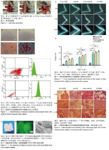
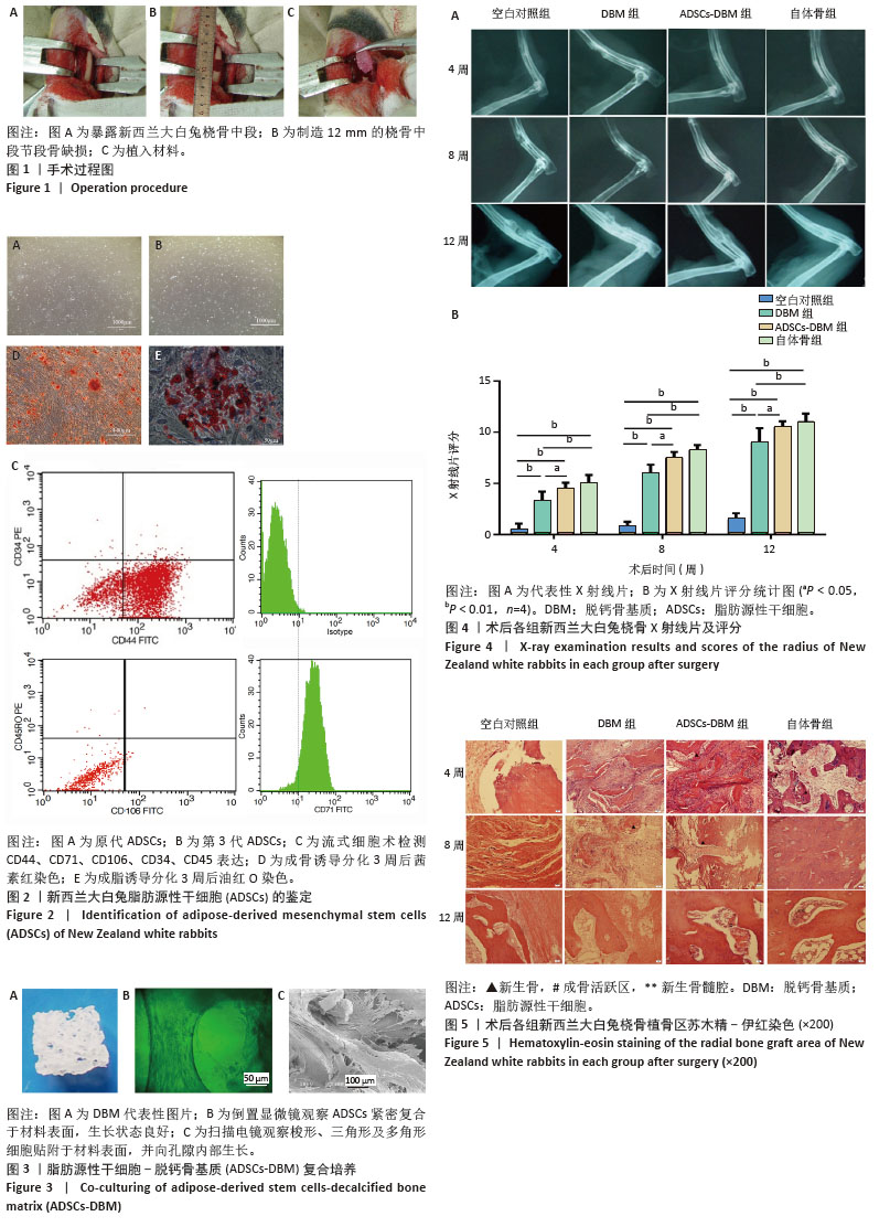
2.1 ADSCs鉴定结果 原代培养接种4 h后,细胞开始贴壁生长,但细胞形态不均一,可见红细胞、成纤维细胞等细胞,随着换液次数的增加,贴壁生长的细胞呈椭圆形或梭形(图2A);传代到第3代,ADSCs聚集生长,呈长梭形、成纤维细胞样外观,有较多突起(图2B)。流式细胞术检测结果显示,第3代细胞CD44、CD71高表达,CD34、 CD45、CD106低表达(图2C),符合间充质干细胞的特征。成骨诱导分化培养后,细胞生长旺盛,增殖迅速,逐渐由长梭形变为多边形或立方形,逐渐汇合呈铺路石状,重叠生长,胞质内颗粒状物质增多,胞外基质分泌逐渐增多,有圆形或卵圆形的结节状物出现。茜素红染色可见染成橘红色的矿化结节,结节形状为圆形或不规则型,大小不一(图2D)。成脂诱导分化培养的细胞形状逐渐从长梭形变为方形或圆形,细胞质内开始出现高折光度的圆形脂滴,聚集成多房型。油红O染色可见染成鲜红色的脂滴聚集(图2E)。 2.2 ADSCs-DBM复合培养 大体观察DBM为乳白色,呈蜂窝状疏松多孔样结构(图3A);倒置显微镜下观察可见ADSCs紧密复合于材料表面,生长状态良好(图3B);扫描电镜观察可见梭形、三角形及多角形细胞贴附于材料表面,并向孔隙内部生长(图3C)。 2.3 桡骨节段性缺损修复情况 2.3.1 实验动物数量分析 24只新西兰大白兔48侧桡骨参加实验,全部进入结果分析,无脱落。 2.3.2 X射线片观察结果 如图4A所示,术后4周,空白对照组骨缺损两侧骨断端锐利,仅有少量骨痂形成,可见明显骨缺损,截骨端骨髓腔封闭;DBM组植骨部位也可见骨痂形成,但骨生成量明显较ADSCs-DBM组少;ADSCs-DBM组两侧骨端有明显骨痂形成,植入物与骨端有部分骨性连接;自体骨组植入物与骨端有少量连续性骨桥形成。 术后8周,空白对照组骨缺损区缩小,骨端可见少量骨痂形成;DBM组骨缺损两断端在靠近尺骨侧形成高密度骨痂连接,远离尺骨侧由云雾状高密度影填充,且在远端可见明显的骨缺损;ADSCs-DBM组骨缺损区内被高密度影填充,两端基本连接,仅在远端有部分小缺损;自体骨组植入的自体骨与宿主骨连接,骨折线消失,连接处呈现高密度影。 术后12周,空白对照组骨缺损区进一步缩小,但未见连续性骨痂,骨髓腔封闭;DBM组骨缺损两端基本连接,但仍见部分骨缺损,骨髓腔没有再通;ADSCs-DBM组骨缺损区完全修复,皮质骨连续,骨髓腔部分再通;自体骨组植骨区与两端宿主骨无明显界限,皮质骨连续,骨髓腔基本完全再通。 术后不同时间X射线片评分结果显示,ADSCs-DBM组在各个取材时间点X射线片评分低于自体骨组,高于DBM组和空白对照组。统计学分析结果显示,ADSCs-DBM组与自体骨组无统计学差异(P > 0.05),ADSCs-DBM组X射线片评分高于DBM组和空白对照组(P < 0.05);DBM组X射线片评分高于空白对照组(P < 0.01)(图4B)。 2.3.3 组织学观察结果 如图5所示,术后4周,空白对照组骨缺损区内纤维结缔组织长入,骨端成骨不活跃;DBM组宿主骨端可见新生骨形成,但数量明显较ADSCs-DBM组少;ADSCs-DBM组材料表面附着大量成骨细胞,沿材料呈线形排列,成骨活跃,材料周围区域明显可见新骨生成;自体骨组植入骨与宿主骨间有新生骨组织形成,无明显炎症反应。 术后8周,空白对照组骨缺损区仍被纤维组织填充;DBM组材料周围可见大量间充质干细胞,并有新生骨组织向材料长入;ADSCs-DBM组植骨区有大量新生骨组织形成,残存材料被新生骨包绕;自体骨组植骨区可见大量新生编织骨形成,排列无序。 术后12周,空白对照组骨缺损区被纤维组织填充,两端骨髓腔骨化闭合;DBM组可见大量新生骨组织生成,但在新生骨组织周围可见纤维组织长入;ADSCs-DBM组植骨区可见大量排列无序的新生编织骨,成骨活跃,植入材料几乎被完全吸收替代,残存材料被新生编织骨包绕;自体骨组植骨区可见新生编织骨开始改建,有新生骨髓腔形成。 "
| [1] ZHEN C, SHI Y, WANG W, et al. Advancements in gradient bone scaffolds: enhancing bone regeneration in the treatment of various bone disorders. Biofabrication. 2024;16(3):032004. [2] BALDWIN P, LI DJ, AUSTON DA, et al. Autograft, Allograft, and Bone Graft Substitutes: Clinical Evidence and Indications for Use in the Setting of Orthopaedic Trauma Surgery. J Orthop Trauma. 2019;33(4):203-213. [3] BELOTI MM, ROSA AL. Bone Regeneration and Repair Materials. J Funct Biomater. 2024;15(3):78. [4] HARAKAS NK. Demineralized bone-matrix-induced osteogenesis. Clin Orthop Relat Res. 1984;(188):239-251. [5] HONSAWEK S, BUMRUNGPANICHTHAWORN P, THITISET T, et al. Gene expression analysis of demineralized bone matrix-induced osteogenesis in human periosteal cells using cDNA array technology. Genet Mol Res. 2011;10(3):2093-2103. [6] HUANG YZ, HE T, CUI J, et al. Urine-Derived Stem Cells for Regenerative Medicine: Basic Biology, Applications, and Challenges. Tissue Eng Part B Rev. 2022;28(5): 978-994. [7] REN J, LI Z, LIU W, et al. Demineralized bone matrix for repair and regeneration of maxillofacial defects: A narrative review. J Dent. 2024;143:104899. [8] ZHANG H, YANG L, YANG XG, et al. Demineralized Bone Matrix Carriers and their Clinical Applications: An Overview. Orthop Surg. 2019;11(5):725-737. [9] CHEN C, LI Z, XU C, et al. Self-Assembled Nanocomposite Hydrogels as Carriers for Demineralized Bone Matrix Particles and Enhanced Bone Repair. Adv Healthc Mater. 2024;13(10):e2303592. [10] DONOS N, AKCALI A, PADHYE N, et al. Bone regeneration in implant dentistry: Which are the factors affecting the clinical outcome? Periodontol 2000. 2023;93(1):26-55. [11] VERMEULEN S, TAHMASEBI BIRGANI Z, HABIBOVIC P. Biomaterial-induced pathway modulation for bone regeneration. Biomaterials. 2022;283:121431. [12] PATEL D, WAIRKAR S. Bone regeneration in osteoporosis: opportunities and challenges. Drug Deliv Transl Res. 2023;13(2):419-432. [13] SUN Y, ZHAO H, YANG S, et al. Urine-derived stem cells: Promising advancements and applications in regenerative medicine and beyond. Heliyon. 2024;10(6):e27306. [14] 李欢,苗博,韩智慧,等.牙髓和牙周韧带间充质干细胞成骨能力差异分析研究[J].中国骨质疏松杂志,2024,30(8):1102-1107. [15] 颉丽英,刘司麒,吴明芮,等.人脐带间充质干细胞移植治疗心肌肥厚模型小鼠[J].中国组织工程研究,2022,26(30):4826-4833. [16] MISHRA VK, SHIH HH, PARVEEN F, et al. Identifying the Therapeutic Significance of Mesenchymal Stem Cells. Cells. 2020;9(5):1145. [17] HICOK KC, DU LANEY TV, ZHOU YS, et al. Human adipose-derived adult stem cells produce osteoid in vivo. Tissue Eng. 2004;10(3-4):371-380. [18] QIN Y, GE G, YANG P, et al. An Update on Adipose-Derived Stem Cells for Regenerative Medicine: Where Challenge Meets Opportunity. Adv Sci (Weinh). 2023;10(20):e2207334. [19] ALMALKI SG. Adipose-derived mesenchymal stem cells and wound healing: Potential clinical applications in wound repair. Saudi Med J. 2022;43(10):1075-1086. [20] WAGNER JM, REINKEMEIER F, WALLNER C, et al. Adipose-Derived Stromal Cells Are Capable of Restoring Bone Regeneration After Post-Traumatic Osteomyelitis and Modulate B-Cell Response. Stem Cells Transl Med. 2019;8(10):1084-1091. [21] LIU J, ZHOU P, LONG Y, et al. Repair of bone defects in rat radii with a composite of allogeneic adipose-derived stem cells and heterogeneous deproteinized bone. Stem Cell Res Ther. 2018;9(1):79. [22] TORRES-GUZMAN RA, HUAYLLANI MT, AVILA FR, et al. Application of Human Adipose-Derived Stem cells for Bone Regeneration of the Skull in Humans. J Craniofac Surg. 2022;33(1):360-363. [23] MAGLIONE M, SALVADOR E, RUARO ME, et al. Bone regeneration with adipose derived stem cells in a rabbit model. J Biomed Res. 2018;33(1):38-45. [24] GU H, XIONG Z, YIN X, et al. Bone regeneration in a rabbit ulna defect model: use of allogeneic adipose-derivedstem cells with low immunogenicity. Cell Tissue Res. 2014;358(2):453-464. [25] WEN C, YAN H, FU S, et al. Allogeneic adipose-derived stem cells regenerate bone in a critical-sized ulna segmental defect. Exp Biol Med (Maywood). 2016;241(13):1401-1409. [26] ZHAO Y, LIN H, ZHANG J, et al. Crosslinked three-dimensional demineralized bone matrix for the adipose-derived stromal cell proliferation and differentiation. Tissue Eng Part A. 2009;15(1):13-21. [27] ZHANG N, ZHOU M, ZHANG Y, et al. Porcine bone grafts defatted by lipase: efficacy of defatting and assessment of cytocompatibility. Cell Tissue Bank. 2014;15(3):357-367. [28] ZHANG N, MA L, LIU X, et al. In vitro and in vivo evaluation of xenogeneic bone putty with the carrier of hydrogel derived from demineralized bone matrix. Cell Tissue Bank. 2018;19(4):591-601. [29] MCGOVERN JA, GRIFFIN M, HUTMACHER DW. Animal models for bone tissue engineering and modelling disease. Dis Model Mech. 2018;11(4):dmm033084. [30] HIXON KR, MILLER AN. Animal models of impaired long bone healing and tissue engineering- and cell-based in vivo interventions. J Orthop Res. 2022;40(4):767-778. [31] EINHORN TA, LANE JM, BURSTEIN AH, et al. The healing of segmental bone defects induced by demineralized bone matrix. A radiographic and biomechanical study. J Bone Joint Surg Am. 1984;66(2):274-279. [32] THOMAS S, JAGANATHAN BG. Signaling network regulating osteogenesis in mesenchymal stem cells. J Cell Commun Signal. 2022;16(1):47-61. [33] MOHAMED-AHMED S, FRISTAD I, LIE SA, et al. Adipose-derived and bone marrow mesenchymal stem cells: a donor-matched comparison. Stem Cell Res Ther. 2018;9(1):168. [34] MOHAMED-AHMED S, YASSIN MA, RASHAD A, et al. Comparison of bone regenerative capacity of donor-matched human adipose-derived and bone marrow mesenchymal stem cells. Cell Tissue Res. 2021;383(3):1061-1075. [35] KANG BJ, RYU HH, PARK SS, et al. Comparing the osteogenic potential of canine mesenchymal stem cells derived from adipose tissues, bone marrow, umbilical cord blood, and Wharton’s jelly for treating bone defects. J Vet Sci. 2012;13(3):299-310. [36] ZHANG J, WEHRLE E, ADAMEK P, et al. Optimization of mechanical stiffness and cell density of 3D bioprinted cell-laden scaffolds improves extracellular matrix mineralization and cellular organization for bone tissue engineering. Acta Biomater. 2020;114:307-322. [37] GODOY ZANICOTTI D, COATES DE, DUNCAN WJ. In vivo bone regeneration on titanium devices using serum-free grown adipose-derived stem cells, in a sheep femur model. Clin Oral Implants Res. 2017;28(1):64-75. [38] WEN Y, JIANG B, CUI J, et al. Superior osteogenic capacity of different mesenchymal stem cells for bone tissue engineering. Oral Surg Oral Med Oral Pathol Oral Radiol. 2013;116(5):e324-e332. [39] HUSCH JFA, COQUELIN L, CHEVALLIER N, et al. Comparison of Osteogenic Capacity and Osteoinduction of Adipose Tissue-Derived Cell Populations. Tissue Eng Part C Methods. 2023;29(5):216-227. [40] BUNNELL BA. Adipose Tissue-Derived Mesenchymal Stem Cells. Cells. 2021;10(12):3433. |
| [1] | Sun Lei, Zhang Qi, Zhang Yu. Pro-osteoblastic effect of chlorogenic acid protein microsphere/polycaprolactone electrospinning membrane [J]. Chinese Journal of Tissue Engineering Research, 2026, 30(8): 1877-1884. |
| [2] | Hu Xiongke, Liu Shaohua, Tan Qian, Liu Kun, Zhu Guanghui. Shikonin intervention with bone marrow mesenchymal stem cells improves microstructure of femur in aged mice [J]. Chinese Journal of Tissue Engineering Research, 2026, 30(7): 1609-1615. |
| [3] | Cao Wenqi, Feng Xiuzhi, Zhao Yi, Wang Zhimin, Chen Yiran, Yang Xiao, Ren Yanling. Effect of macrophage polarization on osteogenesis-angiogenesis coupling in type 2 diabetic osteoporosis [J]. Chinese Journal of Tissue Engineering Research, 2026, 30(4): 917-925. |
| [4] | Xu Wenhe, Li Xiaobing, Liu Fang. Functionalized biomimetic mineralized collagen modified orthopedic implants [J]. Chinese Journal of Tissue Engineering Research, 2026, 30(2): 516-527. |
| [5] | Zhang Zhaowei, Chen Ouzile, Bai Mingru, Wang Chenglin. Therapeutic potential of bioactive substances secreted by dental mesenchymal stem cells for bone repair [J]. Chinese Journal of Tissue Engineering Research, 2026, 30(1): 163-174. |
| [6] | Liu Nian, Dong Xinyue, Wang Songpeng, Xu Yingjiang, Zhang Xiaoming. Stem cell exosomes and biomaterial-assisted exosomes in bone defect repair [J]. Chinese Journal of Tissue Engineering Research, 2026, 30(1): 175-183. |
| [7] | Zuo Na, Tang Qi, Yu Meng, Tao Kai. Effect of miR-196b-5p in adipose-derived stem cell exosomes on burn wound healing in rats [J]. Chinese Journal of Tissue Engineering Research, 2026, 30(1): 43-49. |
| [8] | Lou Guo, Zhang Min, Fu Changxi. Exercise preconditioning for eight weeks enhances therapeutic effect of adipose-derived stem cells in rats with myocardial infarction [J]. Chinese Journal of Tissue Engineering Research, 2025, 29(7): 1363-1370. |
| [9] | Zhao Zengbo, Li Chenxi, Dou Chenlei, Ma Na, Zhou Guanjun. Anti-inflammatory and osteogenic effects of chitosan/sodium glycerophosphate/sodium alginate/leonurine hydrogel [J]. Chinese Journal of Tissue Engineering Research, 2025, 29(4): 678-685. |
| [10] |
Li Yunzhe, Niu Zefan, Wang Zirou, Ai Chongyi, Chen Gang, Wang Xinxing.
Asperosaponin VI promotes osteogenic differentiation of MC3T3-E1 cells under hypoxia environment #br#
#br#
[J]. Chinese Journal of Tissue Engineering Research, 2025, 29(35): 7481-7489.
|
| [11] | Li Guoliang, Zhao Jianyong, Lyu Deliang, Su Juyue, Liu Qilin, Wang Tieqiang, Wang Xin. Improved 3D printed splint for distal radius fracture based on clinical defects: design and rapid grid-free analysis [J]. Chinese Journal of Tissue Engineering Research, 2025, 29(33): 7123-7129. |
| [12] | Hu Liuchao, Luo Yiwen, Wu Zhifang. Fracture line map characteristics of distal radius fractures involving dorsal articular surface: effective fixation with screws for postoperative displacement [J]. Chinese Journal of Tissue Engineering Research, 2025, 29(3): 524-530. |
| [13] | Liu Haowen, Qiao Weiping, Meng Zhicheng, Li Kaijie, Han Xuan, Shi Pengbo. Regulation of osteogenic effects by bone morphogenetic protein/Wnt signaling pathway: revealing molecular mechanisms of bone formation and remodeling [J]. Chinese Journal of Tissue Engineering Research, 2025, 29(3): 563-571. |
| [14] | Zeng Yu, Xie Chengwei, Hong Yuanqi, Su Shenghui, Dong Xieping. In vitro angiogenesis and osteogenesis properties of copper-doped mesoporous bioactive glass [J]. Chinese Journal of Tissue Engineering Research, 2025, 29(28): 5941-5949. |
| [15] | Shi Chunrong, He Jiaxu, Deng Lishan, Wang Hailan, Zhao Aimin, Yu Yiling, Geng Haixia, Song Weijun. Application of graphene oxide in field of oral implant restoration [J]. Chinese Journal of Tissue Engineering Research, 2025, 29(28): 6118-6126. |
| Viewed | ||||||
|
Full text |
|
|||||
|
Abstract |
|
|||||