[1] DANG J, YANG J, YU Z, et al. Bone marrow mesenchymal stem cells enhance angiogenesis and promote fat retention in fat grafting via polarized macrophages. Stem Cell Res Ther. 2022;13(1):52.
[2] LI PL, WANG YX, ZHAO ZD, et al. Clinical-grade human dental pulp stem cells suppressed the activation of osteoarthritic macrophages and attenuated cartilaginous damage in a rabbit osteoarthritis model. Stem Cell Res Ther. 2021;12(1):260.
[3] YUAN Y, LI L, ZHU L, et al. Mesenchymal stem cells elicit macrophages into M2 phenotype via improving transcription factor EB-mediated autophagy to alleviate diabetic nephropathy. Stem Cells. 2020;38(5): 639-652.
[4] UYSAL CA, TOBITA M, HYAKUSOKU H, et al. The Effect of Bone-Marrow-Derived Stem Cells and Adipose-Derived Stem Cells on Wound Contraction and Epithelization. Adv Wound Care (New Rochelle). 2014; 3(6):405-413.
[5] GAO M, ZHANG J, WANG JZ, et al. Effects of hypoxia-pretreated rat adipose-derived mesenchymal stem cells conditioned medium on wound healing of rats with full-thickness defects. Zhonghua Shao Shang Za Zhi. 2020;36(9):803-812.
[6] FAN L, DONG J, HE X, et al. Bone marrow mesenchymal stem cells-derived exosomes reduce apoptosis and inflammatory response during spinal cord injury by inhibiting the TLR4/MyD88/NF-κB signaling pathway. Hum Exp Toxicol. 2021;40(10):1612-1623.
[7] HE D, ZHAO F, JIANG H, et al. LOXL2 from human amniotic mesenchymal stem cells accelerates wound epithelialization by promoting differentiation and migration of keratinocytes. Aging (Albany NY). 2020;12(13):12960-12986.
[8] LIU Y, CHEN J, LIANG H, et al. Human umbilical cord-derived mesenchymal stem cells not only ameliorate blood glucose but also protect vascular endothelium from diabetic damage through a paracrine mechanism mediated by MAPK/ERK signaling. Stem Cell Res Ther. 2022;13(1):258.
[9] MATHEW SA, NAIK C, CAHILL PA, et al. Placental mesenchymal stromal cells as an alternative tool for therapeutic angiogenesis. Cell Mol Life Sci. 2020;77(2):253-265.
[10] MAKHOUL G, CHIU RC, CECERE R. Placental mesenchymal stem cells: a unique source for cellular cardiomyoplasty. Ann Thorac Surg. 2013;95(5):1827-1833.
[11] XU L, LIU Y, SUN Y, et al. Tissue source determines the differentiation potentials of mesenchymal stem cells: a comparative study of human mesenchymal stem cells from bone marrow and adipose tissue. Stem Cell Res Ther. 2017;8(1):275.
[12] LI X, BAI J, JI X, et al. Comprehensive characterization of four different populations of human mesenchymal stem cells as regards their immune properties, proliferation and differentiation. Int J Mol Med. 2014;34(3):695-704.
[13] HUANG Q, YANG Y, LUO C, et al. An efficient protocol to generate placental chorionic plate-derived mesenchymal stem cells with superior proliferative and immunomodulatory properties. Stem Cell Res Ther. 2019;10(1):301.
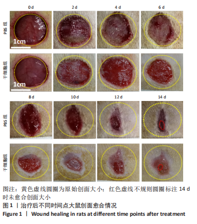
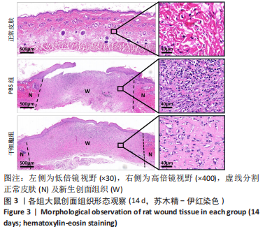
[14] 赵京禹,付小兵,雷永红,等.大鼠小面积全层皮肤缺损创面模型的制备[J].感染、炎症、修复,2008,9(1):64.
[15] ZHANG J, GUAN J, NIU X, et al. Exosomes released from human induced pluripotent stem cells-derived MSCs facilitate cutaneous wound healing by promoting collagen synthesis and angiogenesis. J Transl Med. 2015;13:49.
[16] HASS R, OTTE A. Mesenchymal stem cells as all-round supporters in a normal and neoplastic microenvironment. Cell Commun Signal. 2012;10(1):26.
[17] TSUCHIYA A, TAKEUCHI S, WATANABE T, et al. Mesenchymal stem cell therapies for liver cirrhosis: MSCs as “conducting cells” for improvement of liver fibrosis and regeneration. Inflamm Regen. 2019; 39:18.
[18] YAO Y, XIA Z, CHENG F, et al. Human placental mesenchymal stem cells ameliorate liver fibrosis in mice by upregulation of Caveolin1 in hepatic stellate cells. Stem Cell Res Ther. 2021;12(1):294.
[19] LI Z, WANG H, YANG B, et al. Three-dimensional graphene foams loaded with bone marrow derived mesenchymal stem cells promote skin wound healing with reduced scarring. Mater Sci Eng C Mater Biol Appl. 2015;57:181-188.
[20] OCHIAI D, ABE Y, FUKUTAKE M, et al. Cell sheets using human amniotic fluid stem cells reduce tissue fibrosis in murine full-thickness skin wounds. Tissue Cell. 2021;68:101472.
[21] HEO JS, KIM S. Human adipose mesenchymal stem cells modulate inflammation and angiogenesis through exosomes. Sci Rep. 2022; 12(1):2776.
[22] AFZALI MF, PANNONE SC, MARTINEZ RB, et al. Intravenous injection of adipose-derived mesenchymal stromal cells benefits gait and inflammation in a spontaneous osteoarthritis model. J Orthop Res. 2023;41(4):902-912.
[23] TIAN G, LIU C, WANG H, et al. Human umbilical cord mesenchymal stem cells prevent glucocorticoid-induced osteonecrosis of the femoral head by promoting angiogenesis. J Plast Surg Hand Surg. 2023;57(1-6): 71-77.
[24] NOTODIHARDJO SC, MORIMOTO N, KAKUDO N, et al. Efficacy of Gelatin Hydrogel Impregnated With Concentrated Platelet Lysate in Murine Wound Healing. J Surg Res. 2019;234:190-201.
[25] CHEN TJ, YEH YT, PENG FS, et al. S100A8/A9 Enhances Immunomodulatory and Tissue-Repairing Properties of Human Amniotic Mesenchymal Stem Cells in Myocardial Ischemia-Reperfusion Injury. Int J Mol Sci. 2021;22(20):11175.
[26] HUANG Q, CHENG X, LUO C, et al. Placental chorionic plate-derived mesenchymal stem cells ameliorate severe acute pancreatitis by regulating macrophage polarization via secreting TSG-6. Stem Cell Res Ther. 2021;12(1):337.
[27] LIU J, QIU X, LV Y, et al. Apoptotic bodies derived from mesenchymal stem cells promote cutaneous wound healing via regulating the functions of macrophages. Stem Cell Res Ther. 2020;11(1):507.
|