[1] SCHOENFELD BJ. Does exercise-induced muscle damage play a role in skeletal muscle hypertrophy?. J Strength Cond Res. 2012;26(5):1441-1453.
[2] 袁建琴,王瑞元,李肃反. 离心运动对大鼠骨骼肌结蛋白分布和表达的影响——对骨骼肌损伤机制的研究[J]. 体育科学,2005,25(6): 63-66.
[3] 李俊平,徐玉明,高晓娟,等. 一次低氧运动后快肌和慢肌Titin和Nebulin蛋白含量的变化[J].北京体育大学学报,2012,35(6):47-50.
[4] PEAKE JM, NEUBAUER O, DELLA GATTA PA, et al. Muscle damage and inflammation during recovery from exercise. J Appl Physiol (1985). 2017;122(3):559-570.
[5] SWEENEY HL, HAMMERS DW. Muscle Contraction. Cold Spring Harb Perspect Biol. Cold Spring Harb Perspect Biol. 2018;10(2):a023200.
[6] ZHU P, HAMLISH NX, THAKKAR AV, et al. BMAL1 drives muscle repair through control of hypoxic NAD(+) regeneration in satellite cells. Genes Dev. 2022;36(3-4):149-166.
[7] RUAN W, YUAN X, ELTZSCHIG HK. Circadian rhythm as a therapeutic target. Nat Rev Drug Discov. 2021;20(4):287-307.
[8] CHEN G, TANG Q, YU S, et al. The biological function of BMAL1 in skeleton development and disorders. Life Sci. 2020;253:117636.
[9] CHATTERJEE S, MA K. Circadian clock regulation of skeletal muscle growth and repair. F1000Res. 2016;5:1549.
[10] MURE LS, LE HD, BENEGIAMO G, et al. Diurnal transcriptome atlas of a primate across major neural and peripheral tissues. Science. 2018; 359(6381):eaao0318.
[11] KASSAR-DUCHOSSOY L, GAYRAUD-MOREL B, GOMÈS D, et al. Mrf4 determines skeletal muscle identity in Myf5:Myod double-mutant mice. Nature. 2004;431(7007):466-471.
[12] ANDREWS JL, ZHANG X, MCCARTHY JJ, et al. CLOCK and BMAL1 regulate MyoD and are necessary for maintenance of skeletal muscle phenotype and function. Proc Natl Acad Sci U S A. 2010;107(44): 19090-19095.
[13] HODGE BA, ZHANG X, GUTIERREZ-MONREAL MA, et al. MYOD1 functions as a clock amplifier as well as a critical co-factor for downstream circadian gene expression in muscle. Elife. 2019;8:e43017.
[14] ARMSTRONG RB, OGILVIE RW, SCHWANE JA. Eccentric exercise-induced injury to rat skeletal muscle. J Appl Physiol Respir Environ Exerc Physiol. 1983;54(1):80-93.
[15] HODY S, CROISIER JL, BURY T, et al. Eccentric Muscle Contractions: Risks and Benefits. Front Physiol. 2019;10:536.
[16] KANAZAWA Y, NAGANO M, KOINUMA S, et al. Basement membrane recovery process in rat soleus muscle after exercise-induced muscle injury. Connect Tissue Res. 2021;62(5):519-530.
[17] 丁海丽,黄增浩,任在方,等. 针刺对大鼠运动性骨骼肌损伤内质网应激的干预作用及机制[J]. 中国应用生理学杂志,2021,37(4): 359-364.
[18] MIYAZAKI M, SCHRODER E, EDELMANN SE, et al. Age-associated disruption of molecular clock expression in skeletal muscle of the spontaneously hypertensive rat. PLoS One. 2011;6(11):e27168.
[19] 林雪薇. miR-133a及其相关调节因子在大鼠运动性骨骼肌损伤修复过程中的表达变化[D].上海:上海体育学院,2019.
[20] NOY T, SUAD O, TAGLICHT D, et al. HUWE1 ubiquitinates MyoD and targets it for proteasomal degradation. Biochem Biophys Res Commun. 2012;418(2):408-413.
[21] NEDERGAARD A, VISSING K, OVERGAARD K, et al. Expression patterns of atrogenic and ubiquitin proteasome component genes with exercise: effect of different loading patterns and repeated exercise bouts. J Appl Physiol (1985). 2007;103(5):1513-1522.
[22] ABOU-KHALIL R, YANG F, LIEU S, et al. Role of muscle stem cells during skeletal regeneration. Stem Cells. 2015;33(5):1501-1511.
[23] MCDEARMON EL, PATEL KN, KO CH, et al. Dissecting the functions of the mammalian clock protein BMAL1 by tissue-specific rescue in mice. Science. 2006;314(5803):1304-1308.
[24] DYAR KA, CICILIOT S, WRIGHT LE, et al. Muscle insulin sensitivity and glucose metabolism are controlled by the intrinsic muscle clock. Mol Metab. 2013;3(1):29-41.
[25] CHATTERJEE S, YIN H, NAM D, et al. Brain and muscle Arnt-like 1 promotes skeletal muscle regeneration through satellite cell expansion. Exp Cell Res. 2015;331(1):200-210.
[26] KEMLER D, WOLFF CA, ESSER KA. Time-of-day dependent effects of contractile activity on the phase of the skeletal muscle clock. J Physiol. 2020;598(17):3631-3644.
[27] CHOI Y, CHO J, NO MH, et al. Re-Setting the Circadian Clock Using Exercise against Sarcopenia. Int J Mol Sci. 2020;21(9):3106.
[28] PASTORE S, HOOD DA. Endurance training ameliorates the metabolic and performance characteristics of circadian Clock mutant mice. J Appl Physiol (1985). 2013;114(8):1076-1084.
[29] BRAGER AJ, HEEMSTRA L, BHAMBRA R, et al. Homeostatic effects of exercise and sleep on metabolic processes in mice with an overexpressed skeletal muscle clock. Biochimie. 2017;132:161-165.
[30] ZAMBON AC, MCDEARMON EL, SALOMONIS N, et al. Time- and exercise-dependent gene regulation in human skeletal muscle. Genome Biol. 2003;4(10):R61.
[31] LABEIT S, KOLMERER B. Titins: giant proteins in charge of muscle ultrastructure and elasticity. Science. 1995;270(5234):293-296.
[32] PODOBED PS, ALIBHAI FJ, CHOW CW, et al. Circadian regulation of myocardial sarcomeric Titin-cap (Tcap, telethonin): identification of cardiac clock-controlled genes using open access bioinformatics data. PLoS One. 2014;9(8):e104907.
[33] YU JG, FÜRST DO, THORNELL LE. The mode of myofibril remodelling in human skeletal muscle affected by DOMS induced by eccentric contractions. Histochem Cell Biol. 2003;119(5):383-393.
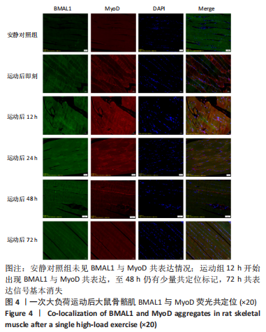
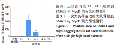
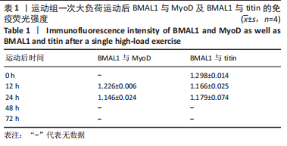
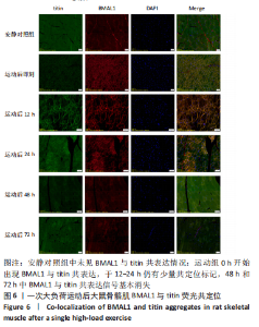
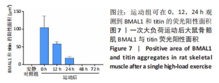
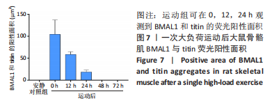
|