[1] GHARIBEH N, AGHEBATI-MALEKI L, MADANI J, et al. Cell-based therapy in thin endometrium and Asherman syndrome. Stem Cell Res The. 2022;13(1):33.
[2] 韩笑,黄晓武.干细胞在子宫内膜损伤后修复的研究进展[J].国际妇产科学杂志,2019,46(4):365-369.
[3] GAO Y, WU G, XU Y, et al. Stem cell-based therapy for asherman syndrome: promises and challenges. Cell Transplant. 2021;30: 09636897211020734.
[4] MA J, ZHAN H, LI W, et al. Recent trends in therapeutic strategies for repairing endometrial tissue in intrauterine adhesion. Biomater Res. 2021;25(1):40.
[5] 段璇,李维宏.干细胞治疗中重度宫腔粘连的临床研究进展[J].现代医药卫生,2021,37(13):2213-2217.
[6] YU D, WONG YM, CHEONG Y, et al. Asherman syndrome-one century later. Fertil Steril. 2008;89(4):759-779.
[7] 周新艳.归肾丸加味治疗子宫内膜偏薄不孕症的临床效果观察[J].中国医药指南,2018,16(16):197-198.
[8] 徐嵘,黄丽,袁芬,等.加味归肾丸治疗薄型子宫内膜肾虚血瘀证50例临床观察[J].甘肃中医药大学学报,2021,38(2):51-55.
[9] 刘燕,关永格,宋阳.归肾丸水提物干预SD大鼠骨髓间充质干细胞增殖及PI3K,AKT蛋白的表达[J].中国组织工程研究,2020,24(13): 1983-1988.
[10] 邵芷若,关永格,宋阳,等.基于网络药理学研究归肾丸对薄型子宫内膜大鼠的治疗作用机制[J].中国实验方剂学杂志,2021,27(17): 168-177.
[11] SHI Y, QIU X, DAI M, et al. Hyperoside attenuates hepatic ischemia-reperfusion injury by suppressing oxidative stress and inhibiting apoptosis in rats. Transplant Proc. 2019;51(6):2051-2059.
[12] HUANG J, TONG X, ZHANG L, et al. Hyperoside Attenuates bleomycin-induced pulmonary fibrosis development in mice. Front Pharmacol. 2020;11:550955.
[13] SUN K, LUO J, JING X, et al. Hyperoside ameliorates the progression of osteoarthritis: An in vitro and in vivo study. Phytomedicine. 2021;80: 153387.
[14] WEI A, SONG Y, NI T, et al. Hyperoside attenuates pregnancy loss through activating autophagy and suppressing inflammation in a rat model. Life Sci. 2020;254:117735.
[15] ZHANG SS, XU XX, XIANG WW, et al. Using 17β‐estradiol heparin‐poloxamer thermosensitive hydrogel to enhance the endometrial regeneration and functional recovery of intrauterine adhesions in a rat model. FASEB J. 2020;34(1):446-457.
[16] XU HL, XU J, ZHANG SS, et al. Temperature-sensitive heparin-modified poloxamer hydrogel with affinity to KGF facilitate the morphologic and functional recovery of the injured rat uterus. Drug Deliv. 2017;24(1): 867-881.
[17] 祖珍玉,罗敏,申东翔,等.脂肪间充质干细胞及普兰尼克 F127 水凝胶复合物对宫腔粘连子宫内膜的修复作用[J].实用医学杂志, 2021,37(19):2437-2441.
[18] HAN Q, DU Y. Advances in the application of biomimetic endometrium interfaces for uterine bioengineering in female infertility. Front Bioeng Biotechnol. 2020;8:153.
[19] 颜洁,关志宇,朱卫丰,等.Box-Behnken 效应面法优化自组装法制备葛根素壳聚糖/海藻酸钠口服纳米粒的处方与工艺研究[J].中草药,2019,50(23):5706-5713.
[20] 李娜,颜洁,关志宇,等.葛根素壳聚糖/海藻酸钠口服纳米粒的制备、表征与药动学研究[J].中草药,2020,51(15):3894-3900.
[21] FENG Q, GAO B, ZHAO X, et al. Establishment of an animal model of intrauterine adhesions after surgical abortion and curettage in pregnant rats. Ann Transl Med. 2020;8(4):56.
[22] 许洁.温敏型KGF-EPL-HP生物黏附水凝胶对子宫内膜损伤的修复作用[D].温州:温州医科大学,2018.
[23] JIANG X, LI X, FEI X, et al. Endometrial membrane organoids from human embryonic stem cell combined with the 3D Matrigel for endometrium regeneration in asherman syndrome. Bioact Mater. 2021;6(11):3935-3946.
[24] LIU F, HU S, YANG H, et al. Hyaluronic acid hydrogel integrated with mesenchymal stem cell‐secretome to treat endometrial injury in a rat model of Asherman’s syndrome. Adv Healthc Mater. 2019;8(14): e1900411.
[25] LIN Y, DONG S, YE X, et al. Synergistic regenerative therapy of thin endometrium by human placenta-derived mesenchymal stem cells encapsulated within hyaluronic acid hydrogels. Stem Cell Res Ther. 2022;13(1):66.
[26] GUO X, ZHU C, LIU X, et al. Hyperoside protects against heart failure-induced liver fibrosis in rats. Acta Histochem. 2019;121(7):804-811.
[27] 郭晓,曲凤霞,辛越,等.金丝桃苷对心力衰竭大鼠肝纤维化的影响及其分子机制[J].山东医药,2021,61(2):40-45.
[28] YE P, YANG X, CHEN X, et al. Hyperoside attenuates OVA-induced allergic airway inflammation by activating Nrf2. Int Immunopharmacol. 2017;44:168-173.
[29] 李淑颖,耿玉萌,崔宇擎,等.菟丝子水提物对子宫内双酚A暴露小鼠的生殖保护作用[J].中国兽医学报,2021,41(4):748-754.
[30] 王琦,宋芳,应康,等.菟丝子对雌性生殖系统保护作用的研究进展[J].包头医学院学报,2021,37(9):117-119.
[31] RUSSO E, VILLA C. Poloxamer hydrogels for biomedical applications. Pharmaceutics. 2019;11(12):671.
[32] ZHANG SS, XIA WT, XU J, et al. Three-dimensional structure micelles of heparin-poloxamer improve the therapeutic effect of 17β-estradiol on endometrial regeneration for intrauterine adhesions in a rat model. Int J Nanomedicine. 2017;12:5643-5657.
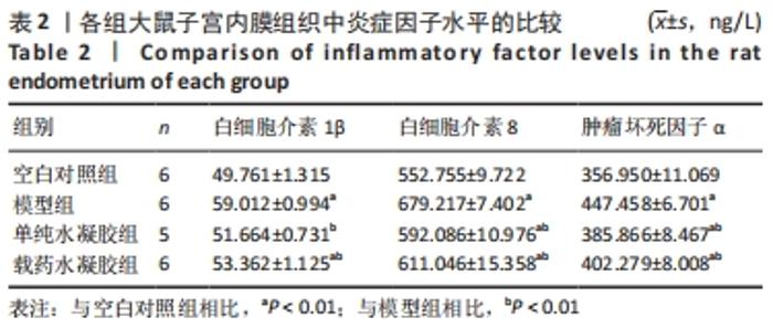
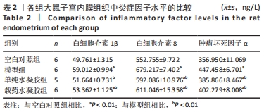
[33] 崔颖,孙大为.白细胞介素与子宫内膜异位症[J].中国妇产科临床杂志,2005(1):74-75+28.
[34] 张莉,吴乃文,杨永华.IL-18、TNF-α、VEGF在宫腔粘连患者子宫内膜中的表达及临床意义[J].中国医药科学,2015,5(5):208-210.
[35] OKADA H, TSUZUKI T, SHINDOH H, et al. Regulation of decidualization and angiogenesis in the human endometrium: mini review. J Obstet Gynaecol Res. 2014;40(5):1180-1187.
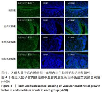
[36] 张莹莹.整合素β3、血管内皮生长因子在薄型子宫内膜植入窗期表达的研究[D].郑州:郑州大学,2012.
[37] 周雨玫,刘雁峰,申萌萌.二补助育汤对胚胎着床障碍小鼠子宫血管生成相关因子表达的影响[J/OL].世界中医药:1-7.[2021-09-23] http://kns.cnki.net/kcms/detail/11.5529.R.20210922.1155.002.html.
[38] ZHANG L, LI Y, GUAN CY, et al. Therapeutic effect of human umbilical cord-derived mesenchymal stem cells on injured rat endometrium during its chronic phase. Stem Cell Res Ther. 2018;9(1):36.
[39] 张汝月,李瑞娇,张引红,等.薄型子宫内膜小鼠模型的免疫组化鉴定与生育力评估[J].中国比较医学杂志,2018,28(12):19-26.
[40] 张奕,胡秀娟,戴志俊,等.多重方法建立大鼠子宫内膜深度损伤模型[J].中华全科医学,2021,19(6):913-916+1012.
|