中国组织工程研究 ›› 2026, Vol. 30 ›› Issue (30): 7971-7978.doi: 10.12307/2026.953
• 组织构建综述 tissue construction review • 上一篇 下一篇
仿生梯度支架修复大段骨缺损:梯度结构-免疫调控-时空控释协同策略
王 浩1,陈仁场1,田轩赫1,梁松林1,曲新田1,郭玉祺1,夏 崴2,吴亚东1,3,李念虎1,4
- 1山东中医药大学第一临床医学院,山东省济南市 250000;2丹阳市中医院,江苏省镇江市 212399;3日照市中医院,山东省日照市 276800;4山东中医药大学附属医院,山东省济南市 250014
-
收稿日期:2026-05-07修回日期:2026-05-30出版日期:2026-10-28发布日期:2026-09-14 -
通讯作者:吴亚东,硕士生导师,副主任医师,山东中医药大学第一临床医学院,山东省济南市 250000;日照市中医院,山东省日照市 276800 共同通讯作者:李念虎,博士生导师,教授,主任医师,山东中医药大学第一临床医学院,山东省济南市 250000;山东中医药大学附属医院,山东省济南市 250014 -
作者简介:王浩,2000年生,汉族,山东中医药大学在读硕士,主要从事脊柱脊髓与骨关节疾病研究。 -
基金资助:山东省自然科学基金项目(ZR2021LZY006),项目负责人:吴亚东;山东省自然科学基金项目(ZR2023MH063),项目负责人:李念虎;山东省医药卫生重点项目(202404071248),项目负责人:吴亚东;北京恒济卫生管理发展基金临床科研专项资助基金(2023HJ002),项目负责人:吴亚东
Repair of large bone defects using bionic gradient scaffolds: a synergistic strategy combining gradient structure, immunomodulation, and spatiotemporal controlled release
Wang Hao1, Chen Renchang1, Tian Xuanhe1, Liang Songlin1, Qu Xintian1, Guo Yuqi1, Xia Wei2, Wu Yadong1, 3, Li Nianhu1, 4
- 1First Clinical Medical College, Shandong University of Traditional Chinese Medicine, Jinan 250000, Shandong Province, China; 2Danyang Hospital of Traditional Chinese Medicine, Zhenjiang 212399, Jiangsu Province, China; 3Rizhao Hospital of Traditional Chinese Medicine, Rizhao 276800, Shandong Province, China; 4Affiliated Hospital of Shandong University of Traditional Chinese Medicine, Jinan 250014, Shandong Province, China
-
Received:2026-05-07Revised:2026-05-30Online:2026-10-28Published:2026-09-14 -
Contact:Wu Yadong, Master’s supervisor, Associate chief physician, First Clinical Medical College, Shandong University of Traditional Chinese Medicine, Jinan 250000, Shandong Province, China; Rizhao Hospital of Traditional Chinese Medicine, Rizhao 276800, Shandong Province, China Co-corresponding author: Li Nianhu, Doctoral supervisor, Professor, Chief physician, First Clinical Medical College, Shandong University of Traditional Chinese Medicine, Jinan 250000, Shandong Province, China; Affiliated Hospital of Shandong University of Traditional Chinese Medicine, Jinan 250014, Shandong -
About author:Wang Hao, MS candidate, First Clinical Medical College, Shandong University of Traditional Chinese Medicine, Jinan 250000, Shandong Province, China -
Supported by:Shandong Provincial Natural Science Foundation, Nos. ZR2021LZY006 (to WYD) and ZR2023MH063 (to LNH); Shandong Provincial Key Project in Medicine and Health, No. 202404071248 (to WYD); Beijing Hengji Health Management Development Fund - Clinical Research Special Grant, No. 2023HJ002 (to WYD)

摘要:

文题释义:
仿生骨梯度支架:是模拟天然骨成分、结构、力学性能等多维度连续梯度特征构建的骨组织工程支架,区别于传统均质支架,可通过光固化3D打印、3D生物打印等技术制备,能实现成分、孔径、力学模量的梯度设计,既为骨缺损提供力学支撑,又可定向调控宿主免疫细胞行为,还能作为活性分子载体实现分区负载,是大段骨缺损修复的核心仿生材料。
免疫时空协同策略:是面向大段骨缺损修复以仿生骨梯度支架为核心载体的协同调控策略,通过支架梯度结构实现空间上的免疫微环境定向调控,结合微球封装、刺激响应性载体等技术实现活性分子在时间上的程序性释放,让免疫调控与活性分子控释在时空维度匹配骨再生的炎症消退、血管生成、成骨分化时序需求,协同重塑利于骨整合的修复微环境。
背景:严重创伤导致的大尺寸骨缺损极难自愈,核心障碍在于缺血性坏死与局部免疫炎症失调。近年来,能主动调控再生微环境的“仿生骨梯度支架”成为破局关键。然而,目前多项研究仍局限于单一优化支架结构或单纯投递药物,缺乏有机协同,导致临床疗效常遇瓶颈。
目的:旨在打破既往单一视角的局限,提出“梯度结构-免疫调控-时空控释”三效协同的理论框架,为研发下一代智能原位骨修复材料提供鲜明的战略指引。
方法:检索PubMed、中国知网等数据库在2017年1月至2026年3月收录的中英文文献。严格筛选并聚焦于仿生骨梯度支架、免疫微环境及多因子时序递送机制,最终提取103篇高水平原始研究与转化案例进行深度剖析。
结果与结论:最新研究表明,利用先进3D打印构建的多维梯度支架,不仅完美提供天然机械支撑,更能定向诱导免疫巨噬细胞向“促愈合”方向转变。更关键的是,将智能控释系统嵌合于支架中,能让辅助再生的活性因子实现“正确时间、正确位置”的释放,完美契合骨再生“早期消炎-中期长血管-晚期成骨”的自然节律。“梯度结构-免疫调控-时空控释”的多元协同是当前修复各类大段骨缺损的最优解。未来该领域的终极趋势是研发具备“缺损区微环境实时感知与动态自适应响应”的极智生物材料。
https://orcid.org/0009-0009-6316-3572 (王浩)
中国组织工程研究杂志出版内容重点:干细胞;骨髓干细胞;造血干细胞;脂肪干细胞;肿瘤干细胞;胚胎干细胞;脐带脐血干细胞;干细胞诱导;干细胞分化;组织工程
中图分类号:
引用本文
王 浩, 陈仁场, 田轩赫, 梁松林, 曲新田, 郭玉祺, 夏 崴, 吴亚东, 李念虎. 仿生梯度支架修复大段骨缺损:梯度结构-免疫调控-时空控释协同策略[J]. 中国组织工程研究, 2026, 30(30): 7971-7978.
Wang Hao, Chen Renchang, Tian Xuanhe, Liang Songlin, Qu Xintian, Guo Yuqi, Xia Wei, Wu Yadong, Li Nianhu. Repair of large bone defects using bionic gradient scaffolds: a synergistic strategy combining gradient structure, immunomodulation, and spatiotemporal controlled release[J]. Chinese Journal of Tissue Engineering Research, 2026, 30(30): 7971-7978.
2.1.1 从单向梯度到“结构-力学-成分”跨尺度耦合的演进 对于严重的创伤性大段骨缺损来说,重建的关键就是从皮质骨到松质骨的复杂异质性。支架设计早期大多数研究只在单一孔隙或者单一成分梯度上进行[7,52-53]。但是多孔材料的力学模量本质上是由孔隙率和拓扑结构来决定的[54],所以单纯追求生物活性的高孔隙率会导致力学崩溃[55-56]。近些年来,高质量的研究已经彻底抛弃了孤立分类的思路,转而采取结构、力学和成分三者相互联系的梯度式综合设计。大段承重骨的“最优梯度构型”已经基本确定,即外周有致密拓扑来提供超过70 MPa的抗压强度抵抗生理载荷,核被层(中心区域)有大孔径高连通性来打破物质传输壁垒,缓解缺血性坏死[7,57]。
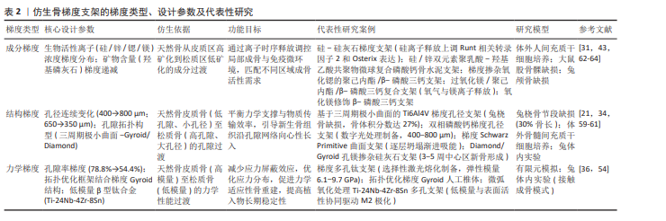
现有研究为支架耦合梯度设计提供了很多的体内外实验依据。从结构力学角度出发,研究者们依靠三周期极小曲面拓扑结构创建出孔径连续改变的梯度支架[21,58],植入兔桡骨大段节段性缺损(近30%骨长),骨体积分数达到27%,而且成功促使新生骨组织沿着梯度孔隙网络由周缘向中心区域进行向心性长入。值得一提的是,不同三周期极小曲面构型在体内的成骨节奏并不一致[59],Diamond和Gyroid孔支架由于具有弯曲孔隙的特点,在术后3-5周内中心区就出现了明显的新生骨,7周后骨组织已经基本填充满了整个孔隙;而片状三周期极小曲面虽然力学性能更好,但是骨再生却较
晚,这就说明孔隙曲率对于成骨的空间分布有着不可小视的作用。另外,为了满足高负荷承载的要求,YANG等[9]构建搭载重组人骨形态发生蛋白2控释系统的3D打印聚合物/陶瓷复合支架,植入羊跖骨5 cm临界缺损模型,术后24周所有实验动物都达到了承重皮质骨的桥接,骨体积比单纯使用重组人骨形态发生蛋白2海绵的对照组明显提高。
与此同时,LIU等[34]从力学形变和物质输运的耦合角度对仿生骨梯度支架的优异成骨性进行了详细的分析,微型CT和体外力学测试研究发现,相比于均质支架,仿生梯度支架具有连续孔径梯度结构,在保持宏观框架强度的同时,可以利用孔径变化来构建差异化的细胞微环境,从而达到力学支撑和生物活性的平衡。
在双相磷酸钙体系中,用数字光处理3D打印技术制备了400-800 μm的梯度孔径支架,结果显示梯度孔槽可以加快新骨沉积,也可以促进血管化的网络重塑,从而达到骨与血管共同新生的目的[60]。而梯度施瓦茨本原曲面(Schwarz Primitive)支架对于力学失效模式的分析又进一步说明了它的优点[61],该结构在压缩载荷下展现出逐层坍塌的渐进吸能失效模式,这种特性对于植入物在体内的长期稳定性至关重要,它能够有效吸收生理活动中的冲击能量,避免了传统支架的突发性断裂风险,为缺损核心区的血供重建提供了决定性的物理基础。
除了物理结构维度的改善之外,生化微环境梯度的加入对天然骨无机、有机组分的空间分布做了更加精细的模拟,现有的方法也由原来的双相掺杂发展成了精确到时空层次的排布。采用选择性激光烧结制备的硅-硅灰石梯度支架,抗压强度达到71 MPa,远高于均质支架,降解后产生的硅离子会特异性上调成骨关键转录因子(Runt相关转录因子2和Osterix)的表达[31]。在多元素协同上,LIANG等[43]把负载有硅和锌的聚乳酸-羟基乙酸共聚物微球与磷酸钙水泥支架结合在一起,利用离子的时序释放来协同改善免疫和成骨微环境;另外还有研究者用3D打印技术制备了含有氧化锶梯度掺杂的聚己内酯/β-磷酸三钙支架[62],实现了锶离子的持续释放,从而重塑局部成骨微环境。镁离子也受到了人们的关注。有研究将过氧化镁加入到聚己内酯/β-磷酸三钙体系当中,依靠它持续释放的氧气和镁离子来维持骨髓间充质干细胞的存活以及成骨分化,在大鼠股骨髁缺损上表现出明显的新骨形成增多现象[63]。另有研究利用水热法制备出含有氧化镁的β-磷酸三钙,从而提高生物相容性,兔颅骨缺损修复实验表明该材料骨再生效果明显优于纯β-磷酸三钙对照组[64]。
在精细制备技术上,有研究用顺序旋涂含有递减浓度羟基磷灰石纳米颗粒的悬浮液来构建连续矿物含量梯度,并且利用激光加工得到漏斗形微通道阵列,给细胞深入浸润提供足够的空间[65]。除此之外,采用有限元分析和机器学习的方式,可以依靠几何特征和成分分布来准确预估支架的机械性能和生物学效果,进而对支架的宏观结构参数进行反向优化,显著缩减了依靠经验式调整的进程[66-67]。
为了方便横向比较不同梯度类型的设计逻辑和研究进展,表2从成分梯度、结构梯度、力学梯度3个方面对各个梯度核心设计参数、仿生依据、功能目标、代表性研究案例、实验模型进行了归纳。从上述证据中可以看出,生化梯度可以准确地引导细胞的行为,而且它和物理结构梯度之间还可以产生相互促进的效应,即所谓的力学和生物学增益。但是目前大多数研究还停留在单个梯度的概念验证上,怎样在十几厘米大的承重支架里将宏观的力学梯度和微观的多种离子成分梯度结合起来,仍然是当前仿生设计中最大的技术难题。
2.1.2 复合制造工艺向临床转化的边界缺陷 伴随着以机器学习为辅助的复合增材制造技术(材料挤出、粉末床熔融等)的发展,建立三周期极小曲面或者Voronoi等复杂的几何梯度也成为常规手段[66,68-71]。目前,可以实现梯度结构精准构建的制备技术正处在多元化的发展阶段。表3整理了选择性激光熔化/烧结、数字光处理、熔融沉积成型结合近场直写静电纺丝、冷冻干燥、机器学习引导的混合制造等主流技术的梯度构建能力、核心优势、目前存在的不足以及典型的应用案例。但是单一种类的材料对于大段负重骨来说存在固有脆性以及生物活性不能兼顾的矛盾,因此混合制造技术应运而生。有团队提出用3D打印聚己内酯骨架(力学支撑)和梯度载细胞聚乙二醇水凝胶(生物活性)相结合的个性化混合梯度策略[69]。更精细的方案就是用熔融沉积成型和近场直写静电纺丝相结合,制备出具有硬质骨架和软质引导网络的多尺度复合支架[70]。
虽然复合制造被视作破局之策,但是对异质界面的深入剖析也暴露出一个致命的缺陷[70],即异质材料的界面力学匹配很难控制。在真实的体内负重环境中,伴随着数万次的生理循环步态载荷,这些软硬交界面的微观撕裂、层间剥离就是造成支架晚期失效的主要原因。复杂生理载荷下长时间疲劳表现的梯度支架技术“黑箱”,是目前梯度支架由“实验室可制造”向“临床可靠制造”过渡所要跨越的鸿沟。
2.2 支架理化信号主导的免疫重塑与“多细胞协同”网络
2.2.1 物理、化学与机电微环境对固有免疫的早期极化诱导 大段骨缺损修复失败的典型病理结局一般是由持续性无菌炎症造成的纤维囊包裹所引起的。相比传统的生物惰性替代物来说,仿生梯度支架的先进之处在于它能够自身产生物理、化学和电学等各方面的信号,从而主动地改变局部免疫生态。大量的机制研究发现,巨噬细胞以及固有免疫防线对于植入物的微观信号具备极高的敏感性[72-75]。
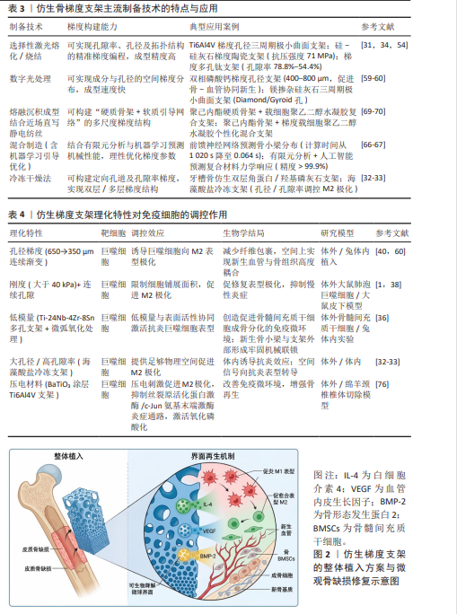
机械拓扑和刚度起着直接的物理极化作用。体外研究表明,当支架局部刚度阈值大于40 kPa并且具有特异连续孔隙的时候,可以限制细胞铺展面积,激活RhoA、ROCK等力学转导通路,使巨噬细胞被强制转化为促修复的M2表型[1,38]。按照同样的思路,有研究利用3D打印制造出Ti-24Nb-4Zr-8Sn多孔支架来主动削减结构模量,再结合微弧氧化赋予表面亲水微孔特性,从而促使巨噬细胞趋向于抗炎表型。兔体内实验证明新生骨小梁和支架外侧形成了牢固的机械联结,植入稳定[36]。孔隙空间自身也是不能忽略的调控因素。一项关于海藻酸盐冷冻支架的研究发现,大孔径或者高孔隙率的结构在体内可以有效地诱导M2极化,小孔径低孔隙率的结构会阻碍这一过程[32-33]。体内研究也得到了证实,在植入650-350 μm孔径连续梯度的钛支架之后,内源性诱导出更多的M2极化,不但显著减少了纤维包绕,而且在空间上使新生组织高度耦合[76]。
其次,物理信号和化学、机电属性混杂给免疫调节带来新的角度。除生物陶瓷降解释
放特定离子的传统方法外,最近的研究如MA等[47]利用包裹白藜芦醇脂质体(Res-liposomes)的聚乙烯醇-壳聚糖多级纤维支架,不但可以清除自由基,而且可以分层释放来促进巨噬细胞向M2型极化,从而改善炎症微环境。机电微环境方面,新兴的3D打印导电水凝胶可以激活整合素、黏着斑激酶、细胞外调节蛋白激酶以及Piezo1、Piezo2介导的钙内流来干预细胞行为[70];同时,由BaTiO3等组成的压电智能生物材料在承受生理机械应力的时候,会把宏观形变转变成微观内源性的电生理信号,这种仿生生理电位被证明可以加快巨噬细胞向愈合表型极化[77]。这种拓扑结构、物理刚度以及内源性电信号的叠加,使支架在不需要外源性药物的情况下,对早期骨免疫微环境进行定标调节创造了条件。表4总结了各种理化性质对于巨噬细胞极化行为以及下游信号通路的影响。
2.2.2 免疫-血管-成骨级联与适应性多细胞协同网络 固有免疫的极化只是开始,优化的早期微环境还会产生促进血管生成和成骨的多细胞协同网络。研究表明,梯度支架诱导的M2极化不但可以分泌转化生长因子β、血管内皮生长因子和骨形态发生蛋白2等旁分泌因子来促进内皮细胞形成管状网、招募并分化出骨髓间充质干细胞[78-79],还可以通过自噬途径促进骨髓间充质干细胞的基质矿化[47],而且伴随着局部导电和微地形网络,实现神经微血管系统的整合[70]。
更前沿的证据链已经冲破了单个单核巨噬系统所限定的范围,塑造出一个由梯度支架激发起来的、向着适应性免疫转变的级联放大网络。已有研究表明,M2型巨噬细胞产生的外泌体不但可以提高骨髓间充质干细胞的迁移能力,而且是跨系统传递信号的桥梁,可以向组织深处游离的适应性免疫细胞(调节性T细胞、树突状细胞)发出定向募集信号[80-82]。内源性离子的动态调节也被证明是增强免疫-成骨级联信号的一种方式。有研究用纳米羟基磷灰石、胶原纤维和贻贝黏附蛋白的多级组装来构建出可以高效捕获内源性金属离子的仿生骨支架;经离子-支架复合物诱导的M2型巨噬细胞,再通过趋化因子配体5/趋化因子受体5/信号转导及转录激活因子3信号轴募集内源性瘦素受体阳性骨骼干细胞并促进其成骨分化[83]。细胞外基质的免疫调节性沉积给血管化、骨组织长入提供了一个引导框架,最后达到免疫微环境重塑和组织再生的正向耦合(图2)。
以上发现显著拓宽了学界对于“免疫-血管-神经-成骨”复杂网络的认识范围。但是目前发现梯度复合支架可以长期、动态调节适应性免疫细胞的活体证据还很单薄,在大尺寸植入物引发的复杂全身免疫级联反应中,支架能否在长达数月的降解演化期内始终维持长效的局部免疫耐受,以此为髓腔内的多组织协同网络赢得时间窗口,仍然是该策略向临床转化急需解决的机制空白。
2.3 整合时空控释功能的梯度支架协同与技术反思
2.3.1 从“超载堆叠”到“按需释药”的策略迭代逻辑 大段骨缺损自然愈合遵循的是血肿
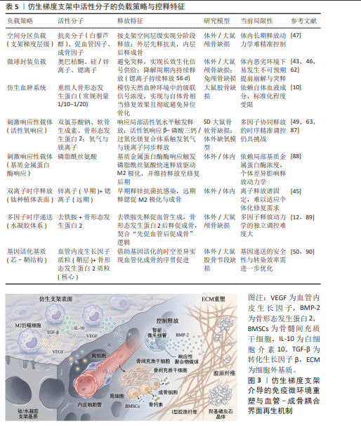
炎症、血管痂和硬骨重塑的时序过程,传统支架大多使用了大量超载的生长因子(骨形态发生蛋白2)。但是临床研究表明,大剂量重组人骨形态发生蛋白2会引发严重的神经根炎、椎体骨溶解、异位骨化等严重并发症[84],这就意味着因子递送策略要从“超载堆叠”变为“按需释药”。控释的核心思想就是模仿天然血肿环境中级联信号浓度[10]。研究发现用仿生血肿系统给大鼠注射常规剂量1/10到1/20的超低剂量重组人骨形态发生蛋白2,在大鼠股骨缺损模型中可以得到和自体骨相当的修复效果,并且没有出现异位骨化的现象[10]。
基于聚合物理化降解的“被动缓释”容易出现初期突释[85-86],已被证明在对抗体内巨噬细胞蛋白酶侵袭方面能力有限。目前公认的最有前景的途径就是创建智能响应性时空递送网络。以3D打印形成的软硬双层结构为载体,用活性氧响应型纳米水凝胶复合支架实现双氯芬酸钠、软骨生成素和骨形态发生蛋白2的分步释放,在骨和软骨缺损模型中可以诱导出抗炎、软骨形成和成骨的连续反应[49];另一种活性氧响应体系将β-磷酸三钙和过氧化镁结合起来,在炎症微环境中同时产生氧气和镁离子,从而达到改善局部缺氧状况并推进成骨目的[87]。基质金属蛋白酶响应性可注射水凝胶将基质金属蛋白酶可裂解的肽段整合进来,炎症刺激时快速释放磷脂酰丝氨酸来驱动巨噬细胞向M2表型转变,并维持药物释放到修复后
期[88]。时序逻辑设计中,由丝素纳米纤维和羟基磷灰石组成的仿生水凝胶,分别负载去铁胺和骨形态发生蛋白2,并且独立调节释放速率,很好地符合了“先促血管后促成骨”的生理规律[89]。另外一项研究用同轴静电纺丝制备了芯-鞘结构纤维水凝胶复合支架,在核心和鞘层分别递送血管内皮生长因子和骨形态发生蛋白2质粒,利用基因活化的时空不同来促进大鼠股骨节段缺损处的血管化成骨[50,90]。除此之外,针对感染性骨缺损这种更为复杂的病理情况,一种带有径向梯度孔径的3D打印钛合金支架也被研制出来,其外层大孔径区域复合药物负载的聚乳酸-羟基乙酸共聚物微球来实现利福平和莫西沙星的长效缓释,而内层小孔径区域则装载骨形态发生蛋白2,从而形成了从早期感染控制到后期骨再生的序贯治疗闭环[91]。从离子调控角度来讲,采用锌和锶双离子体系在钛种植体表面构建出的时序释放平台,依靠锌的早期抗菌效果和锶的远期免疫成骨作用,也体现出分阶段调节的实用意义[45]。目前智能响应性时空递送网络的构建策略已经呈现多样化趋势,空间分区负载、微球封装、仿生血肿系统、刺激响应性载体、双离子时序释放、多因子独立调控、基因活化基质等都是智能响应性时空递送网络的技术路径。表5从负载策略、控释类型、活性分子种类、释放特征、研究模型和目前的局限性等方面对上述策略进行了归纳和横向比较。图3为仿生梯度支架介导的免疫微环境重塑与血管-成骨耦合界面再生机制。
2.3.2 严苛体内微环境导致的释放动力学失效场景分析 尽管这些智能控释系统在实验室条件下获得了符合预期的顺序释放曲线,但将它直接推向临床仍存在落差。临床严重大段骨缺损区伴随着大出血冲刷、血肿机化以及极高浓度的巨噬细胞蛋白酶环境,依赖化学键断裂或材料溶胀的微球往往会发生不可预期的提前崩解与突释。如何抵抗体内恶劣环境干扰,实现原位释放动力学的绝对精准控制,是从理论迈向应用必须跨越的技术鸿沟。
2.4 大段骨缺损证据层级剖析与转化失败场景探讨
2.4.1 “小动物陷阱”与大段承重骨验证的断崖式落差 据估计,全世界每年因为创伤或者疾病导致的骨移植手术需要量达到220万次以上,但是自体骨移植由于供区并发症、来源受限等“金标准”的缺点,急需新一代仿生生物材料的介入[12-13,15,92]。目前对于啮齿类小型动物的非绝对承重模型(大鼠颅骨、兔桡骨等),无论是大鼠颅骨模型中先免疫后成骨的机制验证[47,77,93],还是奥巴枯酮缓释支架的成骨效果验证[46],亦或是负载抗菌肽支架对抗创伤性骨髓炎模型的研究[94],在概念验证层面都取得了很好的成果。
啮齿类动物模型虽然方便于机制研究,但是其骨骼大小、力学载荷环境以及愈合速度都与人类存在较大差别,大动物模型才是验证承重骨修复策略的金标准。大段负重长骨不但要保证材料和皮质骨具有相近的弹性模量[54],而且要承受很长的生理载荷疲劳期[95]。已有基于19 cm3超大体积羊胫骨缺损模型以及长时间的羊植入物表征库研究[96-98],很容易出现支架疲劳断裂或者引起严重的骨吸收等“失败场景”。除极少数用高强度刚性骨架复合高剂量生长因子的情况之外,临床级别承重验证很少。小动物效能数据不可靠的外推,才是造成目前临床转化停滞的最大假象。
2.4.2 临床转化所面临的病理特异性与系统安全性障碍 尽管实验室层面的梯度支架具有很大的机制潜力,但是要实现临床转化还存在着三大系统性障碍。首要的难题就是“规模生物制造”和“力学-生物活性平衡”的工程矛盾。尽管新兴的3D打印和复合纺丝技术可以提供解决方案,但是怎样在不破坏脆弱的生物活性成分(如脂质体药物[47]、导电互联网络[70])的前提下,使支架在功能机械负荷下保持长期可靠性,实现工业级的规模化生产,仍然是一个重大的转化壁垒[77,99];其次就是患者的病理微环境异质性给植入物效能造成的威胁。实验室得到的最佳体外释放曲线或者孔径参数,大多是在健康动物微环境中测定出来的,一旦将它植入到伴有糖尿病或者严重衰老的宿主体内,病灶区积聚的衰老细胞会分泌出大量的衰老相关分泌表型因子,使得原本设计好的M2免疫调控支架完全失效[100]。针对性的解决办法也层出不穷,有研究者研制出以控释淫羊藿苷、Mg2?为载体的支架来促进骨整合,也有研究者制备出以黑磷光热水凝胶为载体的方案,用来逆转糖尿病所造成的骨稳态失衡,为病理状态下修复提供新思路[101],这就意味着同一种支架在健康人和患者身上会产生完全不同的修复结果。对于特殊但是普遍的病理场景进行支架免疫设计的定制化,是目前领域内需要解决的“盲区”。最后,由于缺少长期系统性的安全性数据,所以监管批准很难被实现。加速降解研究显示,不同的材料组成、孔径会造成非常复杂的降解动力学[102];对纳米硅体内外降解的追踪表明,材料表面化学状态会对它在体内归转产生很大影响[103]。目前对于复杂的多相梯度支架,在体评估大多只停留在3-6个月,缺少长达1年以上的长期安全性评价。在没有建立起包含力学疲劳、病理微环境反应以及长时间毒理学评价的完整闭环之前,大规模临床应用还存在很大困难。
| [1] YU Y, CUI H, ZHANG C, et al. Human nail bed extracellular matrix facilitates bone regeneration via macrophage polarization mediated by the JAK2/STAT3 pathway. J Mater Chem B. 2020;8(18): 4067-4079. [2] COOKE ME, RAMIREZ-GARCIALUNA JL, RANGEL-BERRIDI K, et al. 3D Printed Polyurethane Scaffolds for the Repair of Bone Defects. Front Bioeng Biotechnol. 2020;8:557215. [3] TETSWORTH KD, BURNAND HG, HOHMANN E, et al. Classification of Bone Defects: An Extension of the Orthopaedic Trauma Association Open Fracture Classification. J Orthop Trauma. 2021;35(2):71-76. [4] YU D, SHEN W, DAI J, et al. Treatment of large bone defects in load-bearing bone: traditional and novel bone grafts. J Zhejiang Univ Sci B. 2025;26(5):421-447. [5] HUBBE U, BEISER S, KUHN S, et al. A fully ingrowing implant for cranial reconstruction: Results in critical size defects in sheep using 3D-printed titanium scaffold. Biomater Adv. 2022; 136:212754. [6] LEI W, WU Y, YUAN H, et al. Establishing rabbit critical-size bone defects to evaluate the bone-regeneration potential of porous calcium phosphate ceramics. Front Bioeng Biotechnol. 2025;12:1524133. [7] LIU H, LIU J, SUN C, et al. Design and Fabrication of Biomimetic Gradient Bone Tissue Engineering Scaffolds: Evolution from Single-Gradient to Multi-Gradient. Gels. 2026;12(2):131. [8] KOBBE P, LAUBACH M, HUTMACHER DW, et al. Convergence of scaffold-guided bone regeneration and RIA bone grafting for the treatment of a critical-sized bone defect of the femoral shaft. Eur J Med Res. 2020;25(1):70. [9] YANG YP, LABUS KM, GADOMSKI BC, et al. Osteoinductive 3D printed scaffold healed 5 cm segmental bone defects in the ovine metatarsus. Sci Rep. 2021;11(1):6704. [10] WOLOSZYK A, AGUILAR L, PEREZ L, et al. Biomimetic hematoma delivers an ultra-low dose of rhBMP-2 to successfully regenerate large femoral bone defects in rats. Biomater Adv. 2023;148:213366. [11] WANG Q, HUANG Z, HUANG X, et al. Reparative effect of super active platelet combined with allogeneic bone for large bone defects. Artif Organs. 2021;45(10):1219-1228. [12] 周文洁,王凤,吴轶群.上颌窦底提升骨增量材料的专家共识:骨代用品[J].中国口腔种植学杂志,2023, 28(1):3-8. [13] SCHMIDT AH. Autologous bone graft: Is it still the gold standard? Injury. 2021;52 Suppl 2:S18-S22. [14] 蔡元庆,刘谟震,李忠海.同种异体骨移植材料在脊柱融合中的应用[J].中国组织工程研究,2023,27(16): 2571-2579. [15] 唐国柯,文根,刘彦斌,等.骨缺损修复生物材料的研究进展[J].中华骨与关节外科杂志,2023,16(2):185-192. [16] 郭学德,孟红亚,王辞山,等.骨搬移术中植入硫酸钙对促进对合端愈合的效果观察[J].中国骨伤,2022, 35(1):85-89. [17] WU C, LIU K, REN S, et al. Outcome of limb lengthening as a treatment for shortening following successful replantation of traumatic leg amputation: experience with 21 patients. Front Surg. 2025;12:1686865. [18] CASTRISOS G, GONZALEZ MATHEUS I, SPARKS D, et al. Regenerative matching axial vascularisation of absorbable 3D-printed scaffold for large bone defects: A first in human series. J Plast Reconstr Aesthet Surg. 2022;75(7):2108-2118. [19] XIE X, WANG Y, LI Z, et al. Recent advances in gradient biomimetic scaffolds for tendon-bone interface regeneration. Front Bioeng Biotechnol. 2025;13:1629816. [20] FANG L, LIN X, XU R, et al. Advances in the Development of Gradient Scaffolds Made of Nano-Micromaterials for Musculoskeletal Tissue Regeneration. Nanomicro Lett. 2024;17(1):75. [21] XIAO F, YE JH, HUANG CX, et al. Gradient gyroid Ti6Al4V scaffolds with TiO2 surface modification: Promising approach for large bone defect repair. Biomater Adv. 2024;161:213899. [22] LI C, OUYANG L, ARMSTRONG JPK, et al. Advances in the Fabrication of Biomaterials for Gradient Tissue Engineering. Trends Biotechnol. 2021;39(2):150-164. [23] 张凯静,李春年,李一卓,等.不同骨修复材料对根尖骨缺损治疗效果的网状Meta分析[J].中国组织工程研究,2026,30(26):6946-6951. [24] KOUSHIK TM, MILLER CM, ANTUNES E. Bone Tissue Engineering Scaffolds: Function of Multi-Material Hierarchically Structured Scaffolds. Adv Healthc Mater. 2023;12(9):e2202766. [25] TONTI OR, LARSON H, LIPP SN, et al. Tissue-specific parameters for the design of ECM-mimetic biomaterials. Acta Biomater. 2021;132:83-102. [26] ZAMANI Y, AMOABEDINY G, MOHAMMADI J, et al. 3D-printed poly(Ɛ-caprolactone) scaffold with gradient mechanical properties according to force distribution in the mandible for mandibular bone tissue engineering. J Mech Behav Biomed Mater. 2020;104:103638. [27] SARIKHANI B, IMANI R, RAZFAR MR. A review on gradient and hierarchical scaffolds: mechanical design and fabrication via electrospinning and extrusion techniques. Biomed Mater. 2026;21(1):012004. [28] ZHONG Y, CAO X, HUANG M, et al. Biomimetic bone cartilage scaffolds based on trilayer methacrylated hydroxyapatite/GelMA composites for full-thickness osteochondral regeneration. Int J Biol Macromol. 2025; 298:139860. [29] LIU X, WANG B, MA J, et al. 3D-printed heterogeneous biomimetic scaffold utilizing TEMPO-oxidized and mineralized bacterial cellulose nanofibers for osteochondral regeneration. Carbohydr Polym. 2025;370: 124366. [30] 姜洪涛,薛鑫鑫,聂明洋,等.3D打印辅助自体髂骨移植修复足部骨缺损[J].中国矫形外科杂志,2026, 34(3):274-277. [31] KAMBOJ N, KAZANTSEVA J, RAHMANI R, et al. Selective laser sintered bio-inspired silicon-wollastonite scaffolds for bone tissue engineering. Mater Sci Eng C Mater Biol Appl. 2020;116:111223. [32] LIU ZZ, XU NY, WANG ML, et al. Physical confinement in alginate cryogels determines macrophage polarization to a M2 phenotype by regulating a STAT-related mRNA transcription pathway. Biomater Sci. 2022;10(9):2315-2327. [33] FEROZ S, MUHAMMAD N, ULLAH R, et al. Mechanical properties, and in vitro biocompatibility assessment of biomimetic dual layered keratin/ hydroxyapatite scaffolds. Front Bioeng Biotechnol. 2023;11:1304147. [34] LIU F, RAN Q, ZHAO M, et al. Additively Manufactured Continuous Cell-Size Gradient Porous Scaffolds: Pore Characteristics, Mechanical Properties and Biological Responses In Vitro. Materials (Basel). 2020;13(11):2589. [35] WANG W, XIONG Y, ZHAO R, et al. A novel hierarchical biofunctionalized 3D-printed porous Ti6Al4V scaffold with enhanced osteoporotic osseointegration through osteoimmunomodulation. J Nanobiotechnology. 2022;20(1):68. [36] YANG X, WU L, LI C, et al. Synergistic Amelioration of Osseointegration and Osteoimmunomodulation with a Microarc Oxidation-Treated Three-Dimensionally Printed Ti-24Nb-4Zr-8Sn Scaffold via Surface Activity and Low Elastic Modulus. ACS Appl Mater Interfaces. 2024;16(3):3171-3186. [37] SONG X, LIU F, QIU C, et al. Nanosurfacing Ti alloy by weak alkalinity-activated solid-state dewetting (AAD) and its biointerfacial enhancement effect. Mater Horiz. 2021;8(3):912-924. [38] CAMARERO-ESPINOSA S, CARLOS-OLIVEIRA M, LIU H, et al. 3D Printed Dual-Porosity Scaffolds: The Combined Effect of Stiffness and Porosity in the Modulation of Macrophage Polarization. Adv Healthc Mater. 2022;11(1):e2101415. [39] MOORE MJ, LAM YT, SANTOS M, et al. Evaluation of the Immune Response to Chitosan-graft-poly(caprolactone) Biopolymer Scaffolds. ACS Biomater Sci Eng. 2023;9(6): 3320-3334. [40] WEN Z, LI S, LIU Y, et al. An engineered M2 macrophage-derived exosomes-loaded electrospun biomimetic periosteum promotes cell recruitment, immunoregulation, and angiogenesis in bone regeneration. Bioact Mater. 2025;50:95-115. [41] LI M, WU H, GAO K, et al. Smart Implantable Hydrogel for Large Segmental Bone Regeneration. Adv Healthc Mater. 2024;13(32):e2402916. [42] ZHANG H, CAI Q, ZHU Y, et al. A simple hydrogel scaffold with injectability, adhesivity and osteogenic activity for bone regeneration. Biomater Sci. 2021;9(3):960-972. [43] LIANG W, GAO M, LOU J, et al. Integrating silicon/zinc dual elements with PLGA microspheres in calcium phosphate cement scaffolds synergistically enhances bone regeneration. J Mater Chem B. 2020;8(15):3038-3049. [44] LEI L, WEI Y, WANG Z, et al. Core-Shell Bioactive Ceramic Robocasting: Tuning Component Distribution Beneficial for Highly Efficient Alveolar Bone Regeneration and Repair. ACS Biomater Sci Eng. 2020;6(4):2376-2387. [45] WU B, TANG Y, YAO K, et al. Ion-incorporated titanium implants for staged regulation of antibacterial activity and immunoregulation-mediated osteogenesis. Nanoscale. 2024;16(14): 7167-7184. [46] YUAN X, YAO F, LI D, et al. Synergistic enhancement of bone regeneration by obacunone-loaded peg-based composite scaffolds. J Biomater Appl. 2026;40(9):1153-1164. [47] MA T, LI Y, LI A, et al. Resveratrol-liposomes loaded polyvinyl alcohol-chitosan fiber scaffolds mediate bone regeneration via autophagy: synergizing angiogenesis and immunomodulation. Carbohydr Polym. 2026;378:124945. [48] WANG W, LI Z, ZHANG S, et al. A Pseudo-Mytilus Edulis Foot Protein-Based Hydrogel Adhesive with Osteo-Vascular-Immune Coupling Effects for Osteoporotic Bone-Implant Integration. Adv Mater. 2026;38(6):e11840. [49] WANG X, WU S, LI R, et al. ROS-Activated Nanohydrogel Scaffolds with Multi-Factors Controlled Release for Targeted Dual-Lineage Repair of Osteochondral Defects. Adv Sci (Weinh). 2025;12(20):e2412410. [50] ZHAO P, TIAN M, HE H, et al. Development and in vitro/in vivo evaluation of a triply periodic minimal surface-based 3D-printed anti-infective bone scaffold with spatiotemporally controlled osteogenesis. J Mater Chem B. 2025; 13(47):15397-15412. [51] PERIER-METZ C, CIPITRIA A, HUTMACHER DW, et al. An in silico model predicts the impact of scaffold design in large bone defect regeneration. Acta Biomater. 2022;145:329-341. [52] HOU G, LIU B, TIAN Y, et al. An innovative strategy to treat large metaphyseal segmental femoral bone defect using customized design and 3D printed micro-porous prosthesis: a prospective clinical study. J Mater Sci Mater Med. 2020;31(8):66. [53] BU S, YAN S, WANG R, et al. In Situ Precipitation of Cluster and Acicular Hydroxyapatite onto Porous Poly(γ-benzyl-l-glutamate) Microcarriers for Bone Tissue Engineering. ACS Appl Mater Interfaces. 2020;12(11):12468-12477. [54] 于爱华,徐伟,路新,等.基于选择性激光熔化技术的骨缺损修复用梯度多孔钛支架的研制与表征[J].中国有色金属学报(英文版),2023,33(6):1755-1767. [55] 孟增东,朱斌,张亚楠,等.不同孔隙率多孔ZnO/羟基磷灰石复合材料的力学性能与生物相容性[J].中国组织工程研究,2022,26(22):3498-3504. [56] WANG C, XU D, LI S, et al. Effect of Pore Size on the Physicochemical Properties and Osteogenesis of Ti6Al4V Porous Scaffolds with Bionic Structure. ACS Omega. 2020;5(44):28684-28692. [57] REZAPOURIAN M, KAMBOJ N, JASIUK I, et al. Biomimetic design of implants for long bone critical-sized defects. J Mech Behav Biomed Mater. 2022;134:105370. [58] ZHU H, WANG J, WANG S, et al. Additively manufactured bioceramic scaffolds based on triply periodic minimal surfaces for bone regeneration. J Tissue Eng. 2024;15: 20417314241244997. [59] SHEN M, LI Y, LU F, et al. Bioceramic scaffolds with triply periodic minimal surface architectures guide early-stage bone regeneration. Bioact Mater. 2023;25:374-386. [60] WANG Y, LIU Y, CHEN S, et al. Enhancing bone regeneration through 3D printed biphasic calcium phosphate scaffolds featuring graded pore sizes. Bioact Mater. 2024;46:21-36. [61] XIAO F, REN GH, PAN CY, et al. Design and in vitro evaluation of gradient Schwarz Primitive scaffolds with thin-board integration for bone implants. Biomater Adv. 2025;177:214412. [62] FUXIANG S, LI ZLAD, ZHILI W, et al. Strontium oxide-functionalized 3D-printed polycaprolactone/β-tricalcium phosphate nanocomposite scaffolds with osteogenic microenvironment remodeling for accelerated bone regeneration. J Mech Behav Biomed Mater. 2025;172:107146. [63] PENG Z, WANG C, LIU C, et al. 3D printed polycaprolactone/beta-tricalcium phosphate/magnesium peroxide oxygen releasing scaffold enhances osteogenesis and implanted BMSCs survival in repairing the large bone defect. J Mater Chem B. 2021;9(28):5698-5710. [64] SALAMANCA E, PAN YH, SUN YS, et al. Magnesium Modified β-Tricalcium Phosphate Induces Cell Osteogenic Differentiation In Vitro and Bone Regeneration In Vivo. Int J Mol Sci. 2022;23(3):1717. [65] CHEN Y, HAO M, BOUSSO I, et al. Reliable Fabrication of Mineral-Graded Scaffolds by Spin-Coating and Laser Machining for Use in Tendon-to-Bone Insertion Repair. Adv Healthc Mater. 2024;13(31):e2402531. [66] MICALIZZI S, BEVILACQUA A, STEFANO LD, et al. Hybrid additive manufacturing and data-guided design optimization for graded anterior cruciate ligament engineering. Biomed Mater. 2026;21(1). doi: 10.1088/1748-605X/ae30bc [67] PAIS A, ALVES JL, BELINHA J. Predicting trabecular arrangement in the proximal femur: An artificial neural network approach for varied geometries and load cases. J Biomech. 2023;161:111860. [68] PAZHAMANNIL RV, ALKHEDHER M. Advances in additive manufacturing for bone tissue engineering: materials, design strategies, and applications. Biomed Mater. 2025;20(1):012002. [69] ALTUNBEK M, AFGHAH SF, FALLAH A, et al. Design and 3D Printing of Personalized Hybrid and Gradient Structures for Critical Size Bone Defects. ACS Appl Bio Mater. 2023;6(5):1873-1885. [70] LIU Y, LI L, ZHAN X, et al. 3D-printed conductive hydrogel scaffolds for bone regeneration: Electromechanical coupling, neurovascular integration, and immunomodulatory strategies. Biomater Adv. 2026;179:214497. [71] CHEN H, LIU Y, WANG C, et al. Design and properties of biomimetic irregular scaffolds for bone tissue engineering. Comput Biol Med. 2021;130:104241. [72] MAHON OR, BROWE DC, GONZALEZ-FERNANDEZ T, et al. Nano-particle mediated M2 macrophage polarization enhances bone formation and MSC osteogenesis in an IL-10 dependent manner. Biomaterials. 2020; 239:119833. [73] SHI W, FENG Y, TANG J, et al. A Genetically Engineered “Reinforced Concrete” Scaffold Regulates the N2 Neutrophil Innate Immune Cascade to Repair Bone Defects. Adv Healthc Mater. 2024;13(18):e2304585. [74] XUAN Y, GUO Y, LI L, et al. 3D-printed bredigite scaffolds with ordered arrangement structures promote bone regeneration by inducing macrophage polarization in onlay grafts. J Nanobiotechnology. 2024;22(1):102. [75] XIAO L, LIU H, HUANG H, et al. 3D nanofiber scaffolds from 2D electrospun membranes boost cell penetration and positive host response for regenerative medicine. J Nanobiotechnology. 2024;22(1):322. [76] WU H, DONG H, TANG Z, et al. Electrical stimulation of piezoelectric BaTiO3 coated Ti6Al4V scaffolds promotes anti-inflammatory polarization of macrophages and bone repair via MAPK/JNK inhibition and OXPHOS activation. Biomaterials. 2023;293:121990. [77] SHRESTHA S, JIANG N, AMARAL AC, et al. Piezoelectric Smart Biomaterials for Craniomaxillofacial Bone-Regeneration Application. ACS Biomater Sci Eng. 2026; 12(3):1318-1333. [78] HUANG X, CHEN M, WU H, et al. Macrophage Polarization Mediated by Chitooligosaccharide (COS) and Associated Osteogenic and Angiogenic Activities. ACS Biomater Sci Eng. 2020;6(3):1614-1629. [79] YANG L, LIU Y, YANG Y, et al. Functional hollow porous microfiber scaffold with BMP-2 for macrophages polarization and bone regeneration. Biomater Adv. 2025;177:214425. [80] ZHANG J, TONG D, SONG H, et al. Osteoimmunity-Regulating Biomimetically Hierarchical Scaffold for Augmented Bone Regeneration. Adv Mater. 2022;34(36): e2202044. [81] PENG H, LING T, ZHANG Y, et al. Nanowhiskers Orchestrate Bone Formation and Bone Defect Repair by Modulating Immune Cell Behavior. ACS Appl Mater Interfaces. 2023;15(7):9120-9134. [82] WANG B, XIE X, JIANG W, et al. Osteoinductive micro-nano guided bone regeneration membrane for in situ bone defect repair. Stem Cell Res Ther. 2024;15(1):135. [83] YU M, CHEN L, CAI X, et al. Endogenous Metal Ion-Enriched Immunostimulating Biomimetic Scaffold Improves In Situ Bone Regeneration. Adv Mater. 2026;38(11):e07071. [84] TANNOURY CA, AN HS. Complications with the use of bone morphogenetic protein 2 (BMP-2) in spine surgery. Spine J. 2014;14(3):552-559. [85] YOO J, WON YY. Phenomenology of the Initial Burst Release of Drugs from PLGA Microparticles. ACS Biomater Sci Eng. 2020; 6(11):6053-6062. [86] PEI Y, WANG J, KHALIQ NU, et al. Development of poly(lactide-co-glycolide) microparticles for sustained delivery of meloxicam. J Control Release. 2023;353:823-831. [87] MARTIN JR, HOWARD MT, WANG S, et al. Oxidation-Responsive, Tunable Growth Factor Delivery from Polyelectrolyte-Coated Implants. Adv Healthc Mater. 2021;10(9):e2001941. [88] ZHANG M, YU T, LI J, et al. Matrix Metalloproteinase-Responsive Hydrogel with On-Demand Release of Phosphatidylserine Promotes Bone Regeneration Through Immunomodulation. Adv Sci (Weinh). 2024; 11(20):e2306924. [89] CHENG W, DING Z, ZHENG X, et al. Injectable hydrogel systems with multiple biophysical and biochemical cues for bone regeneration. Biomater Sci. 2020;8(9):2537-2548. [90] HE S, FANG J, ZHONG C, et al. Spatiotemporal Delivery of pBMP2 and pVEGF by a Core-Sheath Structured Fiber-Hydrogel Gene-Activated Matrix Loaded with Peptide-Modified Nanoparticles for Critical-Sized Bone Defect Repair. Adv Healthc Mater. 2022;11(21):e2201096. [91] ARAGÓN J, FEOLI S, IRUSTA S, et al. Composite scaffold obtained by electro-hydrodynamic technique for infection prevention and treatment in bone repair. Int J Pharm. 2019;557:162-169. [92] ARCHUNAN MW, PETRONIS S. Bone Grafts in Trauma and Orthopaedics. Cureus. 2021;13(9):e17705. [93] PAN CT, HSU WH, CHENG YS, et al. A New Design of Porosity Gradient Ti-6Al-4V Encapsulated Hydroxyapatite Dual Materials Composite Scaffold for Bone Defects. Micromachines (Basel). 2021;12(11):1294. [94] ZHANG Y, XU T, LI T, et al. A three-phase strategy of bionic drug reservoir scaffold by 3D printing and layer-by-layer modification for chronic relapse management in traumatic osteomyelitis. Mater Today Bio. 2024;29:101356. [95] BOZZO A, AYSOLA V, YEUNG CM, et al. Distraction Osteogenesis Reconstruction Following Resection of Bone Sarcomas: Surgical, Functional, and Oncological Outcomes from a Prospective Trial Analysis. J Bone Joint Surg Am. 2024;106(13):1205-1211. [96] HENKEL J, MEDEIROS SAVI F, et al. Scaffold-guided bone regeneration in large volume tibial segmental defects. Bone. 2021;153:116163. [97] FINZE R, LAUBACH M, RUSSO SERAFINI M, et al. Histological and Immunohistochemical Characterization of Osteoimmunological Processes in Scaffold-Guided Bone Regeneration in an Ovine Large Segmental Defect Model. Biomedicines. 2023;11(10):2781. [98] SPARKS DS, SAIFZADEH S, SAVI FM, et al. A preclinical large-animal model for the assessment of critical-size load-bearing bone defect reconstruction. Nat Protoc. 2020;15(3): 877-924. [99] GAROT C, BETTEGA G, PICART C. Additive Manufacturing of Material Scaffolds for Bone Regeneration: Toward Application in the Clinics. Adv Funct Mater. 2020;31(5): 2006967. [100] ZHENG J, ZHAO H, DONG E, et al. Additively-manufactured PEEK/HA porous scaffolds with highly-controllable mechanical properties and excellent biocompatibility. Mater Sci Eng C Mater Biol Appl. 2021;128:112333. [101] ZHANG Y, QI G, ZHANG L, et al. Black phosphorus-based photothermal-responsive hydrogel enhanced osteoporotic bone injury regeneration by alleviating oxidative stress and remodeling bone homeostasis. J Nanobiotechnology. 2026;24(1):238. [102] DASKALAKIS E, HASSAN MH, OMAR AM, et al. Accelerated Degradation of Poly-ε-caprolactone Composite Scaffolds for Large Bone Defects. Polymers (Basel). 2023;15(3):670. [103] FATIMA N, SALEHI H, CUETO-DÍAZ EJ, et al. Nanostructured Porous Silicon for Bone Tissue Engineering: Kinetics of Particle Degradation and Si-Controlled Release. J Funct Biomater. 2023; 14(10):493. |
| [1] | 覃国忠, 李 伟, 牛军杰, 李义凯, 林文平, 李丹丹, 李 军, 柳 鑫, 曾凡宇. 聚己内酯/β-磷酸三钙支架联合骨髓间充质干细胞与骨形态发生蛋白2修复股骨缺损[J]. 中国组织工程研究, 2026, 30(30): 7801-7808. |
| [2] | 申 震, 黄梓越, 和智娟, 王艺婷, 陈奇刚, 耿春梅, 黄雅静, 吴祖贵. 骨诱导膜中H型血管动态表达及耦合骨生成修复大段骨缺损[J]. 中国组织工程研究, 2025, 29(28): 5950-5956. |
| [3] | 张治博, 王兆林, 王志刚, 李 朋, 姜建浩, 张 锴, 杨淑野, 杜刚强. Ilizarov骨搬移联合抗生素骨水泥促进胫骨大段骨缺损的对接点愈合[J]. 中国组织工程研究, 2025, 29(10): 2038-2043. |
| [4] | 温星花, 丁焕文, 成 凯, 闫晓楠, 彭元昊, 王宇宁, 刘 康, 张挥武. 比格犬股骨大段骨缺损髓内钉固定方案设计的三维有限元建模分析[J]. 中国组织工程研究, 2023, 27(9): 1371-1376. |
| [5] | 丹尼尔•赛德尔丁, 黄晓夏, 陈江涛, 田 征, 艾克拜尔•尤努斯. 3D打印假体重建骨肿瘤术后大段骨缺损[J]. 中国组织工程研究, 2023, 27(29): 4628-4634. |
| [6] | 申 震, 郭 英, 姜自伟, 张 严, 李紫阁, 陈泽华, 叶翔凌, 陈国茜. 基于骨组织工程技术比较骨碎补总黄酮两种给药方式修复大鼠 大段骨缺损模型的效果[J]. 中国组织工程研究, 2022, 26(27): 4346-4352. |
| [7] | 鲍小刚,许国华. 兔胫骨大段完全骨缺损的解剖学模型构建[J]. 中国组织工程研究, 2016, 20(24): 3508-3515. |
| [8] | 王天胜,滕寿发,王 法,郭 利,丁海蛟,刘 鹏,张英霞,曹 宇,王 湛. 生物陶瓷复合骨髓基质干细胞及骨形态发生蛋白修复兔大段桡骨缺损[J]. 中国组织工程研究, 2015, 19(3): 347-351. |
目前临床上常用的修复方法对于大段骨缺损都存在根本性缺陷。自体骨移植是“金标准”,但来源少、供区并发症风险大[11-13]。同种异体骨一直受到免疫原性及骨整合慢的制约[14-15]。以Ilizarov技术为代表的骨搬移术虽然可以实现骨再生,但是对于长节段缺损的治疗周期很长,患者依从性差,而且对合端不愈合的风险明显增加[16-17]。这些临床困境的主要原因就是现有的方法都不能同时满足大段骨缺损修复对于力学匹配、骨传导、骨诱导以及局部免疫微环境重塑的综合要求。一项临床研究给这个问题提供了新的启示,用3D打印的医用级聚己内酯磷酸三钙(mPCL-TCP)仿生骨梯度支架加上血管化的游离皮质骨膜瓣修复了36 cm长的胫骨大段缺损[18],不但证明了仿生梯度支架在极端尺寸骨缺损重建上的可行性,也突出了结构支撑和血供重建的协同作用。但是该策略的成功很大程度上取决于自体骨膜瓣的活性,其免疫调节和成骨诱导过程都是被动的,缺少对局部微环境主动、精确的干预。
由于传统方法的局限性,新兴的仿生梯度支架策略虽然已经初见端倪,但是仍然存在改进的空间,研究方向应该回归到天然骨组织的精妙设计上来寻找答案。天然骨本身就是一种高度优化的仿生模式,从致密皮质骨到多孔松质骨,在结构、力学模量以及生化组分上都存在着连续的梯度变化[19-20]。多维、各向异性的梯度特征使它在承受高强度载荷的时候,还能保持高效营养代谢和细胞相互作用[21-22]。但是早期研究开发的均质或者多孔结构单一的支架,很难再现由宏观到微观的异质性,从而在力学适配、营养物质传递以及引导特定细胞行为等方面效果较差,不能解决大段骨缺损的修复难题[23-26]。
近年来,随着增材制造等先进加工技术的成熟,具有仿生梯度结构的骨组织工程支架为解决上述物理屏障开辟了新路径[22,27]。通过逐层堆叠[28]、3D打印[2,29-31]、冷冻干燥技术[32-33]、静电纺丝与挤出技术结合等手段[27],研究者已能构建孔隙率、孔径乃至化学成分呈连续或阶梯变化的梯度支架[26,34],一方面较好地解决了力学支撑和传质效率之间的矛盾,另一方面梯度结构本身就具有主动调节宿主免疫反应的功能。多项研究证实,具有特定梯度微孔拓扑结构的钛合金或聚合物支架,能通过影响细胞铺展、力学信号转导,进而促使局部巨噬细胞由促炎(M1)表型向促修复(M2)表型极化,减轻慢性炎症,形成利于血管长入和成骨分化的免疫微环境[35-39]。
天然骨愈合是程序非常精确的级联事件,从早期的血肿形成、炎症反应,到中期的血管生成和软硬骨痂形成,再到晚期的骨重塑,每一个步骤都在准确的时间窗口内被特定的生物信号所推动[40]。为了使修复过程和内在节律相适应,在支架不同层次上负载骨生长因子(如骨形态发生蛋白2[9-10,41])、生物活性离子(如锌、锶、硅[42-45])或小分子药物(如奥巴枯酮[46]、白藜芦醇[47])。研究者用设计出的锌/锶双离子释放系统,通过早期释放锌离子来抗菌,同时长期释放锶离子来促进成骨的协同分阶段调控[45]。这种时空控释功能可以和梯度支架固有的物理引导、免疫调节效应产生深层次的协同,从而精准地控制大段骨缺损愈合过程中炎症消退、血管重建、成骨分化等各个环节的时间顺序[48-49],还可以利用时序策略来完成最初的感染控制,进而达到骨重建的目的[50]。
尽管相关研究方兴未艾,但当前大多数研究把梯度结构仿生、免疫微环境调控和活性因子时空递送看作3个相对独立的模块,没有从一个有机协同的整体论角度出发,对三者之间深层次的级联作用机制进行系统解构[51]。该研究对这3个因素在大段骨缺损修复过程中的时序交互和逻辑闭环进行详细分析,找出破解血供重建、力学适配、微环境失稳3个瓶颈的内在机制,整理出目前研究存在的争议点以及临床转化障碍,希望可以为设计精确调控大段骨缺损再生的智能化仿生支架提供理论框架和可行的研究路线。
中国组织工程研究杂志出版内容重点:干细胞;骨髓干细胞;造血干细胞;脂肪干细胞;肿瘤干细胞;胚胎干细胞;脐带脐血干细胞;干细胞诱导;干细胞分化;组织工程
1.1 资料来源
1.1.1 检索人及检索时间 由王浩和陈仁场于2025年7月至2026年3月完成文献检索。
1.1.2 文献检索时限 2017年1月至2026年3月。
1.1.3 检索数据库 PubMed、Embase和Web of Science等外文数据库及中国知网、万方及维普等中文数据库。
1.1.4 检索词 中文检索词为“仿生骨梯度支架,大段骨缺损,免疫调控,骨再生,微环境重塑,控释系统,骨组织工程”;英文检索词为“Bone tissue engineering,bionic gradient bone scaffold,critical-sized bone defect,immunomodulation,bone regeneration,microenvironment remodeling,controlled release system”。
1.1.5 检索文献类型 论著、综述和病例报告。
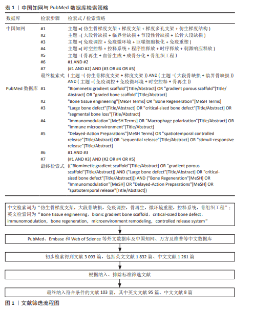
1.1.6 检索策略 以中国知网与PubMed数据库等为示例制定检索策略,通过多检索词的OR、AND逻辑组合筛选相关文献,见表1。
1.1.7 检索文献量 初步检索得到文献3 093篇,包括英文文献1 832篇、中文文献1 261篇。
1.2 入选标准
1.2.1 纳入标准 ①研究主题契合度:文献必须直接涉及到仿生骨梯度支架(成分梯度、结构梯度、力学梯度或者复合梯度)的设计、制备、表征和生物学效应评价,主要应用于大段骨缺损或者临界尺寸骨缺损的修复;② 机制与策略相关性:支架物理化学信号对巨噬细胞极化、免疫微环境重塑的调控作用;支架梯度结构引导血管新生和成骨分化耦合的机制;活性因子、药物、离子在支架中的时空控释策略及对骨再生的时序调控;③研究类型与质量:优先选择研究设计严谨、数据完整、结论可靠的文献,分为原始研究、体外细胞机制研究和临床转化案例,原始研究包括体内动物实验(缺损模型类型、尺寸、观察周期和主要结局指标)、体外细胞机制研究(细胞类型、干预条件和检测指标),高质量综述包括SCI收录期刊或中文核心期刊的系统综述、荟萃分析或者权威述评,临床转化案例为经伦理审批的临床前大动物研究或者早期临床试验报告;④时效性与语言:文献发表时间2017年1月至2026年3月,语言限定为中文或者英文。
1.2.2 排除标准 ①主题偏离:内容只包含非梯度均质支架、单纯骨水泥、金属内固定物、非降解材料,没有梯度结构或者免疫调节的文献;研究对象为非骨组织(软骨、肌腱、神经)修复,没有与骨缺损修复相关联的文献;②设计缺陷与数据不完整:缺少明确的实验分组、样本量过小(每组n < 3)或者统计学方法缺失的原始研究;动物模型不符合大段骨缺损的定义(长骨缺损长度小于2 cm或者没有达到临界尺寸标准,颅骨缺损直径小于3 cm);只报告定性观察而没有定量数据(组织形态学评分、骨体积分数、血管密度等)的研究;③信息重复与低质量文献:同一研究团队在相似模型、干预条件下重复发表的文献,只保留数据最完整的那一篇;发表在非同行评议期刊、会议摘要、学位论文、掠夺性期刊上的文献;经CRAIS或者AIGC检测工具评价,存在明显的学术不端风险或者AI生成内容占比超过期刊限制的文献;④内容陈旧:2017年以前发表的文献(经典里程碑文献除外),后续有更新、更全面的研究已经覆盖其主要观点的文献(经典文献通过手工检索补充,不计入纳入文献总数)。
1.3 文献质量评价和筛选 共检索到3 093篇文献,初筛去除重复文献之后,对剩余文献的标题、摘要进行筛选,不能判断文献相关性的文献则精读全文,选取和主题更加相关的文献;最终纳入符合条件的文献103篇,其中英文文献95篇、中文文献8篇,文献筛选过程严格按照规定的检索与筛选步骤执行(图1)。
1.4 生成式人工智能技术(AIGC)在文中的应用 在论文撰写期间,使用AI模型“nano banana”(纳米香蕉)[开发商:谷歌(Google)]的绘图功能,辅助生成论文中机制可视化图表等。参考提示词:生成一张学术期刊风格的图形摘要(graphical abstract),矢量扁平化信息图风格,白底,极简配色(深灰+蓝+绿+橙点缀),无照片质感,无卡通感,无3D,无主标题。画面横向16∶9。
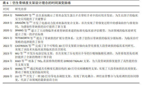
3.1 既往他人在该领域研究的贡献和存在的问题 大段骨缺损修复缺少有效的办法,一直是骨科和再生医学领域的重大临床难题。近些年来,仿生骨梯度支架研究由原来的“被动微环境调节”转向了“主动微环境调节”。以往的研究者在多孔结构设计、巨噬细胞极化诱导以及单个促进骨形成因子递送方面取得了开创性的成果,证明了模仿天然骨的宏微观异质性结构对于引导局部骨组织再生是有益的。但是目前该领域还存在着严重的孤岛化研究倾向,大部分研究只是局部优化物理梯度或者分子递送,没有实现物理结构、免疫微环境、时空控释三者之间的深度协同;其次,梯度参数的设计缺少标准化的工程共识,很难准确地匹配到不同的解剖部位和不同载荷环境下的力学和生物学双重需求;再次,临床病理适配性盲区明显,现有的设计大多是以健康动物模型为基础,没有充分考虑老年、糖尿病等复杂宿主病理状态下局部异常的免疫微环境差异;最后,时空控释系统的体内药代动力学效果也不理想,多因素的序贯精准释放控制较弱,长期降解稳定性差,导致基础成果向临床获批转化的壁垒很高。仿生骨梯度支架设计理念的时间演变脉络见表6。
3.2 该综述区别于他人他篇的特点 此文章的最大特色就是冲破了以往只集中于材料制备工艺或者基于骨免疫机制的传统综述范围,创造性地创建面向严重创伤性大段骨缺损修复的“以梯度结构为根基、以免疫重塑为主导、以时空控释为引擎”的三效协同理论架构。从材料的成分、结构、力学多维梯度特征定向重排局部免疫细胞网络(尤其是巨噬细胞表型变化)的底层逻辑出发,不同于传统综述对“仿生设计”的大而化之,而是把视角深入到临床转化的痛点当中,系统横向比较了3D生物打印等先进制造技术在规模化工程落地上的优势与不足,重点提炼出有高临床转化潜力的经典协同策略案例,较好地弥补了该领域基础理论研究与产业化应用之间的知识断层。
3.3 该综述的局限性 由于检索策略、文献筛选标准和论述篇幅的限制,此文章系统梳理还存在如下不足:在先进制备工艺方面,主要论述了光固化3D打印等目前主流的增材制造技术,对于近几年兴起的“AI材料基因组学驱动设计”“微流控与静电纺丝混合工艺”等前沿的智能制造技术,论述较为简略;在骨免疫学微环境网络剖析方面,论述重点放在了固有免疫系统中巨噬细胞极化机制上,对于T细胞、B细胞等适应性免疫细胞群在梯度支架干预下所发生的协同串扰和级联放大效应研究不够深入;在控释系统评价方面,多从靶向递送策略角度展开分析,对微纳米载体在体内长期降解途径、脱靶效应以及远期脏器毒理学证据追踪的梳理还有待加强。除此之外,此文章主要对近10年内热点突破进行综述,MXene、黑磷基等具有巨大潜力的新型二维智能仿生骨材料的研究还没有被完全涵盖。
3.4 该综述的重要意义 骨科植入物 “动态时序耦合干预”理论可以很好地匹配大段骨缺损愈合级联反应中早期炎症消减、中期血管长入和晚期矿化成骨的自然生理节律,从机制源头上极大提高了骨再生效率,丰富和完善了骨组织工程和骨免疫学交叉前沿理论。
3.5 课题专家组对未来的建议 对于大段骨缺损这种极其复杂的病理微环境来说,目前仿生骨梯度支架研发的主要矛盾就是高端复杂仿生定制化和临床标准化批量生产之间的工程学矛盾,需要经过严格的成本效益、可行性分析。从机制论证角度来讲,专家组强调要加入单细胞测序、空间转录组学以及活体多模态无创示踪技术,创建起长期的“植入物-宿主”动态免疫互作评判体系,从而证实多因子序贯控释在生命体内的真实动力学特性及系统生物安全性。
下一代仿生骨支架理应超越预设的“被动释放”模式,加速向能够实时感知缺损区微环境生化信号(如特定代谢底物浓度、pH值、酶活性或局部应力变化)并触发靶向响应操作的智能闭环系统演进,以期早日将这一前沿学术构想转化为造福大段骨缺损患者的实质性临床工具。
中国组织工程研究杂志出版内容重点:干细胞;骨髓干细胞;造血干细胞;脂肪干细胞;肿瘤干细胞;胚胎干细胞;脐带脐血干细胞;干细胞诱导;干细胞分化;组织工程
目前,严重创伤性大段骨缺损的修复热点已从传统的均质骨替代物,转向具备高度仿生性和主动免疫调节功能的智能骨组织工程支架。现有研究表明,“梯度结构-免疫调控-时空控释”的协同策略是破解缺损中心区坏死、血管化延迟及骨整合不良等临床难题的极佳方案。未来该领域的发展趋势将聚焦于“智能化”突破,即开发具备微环境感知与动态响应能力的新一代原位修复系统。本综述紧扣骨组织工程与骨免疫学的交叉前沿,系统梳理论证了三者协同重塑修复微环境的作用机制,为推动仿生骨梯度支架克服量产壁垒、从实验室迈向临床转化提供了重要的理论支撑与实践指引。
中国组织工程研究杂志出版内容重点:干细胞;骨髓干细胞;造血干细胞;脂肪干细胞;肿瘤干细胞;胚胎干细胞;脐带脐血干细胞;干细胞诱导;干细胞分化;组织工程
| 阅读次数 | ||||||
|
全文 |
|
|||||
|
摘要 |
|
|||||