[1] XIAO PL, CUI AY, HSU CJ, et al. Global, regional prevalence, and risk factors of osteoporosis according to the World Health Organization diagnostic criteria: a systematic review and meta-analysis. Osteoporos Int. 2022;33(10):2137-2153.
[2] 夏维波,余卫,王以朋,等.原发性骨质疏松症社区诊疗指导原则[J].中国全科医学,2019,22(10):1125-1132.
[3] FOESSL I, DIMAI HP, OBERMAYER-PIETSCH B. Long-term and sequential treatment for osteoporosis. Nat Rev Endocrinol. 2023; 19(9):520-533.
[4] 中华医学会骨质疏松和骨矿盐疾病分会.原发性骨质疏松症诊疗指南(2022)[J].中华内分泌代谢杂志,2023,39(5):377-406.
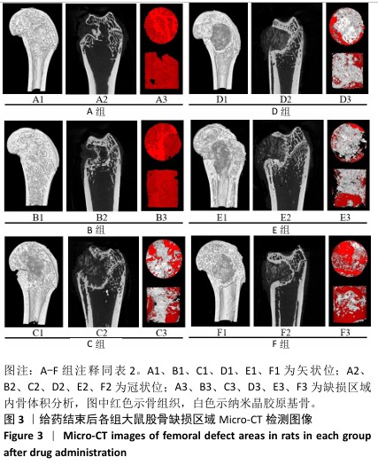
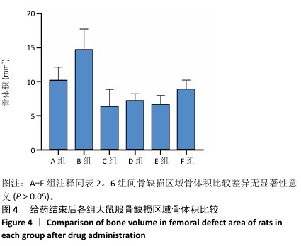
[5] 周世博,关健斌,俞兴,等.股骨骨缺损动物模型制备现状及特点[J].中国组织工程研究,2024,28(4):633-638.
[6] 熊伟,袁灵梅,钱国文,等.“补肾壮骨”中药应用于骨组织工程支架修复节段性骨缺损[J].中国组织工程研究,2023,27(21): 3438-3444.
[7] 周世博,俞兴,仲文庆,等.补肾壮筋汤调控OPG/RANKL/RANK信号通路抗骨质疏松的作用[J].中国实验方剂学杂志,2024,30(16): 44-51
[8] 赵赫.补肾壮筋汤颗粒对腰椎间盘退变大鼠影响的实验研究[D].北京:北京中医药大学,2018.
[9] 赵赫,俞兴,唐向胜,等.现代骨科临床应用补肾壮筋汤的Meta分析[J].世界中医药,2017,12(10):2504-2508.
[10] 俞兴,徐林,毕连涌,等.应用纳米晶胶原基骨材料行腰椎后外侧融合初步效果分析[J].中国矫形外科杂志,2005,13(8):
586-588.
[11] 俞兴,徐林,毕连涌,等.纳米晶胶原基骨材料在腰椎后外侧植骨融合中的临床应用[J].生物骨科材料与临床研究,2004,1(6):6-9.
[12] 俞兴,徐林,崔福斋.纳米晶胶原基骨材料在颈椎前路中的临床应用[J].生物骨科材料与临床研究,2003,1(1):14-16.
[13] YU Y, WANG L, MA P. Nanocrystalline collagen-based bone materials in skeletal optimization in patients. Nano Energy. 2021;22(3):563-569.
[14] HASSANI A, AVCI ÇB, KERDAR SN, et al. Interaction of alginate with nano-hydroxyapatite-collagen using strontium provides suitable osteogenic platform. J Nanobiotechnology. 2022;20(1):310.
[15] 沈耿杨,任辉,张志达,等.龟板联合阿伦磷酸钠对激素性骨质疏松大鼠腰椎Runx2、CTSK表达的影响[J].中华中医药杂志,2017, 32(5):2181-2185.
[16] 张宇楷,田瀚,张银银,等.骨质疏松症的表观遗传学机制及中药干预作用的研究[J].中国骨质疏松杂志,2024,30(10): 1513-1517+1523.
[17] LI N, CORNELISSEN D, SILVERMAN S, et al. An Updated Systematic Review of Cost-Effectiveness Analyses of Drugs for Osteoporosis. Pharmacoeconomics. 2021;39(2):181-209.
[18] NIH Consensus Development Panel on Osteoporosis Prevention, Diagnosis, and Therapy, March 7-29, 2000: highlights of the conference. South Med J. 2001;94(6):569-573.
[19] SALARI N, GHASEMI H, MOHAMMADI L, et al. The global prevalence of osteoporosis in the world: a comprehensive systematic review and meta-analysis. J Orthop Surg Res. 2021;16(1):609.
[20] GBD 2019 RISK FACTORS COLLABORATORS. Global burden of 87 risk factors in 204 countries and territories, 1990-2019: a systematic analysis for the Global Burden of Disease Study 2019. Lancet. 2020; 396(10258):1223-1249.
[21] SUN L, NIU H, WU Y, et al. Bio-integrated scaffold facilitates large bone regeneration dominated by endochondral ossification. Bioact Mater. 2024;35:208-227.
[22] 赵恩哲,吴斗,刘强.骨缺损的治疗现状及研究进展[J].中华实验外科杂志,2022,39(11):2053-2057.
[23] KHALED A, EL-GEBALY O, EL-ROSASY M. Masquelet-Ilizarov technique for the management of bone loss post debridement of infected tibial nonunion. Int Orthop. 2022;46(9):1937-1944.
[24] LI C, OUYANG L, ARMSTRONG JPK, et al. Advances in the Fabrication of Biomaterials for Gradient Tissue Engineering. Trends Biotechnol. 2021;39(2):150-164.
[25] JOHNSON ZM, YUAN Y, LI X, et al. Mesenchymal stem cells and three-dimensional osteoconductive scaffold regenerate calvarial bone in critical size defects in swine. Stem Cells Transl Med. 2021;10(8): 1170-1183.
[26] LI JJ, ROOHANI-ESFAHANI SI, KIM K, et al. Silk coating on a bioactive ceramic scaffold for bone regeneration: effective enhancement of mechanical and in vitro osteogenic properties towards load-bearing applications. J Tissue Eng Regen Med. 2017;11(6):1741-1753.
[27] XU X, LIAO L, TIAN W. Strategies of Prevascularization in Tissue Engineering and Regeneration of Craniofacial Tissues. Tissue Eng Part B Rev. 2022;28(2):464-475.
[28] SARAN U, GEMINI PIPERNI S, CHATTERJEE S. Role of angiogenesis in bone repair. Arch Biochem Biophys. 2014;561:109-117.
[29] YAO S, JIN B, LIU Z, et al. Biomineralization: From Material Tactics to Biological Strategy. Adv Mater. 2017;29(14). doi: 10.1002/adma. 201605903.
[30] 马士卿,王晓婧,彭诚.仿生矿化胶原材料应用于引导骨再生术的研究进展[J].重庆医学,2023,2(7):1072-1077.
[31] HARA ES, OKADA M, NAGAOKA N, et al. Re-Evaluation of Initial Bone Mineralization from an Engineering Perspective. Tissue Eng Part B Rev. 2022;28(1):246-255.
[32] MA L, WANG X, ZHOU Y, et al. Biomimetic Ti-6Al-4V alloy/gelatin methacrylate hybrid scaffold with enhanced osteogenic and angiogenic capabilities for large bone defect restoration. Bioact Mater. 2021;6(10):3437-3448.
[33] ZHANG Y, FAN Z, XING Y, et al. Effect of microtopography on osseointegration of implantable biomaterials and its modification strategies. Front Bioeng Biotechnol. 2022;10:981062.
[34] 张叶,崔元璐.中药在干细胞增殖和组织工程中的应用[J].中国组织工程研究,2016,20(28):4243-4249.
[35] 唐荣穗,周静.中药单体促进骨缺损修复再生的研究进展[J].牙体牙髓牙周病学杂志,2024,29(7):417-421.
[36] 杨斌,王楠,谭睿,等.中药及其单体对骨髓间充质干细胞诱导分化的研究进展[J].中草药,2022,53(24):7915-7924.
[37] GOU Y, HUANG Y, LUO W, et al. Adipose-derived mesenchymal stem cells (MSCs) are a superior cell source for bone tissue engineering. Bioact Mater. 2023;34:51-63.
[38] HOU Y, YAN Z, WU Z. Concise Review; The Recent Methods that Enhance the Osteogenic Differentiation of Human Induced Pluripotent Stem Cells. Curr Stem Cell Res Ther. 2021;16(8):949-957.
[39] XIE L, LIU N, XIAO Y, et al. In Vitro and In Vivo Osteogenesis Induced by Icariin and Bone Morphogenetic Protein-2: A Dynamic Observation. Front Pharmacol. 2020;11:1058.
[40] 王鹏珍,孟庆奇,陈松生,等.淫羊藿苷联合碱性成纤维细胞生长因子促进外周血间充质干细胞自我更新[J].中国生物化学与分子生物学报,2021,37(12):1667-1674.
[41] 钱秀昌.伤科补要[M].5版.上海:上海科学技术出版社,1981:19-45.
[42] KAVITHA SRI A, ARTHI C, NEYA NR, et al. Nano-hydroxyapatite/collagen composite as scaffold material for bone regeneration. Biomed Mater. 2023;18(3):10.1088/1748-605X/acc99e.
[43] ZUO Y, LI Q, XIONG Q, et al. Naringin Release from a Nano-Hydroxyapatite/Collagen Scaffold Promotes Osteogenesis and Bone Tissue Reconstruction. Polymers (Basel). 2022;14(16):3260.
[44] CAO S, LI H, LI K, et al. In vitro mineralization of MC3T3-E1 osteoblast-like cells on collagen/nano-hydroxyapatite scaffolds coated carbon/carbon composites. J Biomed Mater Res A. 2016;104(2):533-543.
[45] 张萌萌.雌激素与雌激素受体骨代谢调节作用[J].中国骨质疏松杂志,2019,25(5):704-708.
[46] 袁雨露,杨桢,丁薇,等.中药抗骨质疏松作用及机制探讨[J].中国实验方剂学杂志,2024,30(4):290-298.
[47] VASIKARAN S, COOPER C, EASTELL R, et al. International Osteoporosis Foundation and International Federation of Clinical Chemistry and Laboratory Medicine position on bone marker standards in osteoporosis. Clin Chem Lab Med. 2011;49(8):1271-1274. |