[1] 于娜,周家为,李霞,等.成人特发性肺纤维化(更新)和进行性肺纤维化临床实践指南(2022版)解读[J].中国现代医学杂志,2023,33(14):1-8.
[2] MARTINEZ FJ, COLLARD HR, PARDO A, et al. Idiopathic pulmonary fibrosis. Nat Rev Dis Primers. 2017;3:17074.
[3] LUPPI F, KALLURI M, FAVERIO P, et al. Idiopathic pulmonary fibrosis beyond the lung: understanding disease mechanisms to improve diagnosis and management. Respir Res. 2021;22(1):109.
[4] SPAGNOLO P, KROPSKI JA, JONES MG, et al. Idiopathic pulmonary fibrosis: Disease mechanisms and drug development. Pharmacol Ther. 2021;222:107798.
[5] 姜秋燕,梁庆,高劭妍,等.特发性肺纤维化治疗药物研究进展[J].中国新药杂志,2021,30(14):1274-1281.
[6] DIAMANTOPOULOS A, WRIGHT E, VLAHOPOULOU K, et al. The Burden of Illness of Idiopathic Pulmonary Fibrosis: A Comprehensive Evidence Review. Pharmacoeconomics. 2018;36(7):779-807.
[7] ZANK DC, BUENO M, MORA AL, et al. Idiopathic Pulmonary Fibrosis: Aging, Mitochondrial Dysfunction, and Cellular Bioenergetics. Front Med (Lausanne). 2018;5:10.
[8] KURITA Y, ARAYA J, MINAGAWA S, et al. Pirfenidone inhibits myofibroblast differentiation and lung fibrosis development during insufficient mitophagy. Respir Res. 2017;18(1):114.
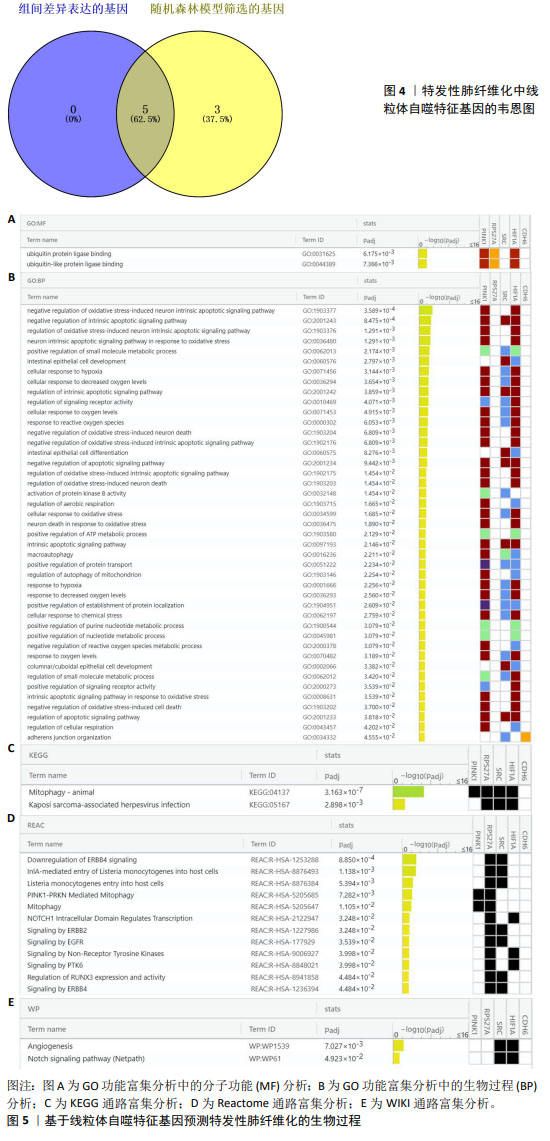
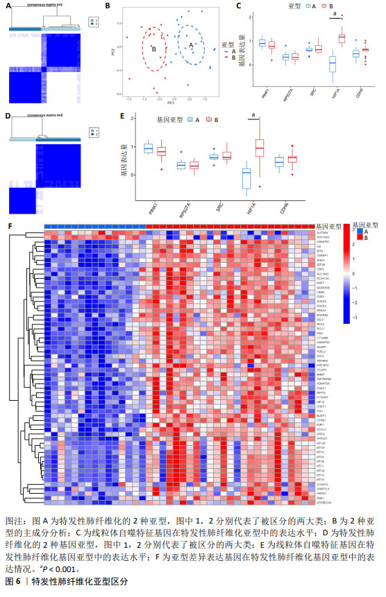
[9] BARRETT T, WILHITE SE, LEDOUX P, et al. NCBI GEO: archive for functional genomics data sets--update. Nucleic Acids Res. 2013;41(Database issue):D991-D995.
[10] SUN K, JING X, GUO J, et al. Mitophagy in degenerative joint diseases. Autophagy. 2021;17(9):2082-2092.
[11] 徐文飞,明春玉,段戡,等.WGCNA和机器学习识别骨关节炎铁死亡特征基因及实验验证[J].中国组织工程研究, 2024,28(30):4909-4914.
[12] KOLBERG L, RAUDVERE U, KUZMIN I, et al. g:Profiler-interoperable web service for functional enrichment analysis and gene identifier mapping (2023 update). Nucleic Acids Res. 2023;51(W1):W207-W212.
[13] LEE SY, AN HJ, KIM JM, et al. PINK1 deficiency impairs osteoblast differentiation through aberrant mitochondrial homeostasis. Stem Cell Res Ther. 2021;12(1):589.
[14] GAN ZY, CALLEGARI S, COBBOLD SA, et al. Activation mechanism of PINK1. Nature. 2022;602(7896):328-335.
[15] BUENO M, LAI YC, ROMERO Y, et al. PINK1 deficiency impairs mitochondrial homeostasis and promotes lung fibrosis. J Clin Invest. 2015;125(2):521-538.
[16] WEI Y, SUN L, LIU C, et al. Naringin regulates endoplasmic reticulum stress and mitophagy through the ATF3/PINK1 signaling axis to alleviate pulmonary fibrosis. Naunyn Schmiedebergs Arch Pharmacol. 2023;396(6):1155-1169.
[17] TWAMLEY-STEIN GM, PEPPERKOK R, Ansorge W, et al. The Src family tyrosine kinases are required for platelet-derived growth factor-mediated signal transduction in NIH 3T3 cells. Proc Natl Acad Sci U S A. 1993;90(16):7696-7700.
[18] AMANCHY R, ZHONG J, HONG R, et al. Identification of c-Src tyrosine kinase substrates in platelet-derived growth factor receptor signaling. Mol Oncol. 2009;3(5-6): 439-450.
[19] BARBAYIANNI I, KANELLOPOULOU P, FANIDIS D, et al. SRC and TKS5 mediated podosome formation in fibroblasts promotes extracellular matrix invasion and pulmonary fibrosis. Nat Commun. 2023;14(1):5882.
[20] PARK SR, KIM HJ, YANG SR, et al. A novel endogenous damage signal, glycyl tRNA synthetase, activates multiple beneficial functions of mesenchymal stem cells. Cell Death Differ. 2018;25(11):2023-2036.
[21] ZHOU MS, ZHENG SY, CHEN C, et al. Gene expression analysis to identify mechanisms underlying improvement of myocardial fibrosis by finerenone in SHR. Biochem Pharmacol. 2024;220:115975.
[22] TSUBOUCHI K, ARAYA J, KUWANO K. PINK1-PARK2-mediated mitophagy in COPD and IPF pathogeneses. Inflamm Regen. 2018;38:18.
[23] CHOURASIA AH, TRACY K, FRANKENBERGER C, et al. Mitophagy defects arising from BNip3 loss promote mammary tumor progression to metastasis. EMBO Rep. 2015;16(9):1145-1163.
[24] KHAWAJA AA, CHONG DLW, SAHOTA J, et al. Identification of a Novel HIF-1α-αMβ2 Integrin-NET Axis in Fibrotic Interstitial Lung Disease. Front Immunol. 2020;11:2190.
[25] PHILIP K, MILLS TW, DAVIES J, et al. HIF1A up-regulates the ADORA2B receptor on alternatively activated macrophages and contributes to pulmonary fibrosis. FASEB J. 2017;31(11):4745-4758.
[26] KOLAHIAN S, ÖZ HH, ZHOU B, et al. The emerging role of myeloid-derived suppressor cells in lung diseases. Eur Respir J. 2016;47(3):967-977.
[27] FERNANDEZ IE, GREIFFO FR, FRANKENBERGER M, et al. Peripheral blood myeloid-derived suppressor cells reflect disease status in idiopathic pulmonary fibrosis. Eur Respir J. 2016;48(4):1171-1183.
[28] LIU T, GONZALEZ DE LOS SANTOS F, RINKE AE, et al. B7H3-dependent myeloid-derived suppressor cell recruitment and activation in pulmonary fibrosis. Front Immunol. 2022;13:901349.
[29] XU Y, LAN P, WANG T. The Role of Immune Cells in the Pathogenesis of Idiopathic Pulmonary Fibrosis. Medicina (Kaunas). 2023;59(11):1984.
[30] ACHAIAH A, RATHNAPALA A, PEREIRA A, et al. Neutrophil lymphocyte ratio as an indicator for disease progression in Idiopathic Pulmonary Fibrosis. BMJ Open Respir Res. 2022;9(1):e001202.
[31] WILSON MS, MADALA SK, RAMALINGAM TR, et al. Bleomycin and IL-1beta-mediated pulmonary fibrosis is IL-17A dependent. J Exp Med. 2010;207(3):535-552.
[32] LI Y, LI S, GU M, et al. Application of network composite module analysis and verification to explore the bidirectional immunomodulatory effect of Zukamu granules on Th1 / Th2 cytokines in lung injury. J Ethnopharmacol. 2022;299:115674.
[33] ANDERSSON CK, ANDERSSON-SJÖLAND A, MORI M, et al. Activated MCTC mast cells infiltrate diseased lung areas in cystic fibrosis and idiopathic pulmonary fibrosis. Respir Res. 2011;12(1):139.
[34] OVERED-SAYER C, RAPLEY L, MUSTELIN T, et al. Are mast cells instrumental for fibrotic diseases? Front Pharmacol. 2014;4:174.
[35] HIRAHARA K, AOKI A, MORIMOTO Y, et al. The immunopathology of lung fibrosis: amphiregulin-producing pathogenic memory T helper-2 cells control the airway fibrotic responses by inducing eosinophils to secrete osteopontin. Semin Immunopathol. 2019;41(3):339-348.
[36] LIU J, WANG J, XIONG A, et al. Mitochondrial quality control in lung diseases: current research and future directions. Front Physiol. 2023;14:1236651.
[37] WANG L, CHEN R, LI G, et al. FBW7 Mediates Senescence and Pulmonary Fibrosis through Telomere Uncapping. Cell Metab. 2020;32(5):860-877.e9.
[38] ZHOU Y, HUANG X, HECKER L, et al. Inhibition of mechanosensitive signaling in myofibroblasts ameliorates experimental pulmonary fibrosis. J Clin Invest. 2013; 123(3):1096-1108.
[39] BODEMPUDI V, HERGERT P, SMITH K, et al. miR-210 promotes IPF fibroblast proliferation in response to hypoxia. Am J Physiol Lung Cell Mol Physiol. 2014;307(4): L283-L294.
[40] YANG L, GILBERTSEN A, XIA H, et al. Hypoxia enhances IPF mesenchymal progenitor cell fibrogenicity via the lactate/GPR81/HIF1α pathway. JCI Insight. 2023;8(4):e163820.
[41] WANG Y, SUN Q, GENG R, et al. Notch intracellular domain regulates glioblastoma proliferation through the Notch1 signaling pathway. Oncol Lett. 2021;21(4):303.
[42] WASNICK R, KORFEI M, PISKULAK K, et al. Notch1 Induces Defective Epithelial Surfactant Processing and Pulmonary Fibrosis. Am J Respir Crit Care Med. 2023; 207(3):283-299.
[43] SHARMA B, SINGH VJ, CHAWLA PA. Epidermal growth factor receptor inhibitors as potential anticancer agents: An update of recent progress. Bioorg Chem. 2021;116: 105393.
[44] PATNAIK SK, CHANDRASEKAR MJN, NAGARJUNA P, et al. Targeting of ErbB1, ErbB2, and their Dual Targeting Using Small Molecules and Natural Peptides: Blocking EGFR Cell Signaling Pathways in Cancer: A Mini-Review. Mini Rev Med Chem. 2022; 22(22):2831-2846.
[45] LIU X, GENG Y, LIANG J, et al. HER2 drives lung fibrosis by activating a metastatic cancer signature in invasive lung fibroblasts. J Exp Med. 2022;219(10):e20220126.
[46] JIANG Y, SHI J, ZHOU J, et al. ErbB4 promotes M2 activation of macrophages in idiopathic pulmonary fibrosis. Open Life Sci. 2023; 18(1):20220692. |