[1] PAKKHESAL M, NAGHAVIALHOSSEINI A, FAALI T, et al. Oral health-related quality of life changes during phase 1 Class II malocclusion treatment using Frankel 2 and Twin-block appliances: A short-term follow-up study. Am J Orthod Dentofacial Orthop. 2023;163(2):191-197.
[2] ABUTALEB MA, LATIEF MHAE, MONTASSER MA. Reflection on patients’ experience with orthodontic appliances wear and its impact on oral health related quality of life: observational comparative study. BMC Oral Health. 2023;23(1):502.
[3] LAZAR L, VLASA A, BERESESCU B, et al. White Spot Lesions (WSLs)-Post-Orthodontic Occurrence, Management and Treatment Alternatives: A Narrative Review. J Clin Med. 2023;12(5):1908.
[4] HÖCHLI D, HERSBERGER-ZURFLUH M, PAPAGEORGIOU S, et al. Interventions for orthodontically induced white spot lesions: a systematic review and meta-analysis. Eur J Orthod. 2017;39(2):122-133.
[5] YAZARLOO S, ARAB S, MIRHASHEMI A, et al. Systematic review of preventive and treatment measures regarding orthodontically induced white spot lesions. Dent Med Probl. 2023;60(3):527-535.
[6] SHUNGIN D, OLSSON AI, PERSSON M. Orthodontic treatment-related white spot lesions: A 14-year prospective quantitative follow-up, including bonding material assessment. Am J Orthod Dentofacial Orthop. 2010;138(2):136.e1-8
[7] PULEIO F, FIORILLO L, GORASSINI F, et al. Systematic Review on White Spot Lesions Treatments. Eur J Dent. 2022;16(1):41-48.
[8] PERDIGÃO J. Resin infiltration of enamel white spot lesions: An ultramorphological analysis. J Esthet Restor Dent. 2020;32(3):317-324.
[9] CHAPMAN JA, ROBERTS WE, ECKERT GJ, et al. Risk factors for incidence and severity of white spot lesions during treatment with fixed orthodontic appliances. Am J Orthod Dentofacial Orthop. 2010; 138(2):188-194.
[10] MALCANGI G, PATANO A, MOROLLA R, et al. Analysis of Dental Enamel Remineralization: A Systematic Review of Technique Comparisons. Bioengineering (Basel). 2023;10(4):472.
[11] GHOLAMI S, BORUZINIAT A, TALEBI A, et al. Effect of Extent of White Spot Lesions on the Esthetic Outcome after Treatment by the Resin Infiltration Technique: A Clinical Trial. Front Dent. 2023;20:40.
[12] LUCCHESE A, GHERLONE E. Prevalence of white-spot lesions before and during orthodontic treatment with fixed appliances. Eur J Orthod. 2013;35(5):664-668.
[13] SOVERAL M, MACHADO V, BOTELHO J, et al. Effect of Resin Infiltration on Enamel: A Systematic Review and Meta-Analysis. J Funct Biomater. 2021;12(3):48.
[14] RAGHAVAN S, ALHAIJA ESA, DUGGAL MS, et al. White spot lesions, plaque accumulation and salivary caries-associated bacteria in clear aligners compared to fixed orthodontic treatment. A systematic review and meta- analysis. BMC Oral Health. 2023;23(1):599.
[15] ROUZI M, ZHANG XQ, JIANG QX, et al. Impact of Clear Aligners on Oral Health and Oral Microbiome During Orthodontic Treatment. Int Dent J. 2023;73(5):603-611.
[16] BISHT S, KHERA AK, RAGHAV P. White spot lesions during orthodontic clear aligner therapy: A scoping review. J Orthod Sci. 2022;11:9
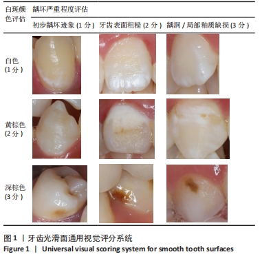
[17] KÜHNISCH J, INKA G , SUSANNE B, et al. Development, methodology and potential of the new universal visual scoring system (univiss) for caries detection and diagnosis. Int J Environ Res Public Health. 2009; 6(9):2500-2509.
[18] 乔斯·W.R.特维斯克. 实用流行病学纵向数据分析方法[M].陈心广,俞斌,王培刚译.北京:人民卫生出版社,2016.
[19] 郭伯良.多水平模型应用[M].北京:北京师范大学出版社,2020.
[20] 张岩波,何大卫.多分类反应变量的多水平多项式模型及其应用[J].中国卫生统计,2000,17(5):130-132.
[21] 郑建中,张岩波.有序分类重复测量数据的多水平模型分析[J].现代预防医学,2003,30(1):6-8.
[22] 杨珉,李晓松.医学和公共卫生研究常用多水平统计模型[M].北京:北京大学医学出版社,2007.
[23] SPATAFORA G, LI YH, HE XS, et al. The Evolving Microbiome of Dental Caries. Microorganisms. 2024;12(1):121.
[24] LIU Y, DANIEL SG, KIM HE, et al. Addition of cariogenic pathogens to complex oral microflora drives significant changes in biofilm compositions and functionalities. Microbiome. 2023;11(1):123.
[25] EDGAR WM. The role of saliva in the control of pH changes in human dental plaque. Caries Res. 1976;10(4):241-254.
[26] TAKAHASHI N. Chapter 11: Future Perspectives in the Study of Dental Caries. Monogr Oral Sci. 2023;31:221-233.
[27] FEATHERSTONE JD. The caries balance: contributing factors and early detection. J Calif Dent Assoc. 2003;31:129-133.
[28] ALSHAHRANI I, SYED S, AMANULLAH M, et al. Changes in Essential Salivary Parameters in Patients Undergoing Fixed Orthodontic Treatment: A Longitudinal Study. Niger J Clin Pract. 2019;22(5):707-712.
[29] MALHI G. Clear aligners vs fixed appliances: which treatment option presents a higher incidence of white spot lesions, plaque accumulation and salivary caries-associated bacteria? Evid Based Dent. 2024;25(1):21-22.
[30] SHOKEEN B, VILORIA E, DUONG E, et al. The impact of fixed orthodontic appliances and clear aligners on the oral microbiome and the association with clinical parameters: A longitudinal comparative study. Am J Orthod Dentofacial Orthop. 2022;161(5):e475-e485.
[31] ROUZI M, JIANG QS, ZHANG HX, et al. Characteristics of oral microbiota and oral health in the patients treated with clear aligners: a prospective study. Clin Oral Investig. 2023;27(11):6725-6734.
[32] MADARIAGA ACP, BUCCI R, RONGO R, et al. Impact of Fixed Orthodontic Appliance and Clear Aligners on the Periodontal Health: A Prospective Clinical Study. Dent J (Basel). 2020;8(1):4.
[33] CHHIBBER A, AGARWAL S, YADAV S, et al. Which orthodontic appliance is best for oral hygiene? A randomized clinical trial. Am J Orthod Dentofacial Orthop. 2018;153(2):175-183.
[34] SIFAKAKIS I, PAPAIOANNOU W, PAPADIMITRIOU A. Salivary levels of cariogenic bacterial species during orthodontic treatment with thermoplastic aligners or fixed appliances: a prospective cohort study. Prog Orthod. 2018;19(1):25.
[35] ALBHAISI Z, AL-KHATEEB SN, ALHAIJA ESA. Enamel demineralization during clear aligner orthodontic treatment compared with fixed appliance therapy, evaluated with quantitative light-induced fluorescence: A randomized clinical trial. Am J Orthod Dentofacial Orthop. 2020;157(5):594-601.
[36] RICHTER AE, ARRUDA AO, PETERS MC, et al. Incidence of caries lesions among patients treated with comprehensive orthodontics. Am J Orthod Dentofacial Orthop. 2011;139(5):657-664.
[37] ROUT T, PATIL A, DESHMUKH S, et al. Remineralizing potential of Calcium Sucrose Phosphate in white spot lesions: A Systematic Review. J Oral Biosci. 2024;66(2):261-271.
[38] BISHT S, KHERA AK, RAGHAV P. White spot lesions during orthodontic clear aligner therapy: A scoping review. J Orthod Sci. 2022;11: 9.
[39] SCHUSTER S, ELIADES G, ZINELIS S, et al. Structural conformation and leaching from in vitro aged and retrieved Invisalign appliances. Am J Orthod Dentofacial Orthop. 2004;126(6):725-728.
[40] PITCHIKA V, KOKEL C, PITCHIKA JA, et al. Longitudinal study of caries progression in 2-and 3-year-old German children. Community Dent Oral Epidemiol. 2016;44(4):354-363. |