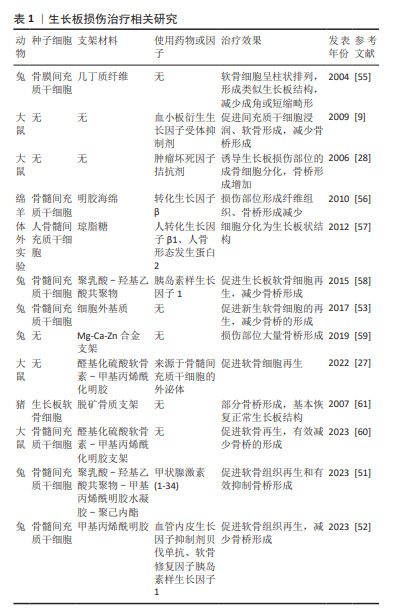
[1] KRONENBERG HM. Developmental regulation of the growth plate. Nature. 2003;423(6937):332-336.
[2] SAMET JD. Pediatric Sports Injuries. Clin Sports Med. 2021;40(4): 781-799.
[3] YAMAMURA MK, CARRY PM, GIBLY RF, et al. Epidemiology of Physeal Fractures and Clinically Significant Growth Disturbances Affecting the Distal Tibia, Proximal Tibia, and Distal Femur: A Retrospective Cohort Study. J Am Acad Orthop Surg. 2023;31(11):e507-e515.
[4] CHUNG R, FOSTER BK, XIAN CJ. Injury responses and repair mechanisms of the injured growth plate. Front Biosci (Schol Ed). 2011;3(1):117-125.
[5] HALLETT SA, ONO W, ONO N. Growth Plate Chondrocytes: Skeletal Development, Growth and Beyond. Int J Mol Sci. 2019;20(23):6009.
[6] STEGEN S, VAN GASTEL N, CARMELIET G. Bringing new life to damaged bone: the importance of angiogenesis in bone repair and regeneration. Bone. 2015;70:19-27.
[7] ZHANG J, PAN J, JING W. Motivating role of type H vessels in bone regeneration. Cell Prolif. 2020;53(9):e12874.
[8] FRIEDLAENDER GE, LIN S, SOLCHAGA LA, et al.The Role of Recombinant Human Platelet-derived Growth Factor-BB (rhPDGF-BB) in Orthopaedic Bone Repair and Regeneration. Curr Pharm Des. 2013;19(19): 3384-3390.
[9] CHUNG R, FOSTER BK, ZANNETTINO AC, et al. Potential roles of growth factor PDGF-BB in the bony repair of injured growth plate. Bone. 2009; 44(5):878-885.
[10] XIE H, CUI Z, WANG L, et al. PDGF-BB secreted by preosteoclasts induces angiogenesis during coupling with osteogenesis. Nat Med. 2014;20(11):1270-1278.
[11] GUÉRIT E, ARTS F, DACHY G, et al. PDGF receptor mutations in human diseases. Cell Mol Life Sci. 2021;78(8):3867-3881.
[12] RAUNIYAR K, BOKHARAIE H, JELTSCH M. Expansion and collapse of VEGF diversity in major clades of the animal kingdom. Angiogenesis. 2023;26(3):437-461.
[13] KAZLAUSKAS A. PDGFs and their receptors. Gene. 2017;614:1-7.
[14] ZAIDI M, LIZNEVA D, YUEN T. The role of PDGF-BB in the bone-vascular relationship during aging. J Clin Invest. 2021;131(20):e153644.
[15] XU B, LUO Y, LIU Y, et al. Platelet-derived growth factor-BB enhances MSC-mediated cardioprotection via suppression of miR-320 expression. Am J Physiol Heart Circ Physiol. 2015;308(9):H980-H989.
[16] CHUNG R, FOSTER BK, XIAN CJ. The potential role of VEGF-induced vascularisation in the bony repair of injured growth plate cartilage. J Endocrinol. 2014;221(1):63-75.
[17] GIANNI-BARRERA R, BUTSCHKAU A, UCCELLI A, et al. PDGF-BB regulates splitting angiogenesis in skeletal muscle by limiting VEGF-induced endothelial proliferation. Angiogenesis. 2018;21(4):883-900.
[18] ZHANG M, YU W, NIIBE K, et al. The Effects of Platelet-Derived Growth Factor-BB on Bone Marrow Stromal Cell-Mediated Vascularized Bone Regeneration. Stem Cells Int. 2018;2018:3272098.
[19] SU W, LIU G, LIU X, et al. Angiogenesis stimulated by elevated PDGF-BB in subchondral bone contributes to osteoarthritis development. JCI Insight. 2020;5(8):e135446.
[20] ZHOU FH, FOSTER BK, SANDER G, et al. Expression of proinflammatory cytokines and growth factors at the injured growth plate cartilage in young rats. Bone. 2004;35:1307-1315.
[21] DEL ROSARIO C, RODRÍGUEZ-ÉVORA M, REYES R, et al. BMP-2, PDGF-BB, and bone marrow mesenchymal cells in a macroporous β-TCP scaffold for critical-size bone defect repair in rats. Biomed Mater. 2015; 10(4):045008.
[22] TAKEUCHI T, YOSHIDA H, TANAKA S. Role of interleukin-6 in bone destruction and bone repair in rheumatoid arthritis. Autoimmun Rev. 2021;20(9):102884.
[23] LIU B, LI J, CHEN B, et al. Dental pulp stem cells induce anti-inflammatory phenotypic transformation of macrophages to enhance osteogenic potential via IL-6/GP130/STAT3 signaling. Ann Transl Med. 2023;11(2):90.
[24] MOREAUX J, HOSE D, KASSAMBARA A, et al. Osteoclast-gene expression profiling reveals osteoclast-derived CCR2 chemokines promoting myeloma cell migration. Blood. 2011;117(4):1280-1290.
[25] SCHWARTZ NB, DOMOWICZ MS. Roles of Chondroitin Sulfate Proteoglycans as Regulators of Skeletal Development. Front Cell Dev Biol. 2022;10:745372.
[26] INDIRAN V, JAGANNATHAN D. Osgood-Schlatter Disease. N Engl J Med. 2018;378(11):e15.
[27] GUAN P, LIU C, XIE D, et al. Exosome-loaded extracellular matrix-mimic hydrogel with anti-inflammatory property Facilitates/promotes growth plate injury repair. Bioact Mater. 2022;10:145-158.
[28] ZHOU FH, FOSTER BK, ZHOU XF, et al. TNF‐α mediates p38 MAP kinase activation and negatively regulates bone formation at the injured growth plate in rats. J Bone Miner Res. 2006;21(7):1075-1088.
[29] ARASAPAM G, SCHERER M, COOL JC, et al. Roles of COX‐2 and iNOS in the bony repair of the injured growth plate cartilage. J Cell Biochem. 2006;99(2):450-461.
[30] CHUNG R, COOL JC, SCHERER MA, et al. Roles of neutrophil-mediated inflammatory response in the bony repair of injured growth plate cartilage in young rats. J Leukoc Biol. 2006;80(6):1272-1280.
[31] LEE MA, NISSEN TP, OTSUKA NY. Utilization of a murine model to investigate the molecular process of transphyseal bone formation. J Pediatr Orthop. 2000;20(6):802-806.
[32] LIU ES, RAIMANN A, CHAE BT, et al. c-Raf promotes angiogenesis during normal growth plate maturation. Development. 2016;143(2): 348-355.
[33] RIBATTI D, D’AMATI A. Bone angiocrine factors. Front Cell Dev Biol. 2023;11:1244372.
[34] SU YW, CHUNG R, RUAN CS, et al. Neurotrophin-3 Induces BMP-2 and VEGF Activities and Promotes the Bony Repair of Injured Growth Plate Cartilage and Bone in Rats. J Bone Miner Res. 2016;31(6):1258-1274.
[35] PREIN C, BEIER F. ECM signaling in cartilage development and endochondral ossification. Curr Top Dev Biol. 2019;133:25-47.
[36] SALHOTRA A, SHAH HN, LEVI B, et al. Mechanisms of bone development and repair. Nat Rev Mol Cell Biol. 2020;21(11):696-711.
[37] TUCKERMANN J, ADAMS RH. The endothelium-bone axis in development, homeostasis and bone and joint disease. Nat Rev Rheumatol. 2021;17(10):608-620.
[38] KUSUMBE AP, RAMASAMY SK, ADAMS RH. Coupling of angiogenesis and osteogenesis by a specific vessel subtype in bone. Nature. 2014; 507(7492):323-328.
[39] ZHANG M, AHN W, KIM S, et al. Endothelial precursor cells stimulate pericyte-like coverage of bone marrow-derived mesenchymal stem cells through platelet-derived growth factor-BB induction, which is enhanced by substance P. Microcirculation. 2017;24(8):e12394.
[40] FANG J, HUANG X, HAN X, et al. Endothelial progenitor cells promote viability and nerve regenerative ability of mesenchymal stem cells through PDGF-BB/PDGFR-β signaling. Aging (Albany NY). 2020;12(1):106-121.
[41] GUZMAN RA, MARUYAMA M, MOEINZADEH S, et al. The effect of genetically modified platelet-derived growth factor-BB over-expressing mesenchymal stromal cells during core decompression for steroid-associated osteonecrosis of the femoral head in rabbits. Stem Cell Res Ther. 2021;12(1):503.
[42] WALIA B, LINGENHELD E, DUONG L, et al. A novel role for cathepsin K in periosteal osteoclast precursors during fracture repair. Ann N Y Acad Sci. 2018;1415(1):57-68.
[43] CUI Z, WU H, XIAO Y, et al. Endothelial PDGF-BB/PDGFR-β signaling promotes osteoarthritis by enhancing angiogenesis-dependent abnormal subchondral bone formation. Bone Res. 2022;10(1):58.
[44] SHEN Z, DONG W, CHEN Z, et al. Total flavonoids of Rhizoma Drynariae enhances CD31hiEmcnhi vessel formation and subsequent bone regeneration in rat models of distraction osteogenesis by activating PDGFBB/VEGF/RUNX2/OSX signaling axis. Int J Mol Med. 2022;50(3):112.
[45] ZHEN G, DAN Y, WANG R, et al. An antibody against Siglec-15 promotes bone formation and fracture healing by increasing TRAP+ mononuclear cells and PDGF-BB secretion. Bone Res. 2021;9(1):47.
[46] GAO BO, DENG R, CHAI YU, et al. Macrophage-lineage TRAP+ cells recruit periosteum-derived cells for periosteal osteogenesis and regeneration. J Clin Investig. 2019;129(6):2578-2594.
[47] SUN FF, HU PF, XIONG Y, et al. Tricetin protects rat chondrocytes against IL-1β-induced inflammation and apoptosis. Oxid Med Cell Longev. 2019;2019:4695381
[48] SHAW N, ERICKSON C, BRYANT SJ, et al. Regenerative medicine approaches for the treatment of pediatric physeal injuries. Tissue Eng Part B Rev. 2018;24(2):85-97.
[49] AZARPIRA MR, SHAHCHERAGHI GH, AYATOLLAHI M, et al.Tissue engineering strategy using mesenchymal stem cell-based chitosan scafolds in growth plate surgery: A preliminary study in rabbits. Orthop Traumatol Surg Res. 2015;101(5):601.
[50] CUNNIFFE GM, DÍAZ-PAYNO PJ, RAMEY JS, et al. Growth plate extracellular matrix-derived scaffolds for large bone defect healing. Eur Cell Mater. 2017:33:130-142.
[51] FAN M, QIANG L, WANG Y, et al. 3D bioprinted hydrogel/polymer scaffold with factor delivery and mechanical support for growth plate injury repair. Front Bioeng Biotechnol. 2023;11:1210786.
[52] QIANG L, FAN M, WANG Y, et al. Injectable hydrogel loaded with bilayer microspheres to inhibit angiogenesis and promote cartilage regeneration for repairing growth plate injury. Front Bioeng Biotechnol. 2023;11:1181580.
[53] LI W, XU R, HUANG J, et al. Treatment of rabbit growth plate injuries with oriented ECM scaffold and autologous BMSCs. Sci Rep. 2017;7: 44140.
[54] 王香港,万谦,刘贺,等.组织工程软骨在生长板损伤修复治疗中的作用及特点[J].中国组织工程研究,2021,25(28):4539-4545.
[55] LI L, HUI JHP, GOH JCH, et al. Chitin as a scaffold for mesenchymal stem cells transfers in the treatment of partial growth arrest. J Pediatr Orthop. 2004;24(2):205-210.
[56] MCCARTY RC, XIAN CJ, GRONTHOS S, et al. Application of autologous bone marrow derived mesenchymal stem cells to an ovine model of growth plate cartilage injury. Open Orthop J. 2010;4:204.
[57] SCHMITT JF, HUA SK, ZHENG Y, et al. Sequential differentiation of mesenchymal stem cells in an agarose scaffold promotes a physis‐like zonal alignment of chondrocytes. J Orthop Res. 2012;30(11):1753-1759.
[58] CLARK A, HILT JZ, MILBRANDT TA, et al. Treating proximal tibial growth plate injuries using poly (lactic-co-glycolic acid) scaffolds. BioResearch Open Access. 2015;4(1):65-74.
[59] SONG MH, YOO WJ, CHO TJ, et al. In vivo response of growth plate to biodegradable Mg-Ca-Zn alloys depending on the surface modification. Int J Mol Sci. 2019;20(15):3761.
[60] GUAN P, JI Y, KANG X, et al. Biodegradable Dual-Cross-Linked Hydrogels with Stem Cell Differentiation Regulatory Properties Promote Growth Plate Injury Repair via Controllable Three-Dimensional Mechanics and a Cartilage-like Extracellular Matrix. ACS Appl Mater Interfaces. 2023;15(7):8986-8998.
[61] VAN DONKELAAR CC, HUISKES R. The PTHrP-Ihh feedback loop in the embryonic growth plate allows PTHrP to control hypertrophy and Ihh to regulate proliferation. Biomech Model Mechanobiol. 2007;6(1-2): 55-62.
[62] WANG W, RIGUEUR D, LYONS KM. TGFβ as a gatekeeper of BMP action in the developing growth plate. Bone. 2020;137:115439.
[63] ZHANG X, SICLARI VA, LAN S, et al. The critical role of the epidermal growth factor receptor in endochondral ossification. J Bone Miner Res. 2011;26(11):2622-2633.
[64] WEI Y, LUO L, GUI T, et al. Targeting cartilage EGFR pathway for osteoarthritis treatment. Sci Transl Med. 2021;13(576):eabb3946.
[65] 姚裕斌,魏刚,丁婕,等.可注射水凝胶微球治疗骨关节炎的实验研究[J].中国修复重建外科杂志,2023,37(8):918-928.
[66] XIONG J, ONAL M, JILKA RL, et al. Matrix-embedded cells control osteoclast formation. Nat Med. 2011;17(10):1235-1241. |
