中国组织工程研究 ›› 2023, Vol. 27 ›› Issue (32): 5203-5208.doi: 10.12307/2023.816

• 口腔组织构建 oral tissue construction • 上一篇    下一篇

前方牵引矫治不同年龄骨性Ⅲ类错牙合颞下颌关节的效果评价

刘亚非1,王雅淋2,左艳萍1,赵利霞1,尉  静1,张  超1,宋  蕾1   

  1. 1河北医科大学第一医院口腔正畸科,河北省石家庄市  050031;2保定市第二医院口腔科,河北省保定市  071000
  • 收稿日期:2022-05-24 接受日期:2022-08-01 出版日期:2023-11-18 发布日期:2023-03-23
  • 通讯作者: 左艳萍,博士,河北医科大学第一医院口腔正畸科,河北省石家庄市 050031
  • 作者简介:刘亚非,女,1975年生,河北省石家庄市人,汉族,2008年河北医科大学毕业,硕士,主要从事口腔正畸专业的研究。
  • 基金资助:
    河北省卫生厅课题编号(20211450):骨性Ⅲ类患者前方牵引矫治后颞下颌关节结构的比较分析,项目负责人:刘亚非

Changes in the temporomandibular joint following maxillary protraction for skeletal class III malocclusion at different ages

Liu Yafei1, Wang Yalin2, Zuo Yanping1, Zhao Lixia1, Wei Jing1, Zhang Chao1, Song Lei1   

  1. 1Department of Orthodontics, The First Hospital of Hebei Medical University, Shijiazhuang 050031, Hebei Province, China; 2Department of Stomatology, Baoding No. 2 Hospital, Baoding 071000, Hebei Province, China
  • Received:2022-05-24 Accepted:2022-08-01 Online:2023-11-18 Published:2023-03-23
  • Contact: Zuo Yanping, MD, Department of Orthodontics, The First Hospital of Hebei Medical University, Shijiazhuang 050031, Hebei Province, China
  • About author:Liu Yafei, Master, Department of Orthodontics, The First Hospital of Hebei Medical University, Shijiazhuang 050031, Hebei Province, China
  • Supported by:
    a grant from the Health Department of Hebei Province, No. 20211450 (to LYF)

摘要:


文题释义:
前方牵引:前方牵引系统有3个基本的组成部分——面罩、口内矫治器和橡皮圈。面罩是一种口外装置,由额垫、颏兜以及连接二者的金属杆组成,支持性金属杆的前端连接一个外弓,通过橡皮圈对上颌骨施加向前向下的牵引力,通过调节矫治器各个部分的螺钉可以调整额垫、颏兜和外弓的位置。口内矫治器将上颌连成一个整体,牵引钩应该位于尖牙区域的牙合平面上方,使牵引力的方向更靠近上颌骨的阻抗中心,防止上颌骨的旋转。橡皮圈提供矫形力,单侧力为300-500 g。根据病例而异,每日应戴足12-14 h以上。
骨性Ⅲ类错牙合:是指由于遗传、环境等因素引起,颌骨的形态、大小位置异常的一类畸形,表现为颌骨是近远中方向上的异常,主要临床特征是前牙反牙合,磨牙近中关系,可能伴有后牙反牙合。是由于下颌发育过度,或下颌前突,或者上颌发育不足,或上颌后缩,或者两者结合导致前牙反牙合,磨牙Ⅲ类咬合关系。临床表现为上颌正常、下颌前突,或上颌后缩、下颌正常,或上颌后缩、下颌前突。

背景:已有研究证实,不同年龄骨性Ⅲ类错牙合患者在前方牵引矫治后牙颌面的改变有显著不同;但是前方牵引矫治不同年龄患者颞下颌关节的改变是否存在差异尚未明确。
目的:探讨年龄对于前方牵引矫治骨性Ⅲ类错牙合对颞下颌关节改变的影响。
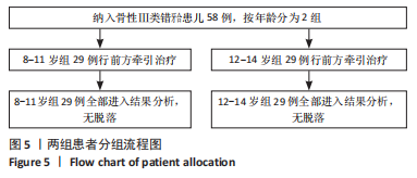
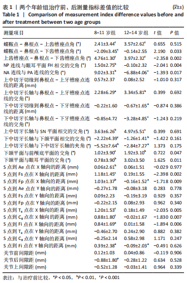
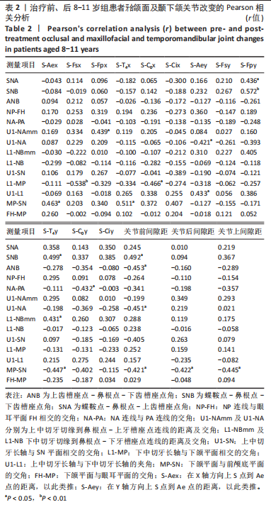
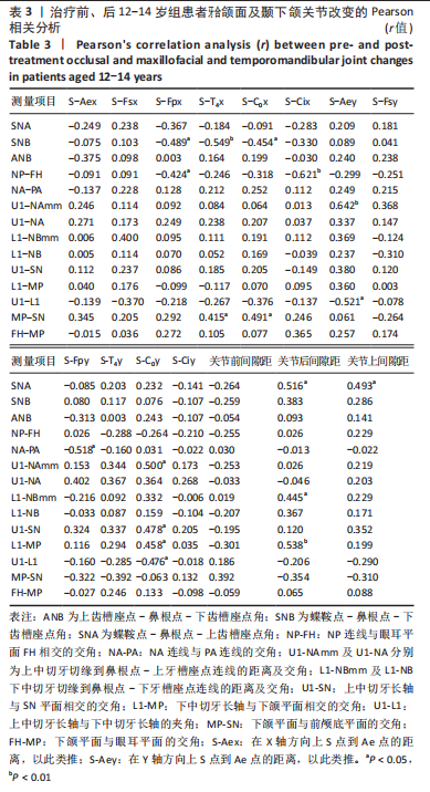
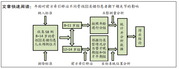
方法:将58例8-14岁的骨性Ⅲ类错牙合患儿分成2组,8-11岁组29例,牙合类型为替牙晚期;12-14岁组29例,牙合类型为恒牙早期。在矫治前、矫治后分别摄X射线头颅侧位片,进行牙合颌面的头影测量分析以及颞下颌关节在坐标系统位置的定量分析。采用配对t检验分别比较两组治疗前后的变化,独立t检验进行两组间治疗前后改变量的比较,Pearson相关分析对两组分别进行牙合颌面改变与颞下颌关节改变的相关性分析。
结果与结论:①2个年龄组矫治后均发生了显著的牙合颌面及颞下颌关节变化;牙合颌面矢状方向上,8-11岁组上齿槽座点-鼻根点-下齿槽座点角(ANB)增加了4.76°,颌突角(NA-PA)增加了9.02°,显著大于12-14岁组的改变3.97°及6.88°(P < 0.05);牙合颌面垂直方向上,8-11岁组下颌平面与前颅底平面的交角(MP-SN)、下颌平面与眼耳平面的交角(FH-MP)分别增加了1.92°及3.02°,明显大于12-14岁组的1.02°及0.78°(P < 0.05)。②颞下颌关节改变在2个年龄组差异有显著性意义,8-11岁组较12-14岁组关节窝上缘位置(S-Fsx)的后移更显著;8-11岁组的关节窝后缘位置(S-Fpx)改变表现为后移,而12-14岁组则为前移;两组的髁状突前缘位置(S-Cix)、后缘位置(S-T4x)均发生后移,但8-11岁组较12-14岁组更明显。③颞下颌关节改变与牙合颌面的改变存在相关关系:8-11岁组的S-Fpx与蝶鞍点-鼻根点-下齿槽座点角(SNB)、NP连线与眼耳平面FH相交的交角(NP-FH)有负相关关系(P < 0.05,r=-0.489;P < 0.05,r=-0.424);12-14岁组的S-Fpx与上中切牙切缘到鼻根点-上牙槽座点连线的距离(U1-NAmm)为正相关关系(P < 0.01,r=0.439);8-11岁S-Cix、S-T4x、髁状突上缘水平向位置(S-Cox)与SNB、NP-FH、MP-SN均呈中等负相关关系。④提示年龄对于前方牵引矫治骨性Ⅲ类错牙合对颞下颌关节改变有较为明显的影响,8-11岁组关节窝及髁状突位置后移明显,髁状突前缘上移,关节后间隙增加;12-14岁组关节窝后缘前移和髁状突前缘下移,关节间隙无改变;不同年龄影响颞下颌关节改变的牙合颌面因素并不相同。
https://orcid.org/0000-0001-5052-189X ( 刘亚非 ) 

中国组织工程研究杂志出版内容重点:组织构建;骨细胞;软骨细胞;细胞培养;成纤维细胞;血管内皮细胞;骨质疏松;组织工程

关键词: 前方牵引, 骨性安氏Ⅲ类错牙合, 颞下颌关节, 年龄, 牙合颌面

Abstract: BACKGROUND: Previous studies have shown that there are significant differences in occlusal and maxillofacial changes in patients with skeletal class III malocclusion of different ages after maxillary protraction treatment. However, the difference of temporomandibular joint changes in patients with different ages remains unknown. 
OBJECTIVE: To study the influence of age on the changes of the temporomandibular joint after maxillary protraction treatment of skeletal class III malocclusion. 
METHODS: Fifty-eight children with skeletal class III malocclusion aged 8-14 years were divided into two groups. Twenty-nine children aged 8-11 years were diagnosed with late mixed dentitions. Twenty-nine children aged 12-14 years were diagnosed with early permanent dentitions. X-ray lateral cephalograms were taken pre- and post-treatment, and cephalometric analysis of occlusal and maxillofacial regions and quantitative analysis of temporomandibular joint position in the coordinate system were carried out. Paired t-test was used for intragroup comparison. Independent t-test was used for intergroup comparison. Pearson correlation analysis was used to analyze the correlation between occlusal and maxillofacial changes and temporomandibular joint changes. 
RESULTS AND CONCLUSION: The occlusal and maxillofacial structure and the temporomandibular joint changed significantly in the two groups after treatment. In the sagittal direction of the occlusal and maxillofacial structure, subspinale-nasion-supramental angle (ANB) in the 8-11-year-old group increased by 4.76°, and nasion-subspinale line and pogonion-subspinale line angle (NA-PA) increased by 9.02°, which was significantly larger than those in the 12-14-year-old group (3.97° and 6.88°) (P < 0.05). In the vertical direction, the mandible plane and sella-nasion line angle (MP-SN) and Frankle plane and sella-nasion line angle (FH-MP) in the 8-11-year-old group increased by 1.92° and 3.02° respectively, which was significantly higher than those in the 12-14-year-old group (1.02° and 0.78°) (P < 0.05). There were significant changes in the temporomandibular joint between the two age groups. Fossa summit on the X axis (S-Fsx) in the 8-11-year-old group moved backward more significantly than that in the 12-14-year-old group. Fossa posterior on the X axis (S-Fpx) in the 8-11-year-old group moved backward, while that in the 12-14-year-old group moved forward. Condyle anterior (S-Cix) and condyle tangent on the X axis (S-T4x) in the two groups both moved backward, which were more significant in the 8-11-year-old group than the 12-14-year-old group. The changes of the temporomandibular joint were correlated with the changes of the occlusal and maxillofacial structure. In the 8-11-year-old group, there was a negative correlation between S-Fpx and sella-nasion-supramental angle (SNB), nasion-pogonion line and Frankle plane angle (NP-FH) (P < 0.05, r=-0.489; P < 0.05, r=-0.424). In the 12-14-year-old group, S-Fpx was positively correlated with the distance from the upper incisor to the nasion-subspinale line (U1-NAmm) (P < 0.01, r=0.439). In the 8-11-year-old group, S-Cix, S-T4x and the condyle superior on the X axis (S-Cox) were negatively correlated with SNB, NP-FH and MP-SN. To conclude, age has obvious effects on the changes of the temporomandibular joint in skeletal class III malocclusion following maxillary protraction. In the 8-11-year-old group, articular fossa and condyle moved backward obviously, fossa anterior moved up, and the posterior joint space increased. In the 12-14-year-old group, the fossa posterior moved forward and the condyle anterior moved down, but the joint space did not change. There are different occlusal and maxillofacial factors affecting temporomandibular joint changes at different ages.

Key words: maxillary protraction, skeletal class III malocclusion, temporomandibular joint, age, maxillofacial

中图分类号: