中国组织工程研究 ›› 2023, Vol. 27 ›› Issue (28): 4462-4467.doi: 10.12307/2023.576

• 组织构建实验造模 experimental modeling in tissue construction • 上一篇    下一篇

七十味珍珠丸干预脑梗死模型大鼠软脑膜微循环及胶质瘢痕的变化

马  辉,孙正启,李岩松   

  1. 西藏民族大学医学院人体形态学教研室,陕西省咸阳市  712000
  • 收稿日期:2022-02-28 接受日期:2022-09-06 出版日期:2023-10-08 发布日期:2023-01-29
  • 通讯作者: 孙正启,教授,西藏民族大学医学院人体形态学教研室,陕西省咸阳市 712000
  • 作者简介:马辉,男,1982年生,河南省安阳市人,汉族,2015年西安交通大学毕业,硕士,讲师,主要从事中枢神经损伤修复方面的研究。
  • 基金资助:
    西藏自治区自然科学基金项目(XZ202101ZR0091G),项目负责人:马辉

Effect of Ratnasampil on pia microcirculation and glial scar in a rat model of cerebral infarction

Ma Hui, Sun Zhengqi, Li Yansong   

  1. Department of Human Morphology, Xizang Minzu University School of Medicine, Xianyang 712000, Shaanxi Province, China
  • Received:2022-02-28 Accepted:2022-09-06 Online:2023-10-08 Published:2023-01-29
  • Contact: Sun Zhengqi, Professor, Department of Human Morphology, Xizang Minzu University School of Medicine, Xianyang 712000, Shaanxi Province, China
  • About author:Ma Hui, Master, Lecturer, Department of Human Morphology, Xizang Minzu University School of Medicine, Xianyang 712000, Shaanxi Province, China
  • Supported by:
    Natural Science Foundation of Tibet Autonomous Region, No. XZ202101ZR0091G (to MH)

摘要:

文题释义:

七十味珍珠丸:是藏药的一种,由珍珠、檀香、降香、九眼石、西红花、牛黄等组成,具有镇静安神、通络活血、开窍醒脑的作用。该药最早记载于藏医经典著作《四部医典》,主要用于治疗白脉病、癫痫、脑溢血、神经性障碍等疾病,目前,该药的药理研究多集中于抗惊厥、改善血液流变学和血流动力学。该药药效温和,不良反应较少。
软脑膜微循环:微循环障碍主要表现为微血管结构、功能、微血流异常等。大部分的脑血管疾病均与软脑膜微循环异常联系密切。脑微循环具有维持脑中枢调节功能的作用,脑部微血管阻塞等会引起脑供血不足,引起一系列脑功能障碍。

背景:改善脑梗死微脑膜循环、抑制瘢痕形成可有效治疗脑梗死疾病,因此寻找安全有效的药物来改善脑微循环及瘢痕形成至关重要。
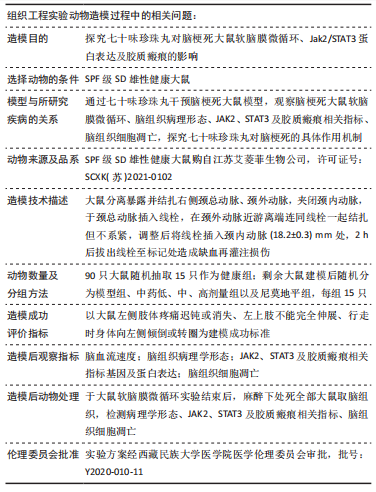
目的:探讨七十味珍珠丸对脑梗死大鼠软脑膜微循环、Janus激酶2(Janus Kinase2,JAK2)/转录激活因子3(signal transducer and activator transcription3,STAT3)蛋白表达及胶质瘢痕的影响。
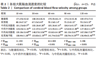
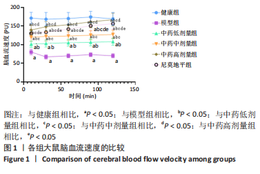
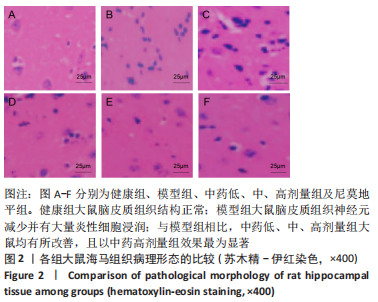
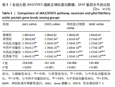
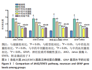
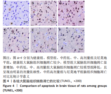
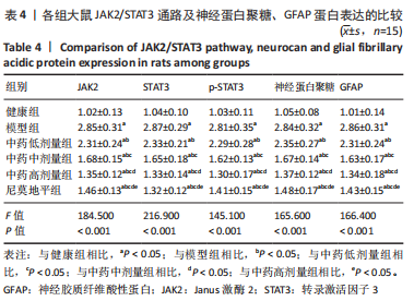
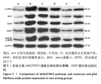
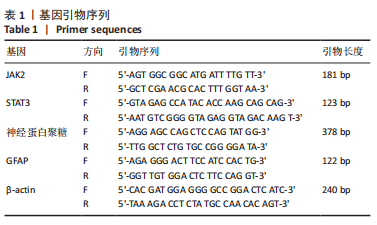
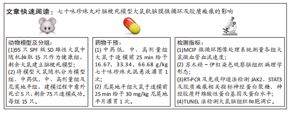
方法:于95只雄性SD大鼠中随机抽取15只作为健康组,剩余大鼠建立脑梗死模型,将模型大鼠随机分为模型组、中药低、中、高剂量组以及尼莫地平组,在建模过程中意外死亡5只,剩余75只建模成功,每组15只。中药低、中、高剂量组分别于造模前25 min给予16.67,33.34,66.68 g/kg七十味珍珠丸混悬液灌胃1次,尼莫地平组于造模前25 min给予 30 mg/kg尼莫地平片灌胃1次。建模后运用MCIP微循环图像处理系统检测脑血流速度,苏木精-伊红染色观察脑组织病理学形态,RT-PCR检测JAK2、STAT3、神经蛋白聚糖、胶质纤维酸性蛋白基因水平,TUNEL技术检测脑组织细胞凋亡情况,免疫印迹检测JAK2、STAT3、p-STAT3、神经蛋白聚糖、胶质纤维酸性蛋白的蛋白表达。

结果与结论:①与健康组相比,模型组不同时间点脑血流速度降低,JAK2、STAT3、p-STAT3、神经蛋白聚糖、胶质纤维酸性蛋白、神经元细胞凋亡率升高(P < 0.05);②与模型组相比,中药低剂量组不同时间点脑血流速度升高,JAK2、STAT3、磷酸化转录激活因子3、神经蛋白聚糖、胶质纤维酸性蛋白、神经元细胞凋亡率降低(P < 0.05);③与中药中剂量组相比,中药高剂量组与尼莫地平组不同时间点脑血流速度升高,JAK2、STAT3、p-STAT3、神经蛋白聚糖、胶质纤维酸性蛋白、神经元细胞凋亡率降低(P < 0.05);④与中药高剂量组相比,尼莫地平组不同时间点脑血流速度降低,JAK2、STAT3、p-STAT3、神经蛋白聚糖、胶质纤维酸性蛋白、神经元细胞凋亡率升高(P < 0.05);⑤健康组大鼠脑皮质组织结构正常;模型组脑皮质组织神经元减少,少数存活的神经元固缩,并有大量炎性细胞浸润;与模型组相比,中药低、中、高剂量组及尼莫地平组均有所改善,且以中药高剂量组效果显著;⑥提示七十味珍珠丸可能通过抑制胶质瘢痕标志蛋白神经蛋白聚糖、神经胶质纤维酸性蛋白水平影响胶质瘢痕的形成,还可改善脑梗死大鼠软脑膜微循环,并通过抑制JAK2/STAT3通路的激活,减少脑组织细胞的凋亡,从而发挥脑保护的作用。

https://orcid.org/0000-0001-6927-3013(马辉)

中国组织工程研究杂志出版内容重点:组织构建;骨细胞;软骨细胞;细胞培养;成纤维细胞;血管内皮细胞;骨质疏松;组织工程

关键词: 七十味珍珠丸, 脑梗死大鼠, 软脑膜微循环, JAK2/STAT3, 胶质瘢痕

Abstract: BACKGROUND: Improving cerebral infarction micromeningeal circulation and inhibiting scar formation can effectively treat cerebral infarction. Therefore, it is very important to develop safe and effective drugs to improve cerebral microcirculation and scar formation.
OBJECTIVE: To investigate the effects of Ratnasampil on pia meningeal microcirculation, Janus kinase 2/signal transducer and activator transcription 3 protein expression and glial scar in rats with cerebral infarction.
METHODS: The 15 of 95 male Sprague-Dawley rats were randomly selected as healthy group and the remaining rats used to establish cerebral infarction models. Five rats died accidentally during the modeling process and the remaining rats were successfully modeled. Model rats were divided into model group, low-dose, medium-dose and high-dose groups of Traditional Chinese medicine and nimodipine group with 15 rats in each group. Low-, medium-and high-dose groups were given 16.67, 33.34 and 66.68 g/kg Ratnasampil suspension once by intragastric administration respectively at 25 minutes before modeling. The nimodipine group was given 30 mg/kg nimodipine tablet once by intragastric administration at 25 minutes before modeling. MCIP microcirculation image processing system was used to detect cerebral blood flow velocity. Hematoxylin-eosin staining was used to observe brain histopathological morphology. Real-time fluorescence quantitative PCR was used to detect gene levels of Janus kinase 2, signal transducer and activator transcription 3, neurocan and glial fibrillary acidic protein. In situ terminal transferase labeling technique was used to measure cell apoptosis. Western blot was used to detect the protein expression of Janus kinase 2, signal transducer and activator transcription 3, phosphorylated signal transducer and activator transcription 3, neurocan and glial fibrillary acidic protein.
RESULTS AND CONCLUSION: Compared with the healthy group, the nimodipine group showed reduced cerebral blood flow velocity, increased expression of Janus kinase 2, signal transducer and activator transcription 3, phosphorylated signal transducer and activator transcription 3, neurocan and glial fibrillary acidic protein, and increased neuronal apoptosis rate at different time points (P < 0.05). Compared with the nimodipine group, the low-dose group showed an increase in cerebral blood flow velocity and a reduction in neuronal apoptosis rate and the expression of Janus kinase 2, signal transducer and activator transcription 3, phosphorylated signal transducer and activator transcription 3, neurocan and glial fibrillary acidic protein (P < 0.05). Compared with the medium-dose group, the high-dose and nimodipine groups showed an increase in cerebral blood flow velocity and a reduction in neuronal apoptosis rate and the expression of Janus kinase 2, signal transducer and activator transcription 3, phosphorylated signal transducer and activator transcription 3, neurocan and glial fibrillary acidic protein (P < 0.05). Compared with the high-dose group, the nimodipine group showed decreased cerebral blood flow velocity, increased neuronal apoptosis rate and elevated expression of Janus kinase 2, signal transducer and activator transcription 3, phosphorylated signal transducer and activator transcription 3, neurocan and glial fibrillary acidic protein at different time points (P < 0.05). The histological structure of the cerebral cortex was normal in the healthy group. In the model group, the number of neurons in the cortex was decreased, some living neurons were pyknotic, and a large number of inflammatory cells were infiltrated. Compared with the model group, these changes were all improved in the low-, medium- and high-dose groups and the nimodipine group, and the high-dose group had a remarkable effect. Overall, these findings suggest that Ratnasampil may affect the formation of glial scar by inhibiting the levels of glial scar marker proteins, including neurocan and glial fibrillary acidic protein, improve the pia meningeal microcirculation, and reduce the apoptosis of brain tissue cells by inhibiting the activation of Janus kinase 2/signal transducer and activator transcription 3 pathway in rats with cerebral infarction, thus playing a role in brain protection.

Key words: Ratnasampil, cerebral infarction rat, pia meningeal microcirculation, JAK2/STAT3, glial scar

中图分类号: