中国组织工程研究 ›› 2022, Vol. 26 ›› Issue (22): 3450-3454.doi: 10.12307/2022.269

• 纳米生物材料 nanobiomaterials • 上一篇    下一篇

一种新型安全近红外发光纳米粒的制备及成像实验

许馨之1,张  悦1,金  颖2,金春香1   

  1. 1吉林大学中日联谊医院超声科,吉林省长春市   130033;2吉林大学第一医院乳腺外科,吉林省长春市   130012
  • 收稿日期:2020-10-21 修回日期:2020-11-28 接受日期:2021-05-10 出版日期:2022-08-08 发布日期:2022-01-10
  • 通讯作者: 金春香,教授,博士生导师,吉林大学中日联谊医院超声科,吉林省长春市 130033
  • 作者简介:许馨之,女,1991年生,新疆维吾尔自治区库尔勒市人,汉族,吉林大学中日联谊医院在读博士,主要从事纳米材料载药/载基因抗肿瘤研究。
  • 基金资助:
    吉林省科技厅国际合作项目(20170414042GH),项目负责人:金春香;吉林省财政厅项目(2019SCZ060),项目负责人:金春香

Preparation and imaging experiment of a new type of safe near-infrared luminescent nanoparticles

Xu Xinzhi1, Zhang Yue1, Jin Ying2, Jin Chunxiang1   

  1. 1Department of Ultrasound, China-Japan Union Hospital, Jilin University, Changchun 130033, Jilin Province, China; 2Department of Breast Surgery, the First Hospital of Jilin University, Changchun 130012, Jilin Province, China
  • Received:2020-10-21 Revised:2020-11-28 Accepted:2021-05-10 Online:2022-08-08 Published:2022-01-10
  • Contact: Jin Chunxiang, Professor, Doctoral supervisor, Department of Ultrasound, China-Japan Union Hospital, Jilin University, Changchun 130033, Jilin Province, China
  • About author:Xu Xinzhi, Doctoral candidate, Department of Ultrasound, China-Japan Union Hospital, Jilin University, Changchun 130033, Jilin Province, China
  • Supported by:
    the International Cooperation Project of Science and Technology Department of Jilin Province, No. 20170414042GH (to JCX); the Project of Jilin Provincial Department of Finance, No. 2019SCZ060 (to JCX)

摘要:

文题释义:
近红外发光碳点:碳点制备简便、成本低、安全性高,是理想的体内示踪剂,但是目前制备的碳点多以蓝绿发光为主,组织穿透性差且无法进行实时追踪,而实验所使用的近红外发光碳点完美解决了上述问题,兼具安全性,且组织穿透性强,是理想的成像剂。
有机-无机杂化介孔二氧化硅:无机二氧化硅虽然具有体表面积大、制备简便、易于修饰等优点,但是其在体内不易降解,可能会造成不必要的循环负担。而将有机、无机硅源结合在一起,制备具有二硫键的杂化二氧化硅纳米粒,不仅具有无机二氧化硅纳米粒的优点,而且显著提高了其生物安全性。


背景:近红外发光碳点具备有蓝绿发光碳点不具备的组织穿透性,是理想的成像剂,但是由于其在体内易降解而无法到达靶点组织,实验将其与纳米粒相结合,使其能通过循环系统到达相应靶点,达到实时成像的目的。
目的:制备具有成像能力且安全性高的近红外成像载碳点介孔有机-无机杂化二氧化硅纳米粒(mesoporous organosilica nanocapsules-carbon nanodots,MON-CDs)。
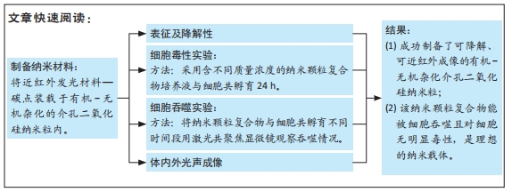
方法:利用胶束/前体共模板组装策略,以原硅酸四乙酯和双[3-(三乙氧基甲硅烷基)丙基]四硫化物为原材料、十六烷基三甲基氯化铵为模板剂、三乙醇胺为碱性催化剂成功制备了有机-无机杂化介孔二氧化硅纳米粒子,并将碳点加入到整个体系中制备MON-CDs。利用透射电镜与荧光光谱仪检测纳米粒的结构、形貌及其加载的荧光强度;利用光声成像仪、扫描激光共聚焦显微镜验证其体外成像能力,并在小鼠乳腺癌模型体内证明其体内光声成像能力;利用CCK-8实验检测不同质量浓度MON-CDs溶液的生物安全性。
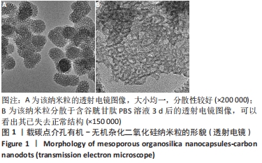
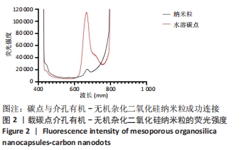
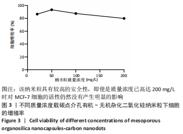
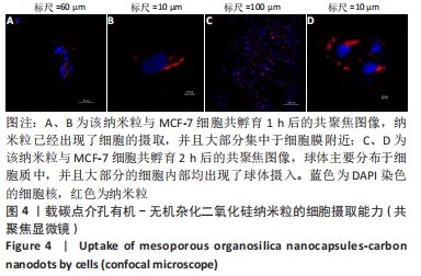
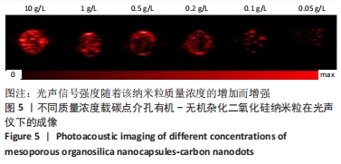
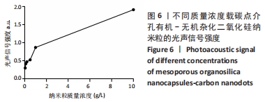
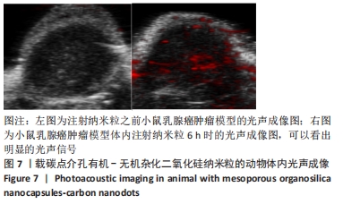
结果与结论:①透射电镜显示,MON-CDs的粒径为(50.0±4.6) nm,呈球形,大小均一且具备良好的分散性,孔道清晰可见,碳点参杂其中;荧光检测显示碳点与介孔有机-无机杂化二氧化硅纳米粒子成功连接;②CCK-8检测显示,当MON-CDs溶液的质量浓度在200 mg/L以内时无明显的细胞毒性;③扫描激光共聚焦显微镜显示,当MON-CDs与MCF-7细胞共孵育1 h时,纳米粒已出现了细胞摄取,并且大部分集中于细胞膜附近;共孵育2 h时,纳米粒累积进入细胞内的量增加,纳米粒主要分布于细胞质中,并且大部分细胞内部均出现了纳米粒的摄入;④光声成像检测显示,随着MON-CDs溶液质量浓度的增加,体外光声信号强度增强;经尾静脉注射MON-CDs溶液6 h后,在乳腺癌小鼠肿瘤组织处观察到了明显的光声信号;⑤结果表明,MON-CDs具有很好的生物安全性且拥有近红外发光,在光声成像仪及激光共聚焦下展现了良好的成像能力。
缩略语:载碳点介孔有机-无机杂化二氧化硅纳米粒:mesoporous organosilica nanocapsules-carbon nanodots,MON-CDs

https://orcid.org/0000-0002-7054-1406 (许馨之) 

中国组织工程研究杂志出版内容重点:生物材料;骨生物材料口腔生物材料纳米材料缓释材料材料相容性;组织工程

关键词: 材料, 纳米粒, 二氧化硅, 示踪剂, 近红外材料, 安全性, 生物成像, 安全降解

Abstract: BACKGROUND: Near-infrared luminescent carbon dots have tissue penetration that blue-green luminescent carbon dots do not have. They are ideal imaging agents. However, they are easily degraded in the body and cannot reach target tissues. This experiment combines them with nanoparticles, and it can reach the target through the circulatory system to achieve the purpose of real-time imaging. 
OBJECTIVE: To prepare mesoporous organosilica nanocapsules-carbon nanodots (MON-CDs) with imaging capability and high safety.
METHODS:  Utilizing the micelle/precursor co-template assembly strategy, using traditional tetraethoxysilane and bis[3-(triethoxysilyl)propyl]tetrasulfide) as the material, cetyltrimethylammonium chloride as the stencil agent, and triethanolamine as the alkaline catalyst, mesoporous organosilica nanoparticle (MON) was successfully prepared and carbon dots were added to the entire system to prepare MON-CDs. Transmission electron microscope and fluorescence spectrometer were applied to detect the structure, morphology and fluorescence intensity of nanoparticles. Photoacoustic image system and scanning laser confocal microscope were utilized to verify imaging ability, and in vivo photoacoustic imaging in mice was proven. CCK-8 was used to test the biosafety of MON-CDs solutions of different mass concentrations.  
RESULTS AND CONCLUSION: (1) The transmission electron microscope showed that the particle size of MON-CDs was (50.0±4.6) nm, which was spherical, uniform in size, and had good dispersibility. The pores were clearly visible and the carbon dots were mixed in it. Fluorescence detection showed that the carbon dots and mesoporous organic-inorganic hybrid silica nanoparticles were successfully connected. (2) CCK-8 detection showed that when the mass concentration of MON-CDs solution was within 200 mg/L, there was no obvious cytotoxicity. (3) Scanning laser confocal microscope showed that when MON-CDs were incubated with MCF-7 cells for 1 hour, the nanoparticles had already been taken up by cells, and most of them were concentrated near the cell membrane. When the CDs were incubated for 2 hours, the amount of nanoparticles accumulated into the cells increased, and the nanoparticles were mainly distributed in the cytoplasm. In addition, the uptake of nanoparticles occurred in most of the cells. (4) Photoacoustic imaging showed that with the proliferation of MON-CDs solution mass concentration, the in vitro photoacoustic signal intensity increased. At 6 hours after MON-CDs solution injection via tail vein, photoacoustic signals were observed at the tumor tissues of breast cancer mice. (5) The results showed that MON-CDs have good biosafety and possess near-infrared luminescence, and demonstrate good imaging capabilities under photoacoustic imager and laser confocal microscope.

Key words: materials, nanoparticles, silica, tracers, near-infrared materials, safety, bioimaging, safe degradation

中图分类号: