中国组织工程研究 ›› 2020, Vol. 24 ›› Issue (20): 3152-3156.doi: 10.3969/j.issn.2095-4344.2610

• 脊柱组织构建 spinal tissue construction • 上一篇    下一篇

基于筋束骨理论建立慢性劳损型上颈椎失稳尸体模型及评价

梁  龙1,于  杰1,2,魏  戌1,2,周帅琪1,3,尹逊路1,2,刘广伟2,谢  瑞1,谢  榕1,3,庄明辉1,朱立国1,2,冯敏山1,2   

  1. 1中国中医科学院望京医院脊柱二科,北京市  100102;2中医正骨技术北京市重点实验室,北京市  100102;3北京中医药大学,北京市  100029
  • 收稿日期:2019-08-29 修回日期:2019-08-31 接受日期:2019-10-24 出版日期:2020-07-18 发布日期:2020-04-13
  • 通讯作者: 朱立国,博士,教授,中国中医科学院望京医院脊柱二科,北京市 100102;中医正骨技术北京市重点实验室,北京市 100102 并列通讯作者:冯敏山,博士,主任医师,中国中医科学院望京医院脊柱二科,北京市 100102;中医正骨技术北京市重点实验室,北京市 100102
  • 作者简介:梁龙,男,1990年生,安徽省全椒县人,汉族,中国中医科学院在读博士,医师,主要从事骨与关节退变研究。
  • 基金资助:
    国家自然科学基金(81173279);国家自然科学基金(81774330);国家科技支撑计划(2014BAI08B06);中医药行业科研专项(201107004);横向课题(HZL2017001);中国中医科学院“十三五”重点领域科研专项(ZZ10-022);中国中医科学院科技创新优势团队(YS1304);国家留学基金委基金(201908110307)

Establishment and evaluation of a cadaveric model of chronic strain-induced upper cervical spine instability based on fascia-bone theory

Liang Long1, Yu Jie1, 2, Wei Xu1, 2, Zhou Shuaiqi1, 3, Yin Xunlu1, 2, Liu Guangwei2, Xie Rui1, Xie Rong1, 3, Zhuang Minghui1, Zhu Liguo1, 2, Feng Minshan1, 2   

  1. 1Second Department of Spine, Wangjing Hospital, China Academy of Chinese Medical Sciences, Beijing 100102, China; 2Beijing Key Laboratory of TCM Bone Setting, Beijing 100102, China; 3Beijing University of Chinese Medicine, Beijing 100029, China
  • Received:2019-08-29 Revised:2019-08-31 Accepted:2019-10-24 Online:2020-07-18 Published:2020-04-13
  • Contact: Zhu Liguo, MD, Professor, Second Department of Spine, Wangjing Hospital, China Academy of Chinese Medical Sciences, Beijing 100102, China; Beijing Key Laboratory of TCM Bone Setting, Beijing 100102, China Feng Minshan, MD, Chief physician, Second Department of Spine, Wangjing Hospital, China Academy of Chinese Medical Sciences, Beijing 100102, China; Beijing Key Laboratory of TCM Bone Setting, Beijing 100102, China
  • About author:Liang Long, MD candidate, Physician, Second Department of Spine, Wangjing Hospital, China Academy of Chinese Medical Sciences, Beijing 100102, China
  • Supported by:
    the National Natural Science Foundation of China, No. 81173279  and 81774330; the National Science and Technology Support Program, No. 2014BAI08B06; Special Research Project of the Chinese Medicine Industry, No. 201107004; Horizontal Project, No. HZL2017001; “Thirteenth Five-Year” Specific Scientific Research Project of China Academy of Chinese Medical Sciences, No. ZZ10-022; Science and Technology Innovation Team of China Academy of Chinese Medical Sciences, No. YS1304; a granted by China Scholarship Council, No. 201908110307

摘要:

文题释义:

慢性劳损所致的上颈椎失稳:是由于长期累积性力引起颈部筋伤,筋不束骨,最终发生“筋出槽,骨错缝”,导致上颈椎活动度的异常改变。

“筋束骨”理论:“诸筋者,皆会于节”“宗筋主束骨而利机关也”等论述均表明筋能聚于关节附近维持关节的稳定性。当筋长期劳损后,出现“筋伤”,进而“束骨”能力下降,最终导致“骨错缝”的发生。

背景:对于上颈椎失稳机制的尸体标本研究多集中于急性暴力模型,尚缺乏慢性劳损型上颈椎失稳模型。

目的:根据“筋束骨”理论构建慢性劳损致上颈椎失稳尸体模型并进行模型评价。 

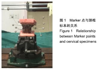
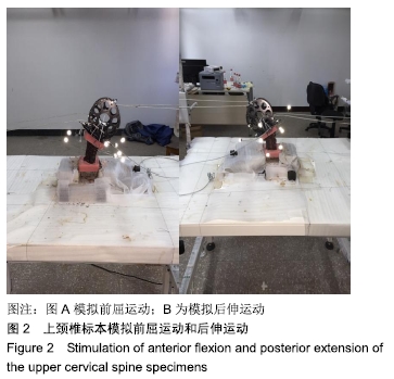
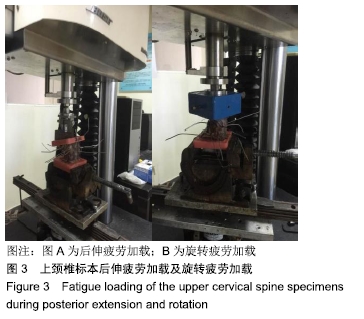
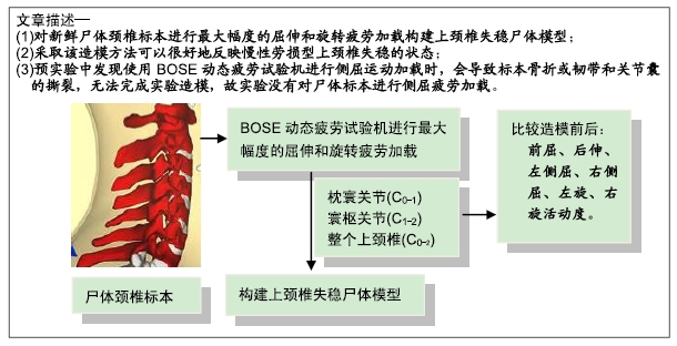
方法:将9具新鲜尸体颈椎标本准备完毕后,先用Motion Analysis运动捕捉系统进行正常椎体活动度的检测,再使用BOSE动态疲劳试验机进行最大幅度的屈伸和旋转疲劳加载构建上颈椎失稳尸体模型,再次进行活动度的检测,比较造模前和造模后的上颈椎模型的枕寰关节、寰枢关节和整个上颈椎结构的前屈、后伸、左侧屈、右侧屈、左旋、右旋活动度。研究方案的实施符合南方医科大学的相关伦理要求,标本供者自愿捐赠。

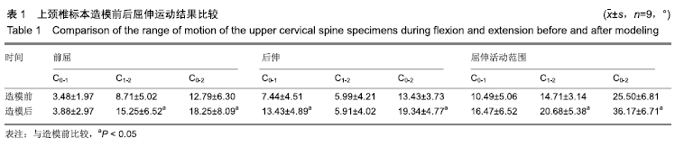
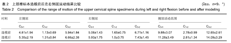
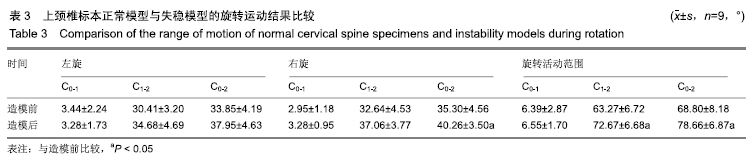
结果与结论:①在前屈运动时,造模后标本的寰枢关节(C1-2)、整个上颈椎(C0-2)活动范围明显大于造模前的标本(P < 0.05);后伸时,造模后标本的寰枕关节(C0-1)、整个上颈椎(C0-2)活动范围明显大于造模前(P < 0.05);在屈伸复合运动时,造模后标本的寰枢关节(C1-2)、整个上颈椎(C0-2)活动范围显著大于造模前标本(P < 0.05);②在侧屈活动中,造模后标本寰枕关节(C0-1)、寰枢关节(C1-2)、整个上颈椎(C0-2)的活动度均较造模前有所增大,但差异无显著性意义(P > 0.05);③在右旋活动时,造模后标本的整个上颈椎(C0-2)的活动度较造模前明显增加(P < 0.05);④在左右旋复合运动时,造模后标本的寰枢关节(C1-2)、整个上颈椎(C0-2)活动范围明显大于造模前的标本(P < 0.05);⑤结果说明,采取该造模方法可以很好地反映慢性劳损型上颈椎失稳的状态。

ORCID: 0000-0001-7193-8547(梁龙)

中国组织工程研究杂志出版内容重点:组织构建;骨细胞;软骨细胞;细胞培养;成纤维细胞;血管内皮细胞;骨质疏松组织工程

关键词: 上颈椎失稳, 慢性劳损, 造模, 活动度, 筋束骨

Abstract:

BACKGROUND: Studies regarding the instability of upper cervical spine in cadaveric specimens are mostly based on an acute violence model, and there is still no chronic strain-induced upper cervical instability model.

OBJECTIVE: To construct and evaluate the cadaveric model of the upper cervical spine instability caused by chronic strain according to the theory of “fascia and bone.”

METHODS: Nine fresh cadaveric cervical spine specimens were prepared and the normal vertebral range of motion was detected by Motion Analysis Motion Capture System. The upper cervical spine instability model was constructed by using BOSE dynamic fatigue testing machine with maximum flexion, extension and rotation fatigue loading. Then, the vertebral range of motion was tested again. The anterior flexion, posterior extension, left flexion, right flexion, left and right rotation of the occipitoatlantoaxial joint, the atlantoaxial joint and the whole upper cervical spine were compared before and after modeling. The implementation of the study protocol complied with the relevant ethical requirements of Southern Medical University, and the specimens were voluntarily donated.

RESULTS AND CONCLUSION: (1) During anterior flexion, the range of motion of the atlantoaxial joint (C1-2) and the entire upper cervical vertebra (C0-2) of the specimens after modeling was significantly larger than that before modeling (P < 0.05). During posterior extension, the range of motion of the atlantooccipital joint (C0-1) and the entire upper cervical vertebra (C0-2) of the specimens after modeling was significantly larger than that before modeling (P < 0.05). During both flexion and extension, the range of motion of the atlantoaxial joint (C1-2) and the entire upper cervical vertebra (C0-2) of the specimens after modeling was significantly larger than that of the pre-modeling specimen (P < 0.05). (2) During lateral flexion, the range of motion of the atlantooccipital joint (C0-1), the atlantoaxial joint (C1-2), and the entire upper cervical vertebra (C0-2) of the specimens after modeling was increased compared with that before modeling. However, there was no significant difference (P > 0.05). (3) During right rotation, the range of motion of the whole upper cervical spine (C0-2) of the specimens after modeling was significantly increased compared with that before modeling (P < 0.05). During both left and right rotation, the range of motion of the atlantoaxial joint (C1-2) and the whole upper cervical spine (C0-2) of the specimens was significantly larger than that of the specimens before modeling (P < 0.05). Therefore, this model can be used to reflect the state of upper cervical instability caused by chronic strain.

Key words: the upper cervical instability, chronic strain, modeling, range of motion, fascia and bone

中图分类号: