中国组织工程研究 ›› 2018, Vol. 22 ›› Issue (14): 2185-2189.doi: 10.3969/j.issn.2095-4344.0795

• 纳米生物材料 nanobiomaterials • 上一篇    下一篇

纯钛表面纳米银涂层构建方法及对金黄色葡萄球菌抗菌性能的影响

王复超,王明伟,李华壮,王善涛,赵光宗   

  1. 潍坊市益都中心医院,山东省潍坊市 262500
  • 收稿日期:2018-03-20 出版日期:2018-05-18 发布日期:2018-05-18
  • 通讯作者: 李华壮,博士,主任医师,潍坊市益都中心医院,山东省潍坊市 262500
  • 作者简介:王复超,男,汉族,1978年生,山东省临沂市沂水县人,2007年潍坊医学院毕业,硕士,主治医师。
  • 基金资助:

    山东省医药卫生科技发展计划项目(2016YX105)

Construction of nano-silver coating on pure titanium surface and its antibacterial effect on Staphylococcus aureus

  • Received:2018-03-20 Online:2018-05-18 Published:2018-05-18
  • Contact: Li Hua-zhuang, M.D., Chief physician, Yidu Central Hospital of Weifang, Weifang 262500, Shandong Province, China
  • About author:Wang Fu-chao, Master, Attending physician, Yidu Central Hospital of Weifang, Weifang 262500, Shandong Province, China

摘要:

文章快速阅读:


文题释义:
金属钛:是一种对人体无害的金属,具有较强的理化性能及化学稳定性,并且其低弹性模量与人体骨骼相仿,已经广泛用于骨科、口腔等医学领域。
纯银及复合材料:纯银是一种具有白色光泽的金属元素,纯银具有较强的韧性、延展性,并且材料导电、导热性能较好。当以20∶1比例的微米羟基磷灰石与微米银球磨经水浴、超声及热处理后能完成纯钛表面纳米银涂层构建,对金黄色葡萄球菌具有良好的抗菌性能,降低骨植入后感染率提供新的方法。
 
 
背景:纳米银是以纳米技术为基础制成的新型抗菌材料,理化性能稳定,且在电学、光学、催化方法性能较高,但是对于金黄色葡萄球菌抗菌性能颇有争议。
目的:探讨纯钛表面纳米银涂层构建方法及对金黄色葡萄球菌抗菌性能的影响。
方法:①纯钛表面纳米银涂层的构建:以羟基磷灰石及银粉末作为研究对象,根据20∶1比例进行充分混合,将混合物进行球磨后保证颗粒能达到纳米级;②抗菌性能观察:以金黄色葡萄球菌为对象,采用最小抑菌浓度试验测定纯钛表面纳米银涂层构建对金黄色葡萄球菌振荡培养时的抑菌浓度值,采用扫描电镜观察经不同质量浓度纯钛表面纳米银涂层处理后葡萄球菌结构。

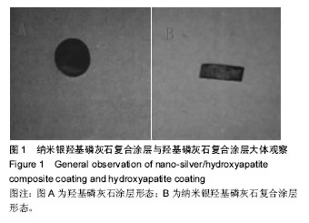
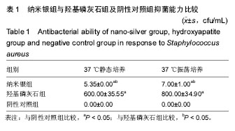
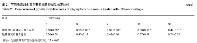
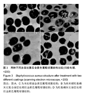
结果和结论:①纳米银羟基磷灰石复合涂层与羟基磷灰石复合涂层一般形态表面涂层相对均匀。纳米银羟基磷灰石复合涂层颜色略呈白色但是略带黄,羟基磷灰石复合涂层钛片呈白色,涂层表面毛糙,结合牢固,均未见涂层剥脱;②纳米银组在37 ℃静态培养与37 ℃振荡培养抑菌能力,均高于羟基磷灰石组(P < 0.05);③纳米银羟基磷灰石复合涂层在7,10,30 h对金黄色葡萄球菌600 nm吸光度值均低于羟基磷灰石复合涂层(P < 0.05);④与羟基磷灰石涂层相比,纳米银羟基磷灰石复合涂层细胞颜色变浅,存在破裂死亡的细胞,并且金黄色葡萄球菌数量减少,存在大量空泡;⑤结果提示:以20∶1比例的微米羟基磷灰石与微米银球磨经水浴、超声及热处理后成功构建纳米银涂层,该涂层在金黄色葡萄球菌中能发挥优越抗菌效果。

ORCID: 0000-0001-6106-1470(李华壮)

关键词: 纳米银, 纳米技术, 抗菌材料, 纯钛表面纳米银涂层, 金黄色球菌, 生物材料, 最小抑菌浓度试验, 烧瓶振荡法, 扫描电镜, 组织工程

Abstract:

BACKGROUND: Nano-silver is a new antibacterial material developed based on nano-technology. It is characterized as stable physical and chemical properties, and good electrical, optical, and catalytic performance, but its antibacterial response to Staphylococcus aureus is controversial.

OBJECTIVE: To explore the preparation methods of nano-silver coating on pure titanium surface and to explore its antibacterial effect on Staphylococcus aureus.
METHODS: (1) Preparation of nano-silver coating on the surface of pure titanium: Hydroxyapatite and silver powder as research objects were fully mixed at a ratio of 20:1. The mixture was ball-milled to ensure the formation of nanosized particles. The mixture of hydroxyapatite and silver powder was put into a stirrer for continuous stirring to ensure the uniform distribution. The mixture was then placed in an ethanol solution followed by insertion of a titanium plate (anode) and a stainless steel plate (cathode). The precipitated powder was obtained on the titanium surface at a voltage of 20 V, and the pure titanium surface with nano-silver coating was prepared after heat treatment in a tube resistance furnace. (2) Observation of antibacterial properties: A minimal inhibitory concentration test was used to determine the antibacterial concentration of the nano-silver coating on the pure titanium surface when shaken with Staphylococcus aureus. A scanning electron microscope was used to observe the structure of Staphylococcus aureus on the pure titanium surface with nano-silver coatings of different mass concentrations.

RESULTS AND CONCLUSION: Nano-silver/hydroxyapatite composite coating and hydroxyapatite coating were relatively uniform. The nano-silver/hydroxyapatite composite coating was white in color but slightly yellowish. The hydroxyapatite coating was white in color. The coating surface was rough and bonded firmly. No peeling of the coating was observed under gross observation. The antibacterial ability of the nano-silver group at 37 static culture and at 37 ℃ shaking culture was significantly higher than that of the hydroxy-apatite group (P < 0.05). The absorbance value of Staphylococcus aureus at 600 nm in the nano-silver group was lower than that of hydroxyapatite group at 7, 10, 30 hours after intervention (P < 0.05). The layer cells on the nano-silver coating become lighter in color compared with those on the hydroxyapatite coating and there were cells that ruptured and died. The number of Staphylococcus aureus on the nano-silver coating was reduced, and a large number of vacuoles were found. These findings indicate that the 20:1 mixture of hydroxyapatite and silver at micron level can be used to prepare nano-silver coating through ball milling, water bath, ultrasound and heat treatment. The prepared coating can exert excellent antibacterial effects on Staphylococcus aureus.

Key words: Nanotechnology, Staphylococcus, Staphylococcaceae

中图分类号: