|
[1] Poli M, Burillon C, Auxenfans C, et al. Immunocytochemical Diagnosis of Limbal Stem Cell Deficiency: Comparative Analysis of Current Corneal and Conjunctival Biomarkers. Cornea. 2015;34(7):817-823.
[2] Casaroli-Marano RP, Nieto-Nicolau N, Martínez-Conesa EM, et al. Potential Role of Induced Pluripotent Stem Cells (IPSCs) for Cell-Based Therapy of the Ocular Surface. J Clin Med. 2015; 4(2):318-342.
[3] deAraujo AL, Gomes JÁ. Corneal stem cells and tissue engineering: Current advances and future perspectives. World J Stem Cells. 2015;7(5):806-814.
[4] Chen C, Shen B, Xiao JJ,et al. Currently Clinical Views on Genetics of Wilson's Disease. Chin Med J (Engl). 2015; 128(13): 1826-1830.
[5] Pedrotti E, Passilongo M, Fasolo A, et al. In Vivo Confocal Microscopy 1 Year after Autologous Cultured Limbal StemCell Grafts. Ophthalmology. 2015;122(8):1660-1668.
[6] Al Arfaj K. Boston keratoprosthesis - Clinical outcomes with wider geographic use and expanding indications - A systematic review.Saudi J Ophthalmol. 2015;29(3):212-221.
[7] Zhang MC, Liu X, Jin Y,et al. Lamellar keratoplasty treatment of fungal corneal ulcers with acellular porcine corneal stroma. Am J Transplant. 2015;15(4):1068-1075.
[8] Koulikovska M, Rafat M, Petrovski G, et al. Enhanced regeneration of corneal tissue via a bioengineered collagen construct implanted by a nondisruptive surgical technique. Tissue Eng Part A.2015;21(5-6):1116-1130.
[9] Li DQ, Wang Z, Yoon KC, et al.Characterization, isolation, expansion and clinical therapy of human corneal epithelial stem/progenitor cells.J Stem Cells. 2014;9(2):79-91.
[10] Samoil? O, Sori??u O, Totu L, et al. Cultivation and characterization of limbal epithelial stem cells in rabbits. Rom J Morphol Embryol.2014;55(1):63-69.
[11] Burman S, Sangwan V. Cultivated limbal stem cell transplantation for ocular surface reconstruction. Clin Ophthalmol. 2008;2(3):489-502.
[12] Fernandes M, Sangwan VS, Rao SK, et al. Limbal stem cell transplantation.Indian J Ophthalmol. 2004;52(1):5-22.
[13] Chen G, Tian F, Li C, et al.In vivo real-time visualization of mesenchymal stem cells tropism for cutaneous regeneration using NIR-II fluorescence imaging. Biomaterials. 2015;53: 265-273.
[14] Yang JW, Zhang YF, Wan CY, et al. Autophagy in SDF-1α- mediated DPSC migration and pulp regeneration. Biomaterials. 2015;44:11-23.
[15] Yuan S, Fan G. Stem cell-based therapy of corneal epithelial and endothelial diseases. Regen Med. 2015;10(4):495-504.
[16] Tsai IL, Hsu CC, Hung KH, et al. Applications of biomaterials in corneal wound healing.J Chin Med Assoc. 2015;78(4):212-217.
[17] Rocca CJ, Kreymerman A, Ur SN, et al.Treatment of Inherited Eye Defects by Systemic Hematopoietic Stem Cell Transplantation. Invest Ophthalmol Vis Sci. 2015;56(12): 7214-7223.
[18] Tanimoto N, Michalakis S, Weber BH, et al. In-Depth Functional Diagnostics of Mouse Models by Single-Flash and Flicker Electroretinograms without Adapting Background Illumination. Adv Exp Med Biol. 2016;854:619-625.
[19] Singh V, Shukla S, Ramachandran C,et al. Science and Art of Cell-Based Ocular Surface Regeneration. Int Rev Cell Mol Biol. 2015;319:45-106.
[20] Sehic A, Utheim ØA, Ommundsen K, et al. Pre-Clinical Cell-Based Therapy for Limbal Stem Cell Deficiency. J Funct Biomater. 2015;6(3):863-888.
[21] Sehic A, Utheim ØA, Ommundsen K, et al.Pre-Clinical Cell-Based Therapy for Limbal Stem Cell Deficiency.J Funct Biomater. 2015;6(3):863-888.
[22] Bardag-Gorce F, Oliva J, Wood A, et al. Carrier-free cultured autologous oral mucosa epithelial cell sheet (CAOMECS) for corneal epithelium reconstruction: a histological study.Ocul Surf. 2015;13(2):150-163.
[23] Zhao Y, Ma L. Systematic review and meta-analysis on transplantation of ex vivo cultivated limbal epithelial stem cell on amniotic membrane in limbal stem cell deficiency.Cornea. 2015; 34(5):592-600.
[24] Sheth R, Neale MH, Shortt AJ, et al. Culture and Characterization of Oral Mucosal Epithelial Cells on a Fibrin Gel for Ocular Surface Reconstruction. Curr Eye Res. 2015;40(11): 1077-1087.
[25] Sareen D, Saghizadeh M, Ornelas L,et al. Differentiation of human limbal-derived induced pluripotent stem cells into limbal-like epithelium. Stem Cells Transl Med. 2014;3(9): 1002-1012.
[26] Levis HJ, Kureshi AK, Massie I, et al. Tissue Engineering the Cornea: The Evolution of RAFT. J Funct Biomater.2015;6(1): 50-65.
[27] Syed-Picard FN, Du Y, Lathrop KL, et al. Dental pulp stem cells: a new cellular resource for corneal stromal regeneration.Stem Cells Transl Med. 2015;4(3):276-285.
[28] Kenyon KR, Tseng SC. Limbal autograft transplantation for ocular surface disorders. Ophthalmology. 1989; 96(5):709-723.
[29] Pellegrini G, Traverso CE, Franzi AT, et al. Long-term restoration of damaged corneal surfaces with autologous cultivated corneal epithelium. Lancet. 1997; 349:990-993.
[30] Ang LP, Sotozono C, Koizumi N, et al. A comparison between cultivated and conventional limbal stem cell transplantation for Stevens-Johnson syndrome. Am J Ophthalmol. 2007; 143(1): 178-180.
[31] Shortt AJ, Bunce C, Levis HJ, et al.Three-year outcomes of cultured limbal epithelial allografts in aniridia and Stevens- Johnson syndrome evaluated using the Clinical Outcome Assessment in Surgical Trials assessment tool.Stem Cells Transl Med. 2014;3(2):265-275.
[32] Meyer-Blazejewska EA, Call MK, Yamanaka O, et al. From hair to cornea: toward the therapeutic use of hair follicle-derived stem cells in the treatment of limbal stem cell deficiency. Stem Cells. 2011;29(1):57-66.
[33] Yang X, Qu L, Wang X, et al.Plasticity of epidermal adult stem cells derived from adult goat ear skin.Mol Reprod Dev. 2007; 74(3):386-96.
[34] Kadar T, Dachir S, Cohen L, et al.Ocular injuries following sulfur mustard exposure--pathological mechanism and potential therapy. Toxicology. 2009;263(1):59-69.
[35] Daya SM, Watson A, Sharpe JR, et al.Outcomes and DNA analysis of ex vivo expanded stem cell allograft for ocular surface reconstruction. Ophthalmology.2005; 112(3):470-477.
[36] Henderson TR, Coster DJ, Williams KA. The long term outcome of limbal allografts: the search for surviving cells. Br J Ophthalmol. 2001; 85(5):604-609.
[37] Rama P, Matuska S, Paganoni G, et al. Limbal stem-cell therapy and long-term corneal regeneration. N Engl J Med. 2010; 363(2):147-155.
[38] Aiuti A, Webb IJ, Bleul C, et al. The chemokine SDF-1 is a chemoattractant for human CD34+ hematopoietic progenitor cells and provides a new mechanism to explain the mobilization of CD34+ progenitors to peripheral blood. J Exp Med. 1997; 185(1):111-120.
[39] Jo DY, Rafii S, Hamada T, et al. Chemotaxis of primitive hematopoietic cells in response to stromal cell-derived factor-1. J Clin Invest. 2000; 105(1):101-111.
[40] Ratajczak MZ, Reca R, Wysoczynski M, et al. Transplantation studies in C3-deficient animals reveal a novel role of the third complement component (C3) in engraftment of bone marrow cells. Leukemia. 2004; 18(9):1482-1490.
[41] Machalińska A, K?os P, Baumert B, et al. Stem Cells are mobilized from the bone marrow into the peripheral circulation in response to retinal pigment epithelium damage--a pathophysiological attempt to induce endogenous regeneration. Curr Eye Res. 2011; 36(7):663-672.
[42] Li Y, Reca RG, Atmaca-Sonmez P, et al.Retinal pigment epithelium damage enhances expression of chemoattractants and migration of bone marrow-derived stem cells. Invest Ophthalmol Vis Sci. 2006; 47(4):1646-1652.
[43] Bourcier T, Berbar T, Paquet S, et al. Characterization and functionality of CXCR4 chemokine receptor and SDF-1 in human corneal fibroblasts. Mol Vis. 2003; 9:96-102.
[44] Xie HT, Chen SY, Li GG, et al. Limbal epithelial stem/progenitor cells attract stromal niche cells by SDF-1/CXCR4 signaling to prevent differentiation. Stem Cells. 2011; 29(11):1874-1885.
|
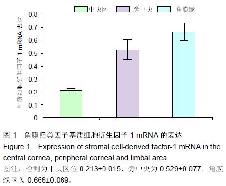
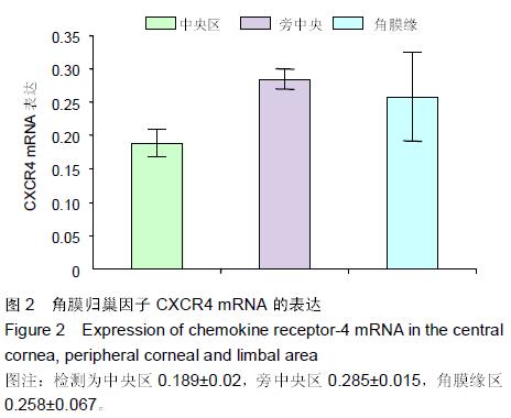
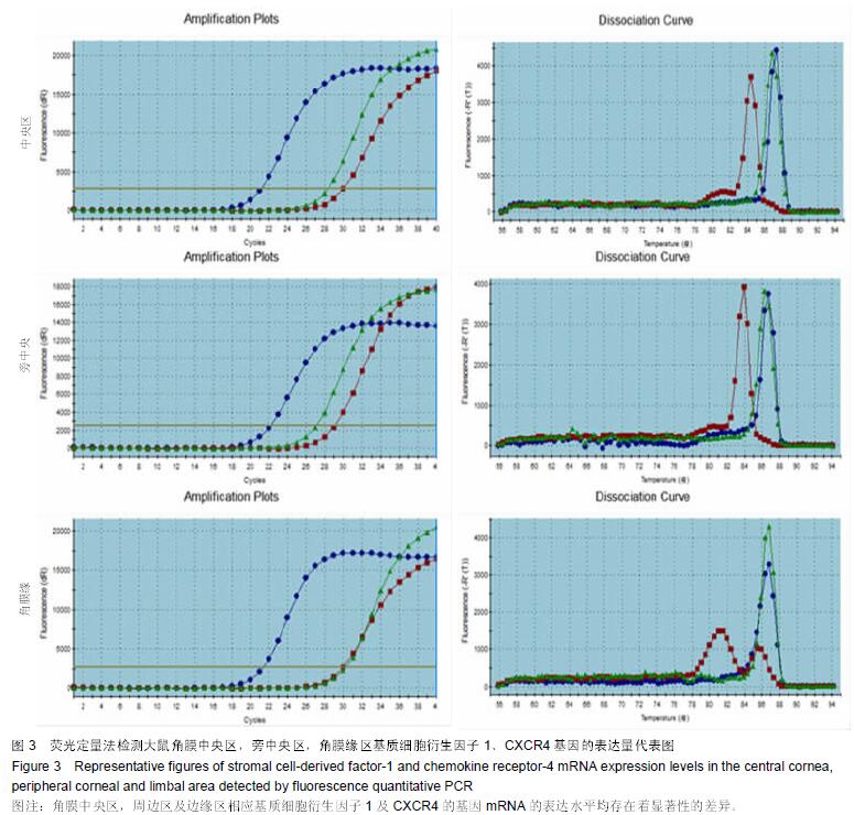
.jpg)