[1] TSAO CW, ADAY AW, ALMARZOOQ ZI, et al. Heart disease and stroke statistics-2023 update: a report from the american heart association. Circulation. 2023;147(8):e93-e621.
[2] KITSUKA T, SHIRAKI A, OYAMA J, et al. A novel soluble epoxide hydrolase vaccine protects murine cardiac muscle against myocardial infarction. Sci Rep. 2022;12(1):6923.
[3] YU H, LU K, ZHU J, et al. Stem cell therapy for ischemic heart diseases. Br Med Bull. 2017; 121(1):135-154.
[4] KITSUKA T, TAKAHASHI F, REINHARDT J, et al. Advances in cardiac tissue engineering. Bioengineering. 2022;9(11):696.
[5] 王颖薇,秦子夕,武征.心肌补片的制备与结构功能特点[J].中国组织工程研究,2015, 19(38):6211-6216.
[6] THUMMARATI P, LAIWATTANAPAISAL W, NITTA R, et al. Recent advances in cell sheet engineering: from fabrication to clinical translation. Bioengineering (Basel). 2023;10(2):211.
[7] 周术奎,张楷乐,王营,等.细胞膜片技术在组织工程中的应用与研究进展[J].中国组织工程研究,2016,20(11):1630-1636.
[8] WANG Z, WANG L, LI T, et al. 3D bioprinting in cardiac tissue engineering. Theranostics. 2021;11(16):7948-7969.
[9] WEI Q, ZHOU J, AN Y, et al. Modification, 3D printing process and application of sodium alginate based hydrogels in soft tissue engineering: a review. Int J Biol Macromol. 2023; 232:123450.
[10] SEKINE H, SHIMIZU T, DOBASHI I, et al. Cardiac cell sheet transplantation improves damaged heart function via superior cell survival in comparison with dissociated cell injection. Tissue Engineering Part A. 2011;17(23-24):2973-2980.
[11] MASUDA N, SEKINE H, NIINAMI H, et al. Engineering of functional cardiac tubes by stepwise transplantation of cardiac cell sheets onto intestinal mesentery. Heart Vessels. 2020;35(6):859-867.
[12] KAYNAK BAYRAK G, GÜMÜŞDERELIOĞLU M. Construction of cardiomyoblast sheets for cardiac tissue repair: comparison of three different approaches. Cytotechnology. 2019; 71(4):819-833.
[13] QI YX, HAN Y, JIANG ZL. Mechanobiology and vascular remodeling: from membrane to nucleus. Adv Exp Med Biol. 2018;1097:69-82.
[14] SHUDO Y, COHEN JE, MACARTHUR JW, et al. A tissue-engineered chondrocyte cell sheet induces extracellular matrix modification to enhance ventricular biomechanics and attenuate myocardial stiffness in ischemic cardiomyopathy. Tissue Engineering Part A. 2015;21(19-20):2515-2525.
[15] VENUGOPAL H, HANNA A, HUMERES C, et al. Properties and functions of fibroblasts and myofibroblasts in myocardial infarction. Cells. 2022;11(9):1386.
[16] CHAUDHURI R, RAMACHANDRAN M, MOHARIL P, et al. Biomaterials and cells for cardiac tissue engineering: current choices. Mater Sci Eng C. 2017;79:950-957.
[17] IWAMIYA T, MATSUURA K, MASUDA S, et al. Cardiac fibroblast-derived VCAM-1 enhances cardiomyocyte proliferation for fabrication of bioengineered cardiac tissue. Regen Ther. 2016;4:92-102.
[18] ZAKHAROVA L, MASTROENI D, MUTLU N, et al. Transplantation of cardiac progenitor cell sheet onto infarcted heart promotes cardiogenesis and improves function. Cardiovasc Res. 2010;87(1):40-49.
[19] THUMMARATI P, KINO-OKA M. Effect of co-culturing fibroblasts in human skeletal muscle cell sheet on angiogenic cytokine balance and angiogenesis. Front Bioeng Biotechnol. 2020,8:578140.
[20] GUO R, MORIMATSU M, FENG T, et al. Stem cell-derived cell sheet transplantation for heart tissue repair in myocardial infarction. Stem Cell Res Ther. 2020;11(1):19.
[21] OLIVA J, FLORENTINO A, BARDAG-GORCE F, et al. Engineering, differentiation and harvesting of human adipose-derived stem cell multilayer cell sheets. Regen Med. 2019; 14(3):151-163.
[22] XIAO Y, CHEN Y, SHAO C, et al. Strategies to improve the therapeutic effect of pluripotent stem cell-derived cardiomyocytes on myocardial infarction. Front Bioeng Biotechnol. 2022;10:973496.
[23] LEOR J, ABOULAFIA-ETZION S, DAR A, et al. Bioengineered cardiac grafts: a new approach to repair the infarcted myocardium? Circulation. 2000;102(19 Suppl 3):I56-I61.
[24] KEHAT I, KENYAGIN-KARSENTI D, SNIR M, et al. Human embryonic stem cells can differentiate into myocytes with structural and functional properties of cardiomyocytes. J Clin Invest. 2001;108(3):407-414.
[25] TAKAHASHI K, TANABE K, OHNUKI M, et al. Induction of pluripotent stem cells from adult human fibroblasts by defined factors. Cell. 2007;131(5):861-872.
[26] STEVENS KR, PABON L, MUSKHELI V, et al. Scaffold-free human cardiac tissue patch created from embryonic stem cells. Tissue Eng Part A. 2009;15(6):1211-1222.
[27] MENASCHÉ P, VANNEAUX V, HAGÈGE A, et al. Human embryonic stem cell-derived cardiac progenitors for severe heart failure treatment: first clinical case report: Figure 1. Eur Heart J. 2015;36(30):2011-2017.
[28] BUDHARAJU H, SUNDARAMURTHI D, SETHURAMAN S. Efficient dual crosslinking of protein–in–polysaccharide bioink for biofabrication of cardiac tissue constructs. Biomat Adv. 2023;152:213486.
[29] SHIN HS, THAKORE A, TADA Y, et al. Angiogenic stem cell delivery platform to augment post-infarction neovasculature and reverse ventricular remodeling. Sci Rep. 2022;12(1): 17605.
[30] ZHANG L, GUO J, ZHANG P, et al. Derivation and high engraftment of patient-specific cardiomyocyte sheet using induced pluripotent stem cells generated from adult cardiac fibroblast. Circ Heart Fail. 2015;8(1):156-166.
[31] TRIESCHMANN J, BETTIN D, HAUSTEIN M, et al. The interaction between adult cardiac fibroblasts and embryonic stem cell-derived cardiomyocytes leads to proarrhythmic changes inin vitro cocultures. Stem Cells Int. 2016;2016:1-12.
[32] MEHANNA RA, ESSAWY MM, BARKAT MA, et al. Cardiac stem cells: current knowledge and future prospects. World J Stem Cells. 2022;14(1):1-40.
[33] DERGILEV K, TSOKOLAEVA Z, MAKAREVICH P, et al. C-kit cardiac progenitor cell based cell sheet improves vascularization and attenuates cardiac remodeling following myocardial infarction in rats. BioMed Res Int. 2018;2018:1-13.
[34] GAETANI R, FEYEN DAM, VERHAGE V, et al. Epicardial application of cardiac progenitor cells in a 3D-printed gelatin/hyaluronic acid patch preserves cardiac function after myocardial infarction. Biomaterials. 2015;61:339-348.
[35] KAMATA S, MIYAGAWA S, FUKUSHIMA S, et al. Improvement of cardiac stem cell sheet therapy for chronic ischemic injury by adding endothelial progenitor cell transplantation: analysis of layer-specific regional cardiac function. Cell Transplantation. 2014,23(10): 1305-1319.
[36] HATA H, MATSUMIYA G, MIYAGAWA S, et al. Grafted skeletal myoblast sheets attenuate myocardial remodeling in pacing-induced canine heart failure model. J Thorac Cardiovasc Surg. 2006;132(4):918-924.
[37] CHANG D, FAN T, GAO S, et al. Application of mesenchymal stem cell sheet to treatment of ischemic heart disease. Stem Cell Res Ther. 2021;12(1):384.
[38] NAKAO M, INANAGA D, NAGASE K, et al. Characteristic differences of cell sheets composed of mesenchymal stem cells with different tissue origins. Regen Ther. 2019;11:34-40.
[39] KIM J, JOO HJ, KIM M, et al. Transplantation of adipose-derived stem cell sheet attenuates adverse cardiac remodeling in acute myocardial infarction. Tissue Engineering Part A. 2017;23(1-2):1-11.
[40] ABBASGHOLIZADEH R, ISLAS JF, NAVRAN S, et al. A highly conductive 3d cardiac patch fabricated using cardiac myocytes reprogrammed from human adipogenic mesenchymal stem cells. Cardiovasc Eng Technol. 2020;11(2):205-218.
[41] HIGUCHI A, HIRAD AH, KUMAR SS, et al. Thermoresponsive surfaces designed for the proliferation and differentiation of human pluripotent stem cells. Acta Biomaterialia. 2020;116:162-173.
[42] MIKI K, UENAKA H, SAITO A, et al. Bioengineered myocardium derived from induced pluripotent stem cells improves cardiac function and attenuates cardiac remodeling following chronic myocardial infarction in rats. Stem Cells Transl Med. 2012;1(5):430-437.
[43] MASUMOTO H, IKUNO T, TAKEDA M, et al. Human iPS cell-engineered cardiac tissue sheets with cardiomyocytes and vascular cells for cardiac regeneration. Sci Rep. 2014;4(1):6716.
[44] ISHIGAMI M, MASUMOTO H, IKUNO T, et al. Human iPS cell-derived cardiac tissue sheets for functional restoration of infarcted porcine hearts. PLoS One. 2018;13(8):e201650.
[45] WANG M, DENG Y, XIE P, et al. Optimal design and biomechanical analysis of a biomimetic lightweight design plate for distal tibial fractures: a finite element analysis. Front Bioeng Biotechnol. 2022;10:820921.
[46] YOSHIDA S, MIYAGAWA S, FUKUSHIMA S, et al. Maturation of human induced pluripotent stem cell-derived cardiomyocytes by soluble factors from human mesenchymal stem cells. Mol Ther. 2018;26(11):2681-2695.
[47] INUI A, SEKINE H, SANO K, et al. Generation of a large-scale vascular bed for the in vitro creation of three-dimensional cardiac tissue. Regen Ther. 2019;11:316-323.
[48] JIA Z, GUO H, XIE H, et al. Harvesting prevascularized smooth muscle cell sheets from common polystyrene culture dishes. PLOS ONE. 2018;13(9):e204677.
[49] MASUDA S, MATSUURA K, ANAZAWA M, et al. Formation of vascular network structures within cardiac cell sheets from mouse embryonic stem cells. Regen Ther. 2015;2:6-16.
[50] GAO L, GREGORICH ZR, ZHU W, et al. Large cardiac muscle patches engineered from human induced-pluripotent stem cell–derived cardiac cells improve recovery from myocardial infarction in swine. Circulation. 2018;137(16):1712-1730.
[51] GAO L, KUPFER ME, JUNG JP, et al. Myocardial tissue engineering with cells derived from human-induced pluripotent stem cells and a native-like, high-resolution, 3-dimensionally printed scaffold. Circ Res. 2017;120(8):1318-1325.
[52] KIM KS, JOO HJ, CHOI SC, et al. Transplantation of 3D bio-printed cardiac mesh improves cardiac function and vessel formation via ANGPT1/Tie2 pathway in rats with acute myocardial infarction. Biofabrication. 2021. doi: 10.1088/1758-5090/ac1e78.
[53] YEUNG E, FUKUNISHI T, BAI Y, et al. Cardiac regeneration using human‐induced pluripotent stem cell‐derived biomaterial‐free 3D‐bioprinted cardiac patch in vivo. J Tissue Eng Regen Med. 2019;13(11):2031-2039.
[54] ARAI K, MURATA D, TAKAO S, et al. Drug response analysis for scaffold-free cardiac constructs fabricated using bio-3D printer. Sci Rep. 2020;10(1):8972.
[55] KAWAI Y, TOHYAMA S, ARAI K, et al. Scaffold-free tubular engineered heart tissue from human induced pluripotent stem cells using bio-3D printing technology in vivo. Front Cardiovasc Med. 2022;8:806215.
[56] LANCASTER JJ, SANCHEZ P, REPETTI GG, et al. Human induced pluripotent stem cell–derived cardiomyocyte patch in rats with heart failure. Ann Thoracic Surg. 2019;108(4): 1169-1177.
[57] ROCHE CD, SHARMA P, ASHTON AW, et al. Printability, durability, contractility and vascular network formation in 3D bioprinted cardiac endothelial cells using alginate–gelatin hydrogels. Front Bioeng Biotechnol. 2021;9:636257.
[58] ALONZO M, EL KHOURY R, NAGIAH N, et al. 3D biofabrication of a cardiac tissue construct for sustained longevity and function. ACS App Mater Interfaces. 2022;14(19):21800-21813.
[59] ANIL KUMAR S, ALONZO M, ALLEN SC, et al. A visible light-cross-linkable, fibrin–gelatin-based bioprinted construct with human cardiomyocytes and fibroblasts. ACS Biomater Sci Eng. 2019;5(9):4551-4563.
[60] GAEBEL R, MA N, LIU J, et al. Patterning human stem cells and endothelial cells with laser printing for cardiac regeneration. Biomaterials. 2011;32(35):9218-9230.
[61] SEKINE H, SHIMIZU T, SAKAGUCHI K, et al. In vitro fabrication of functional three-dimensional tissues with perfusable blood vessels. Nat Commun. 2013;4(1):1399.
[62] SEKINE H, SHIMIZU T, HOBO K, et al. Endothelial cell coculture within tissue-engineered cardiomyocyte sheets enhances neovascularization and improves cardiac function of ischemic hearts. Circulation. 2008;118(14_suppl_1):S145-S152.
[63] TSURUYAMA S, MATSUURA K, SAKAGUCHI K, et al. Pulsatile tubular cardiac tissues fabricated by wrapping human iPS cells-derived cardiomyocyte sheets. Regen Ther. 2019; 11:297-305.
[64] KOBAYASHI H, SHIMIZU T, YAMATO M, et al. Fibroblast sheets co-cultured with endothelial progenitor cells improve cardiac function of infarcted hearts. J Artific Organs. 2008;11(3):141-147.
[65] KAWAMURA M, PAULSEN MJ, GOLDSTONE AB, et al. Tissue-engineered smooth muscle cell and endothelial progenitor cell bi-level cell sheets prevent progression of cardiac dysfunction, microvascular dysfunction, and interstitial fibrosis in a rodent model of type 1 diabetes-induced cardiomyopathy. Cardiovasc Diabetol. 2017;16(1):142.
[66] SHUDO Y, COHEN JE, MACARTHUR JW, et al. Spatially oriented, temporally sequential smooth muscle cell-endothelial progenitor cell bi-level cell sheet neovascularizes ischemic myocardium. Circulation. 2013;128(11_suppl_1):S59-S68.
[67] NOOR N, SHAPIRA A, EDRI R, et al. 3D printing of personalized thick and perfusable cardiac patches and hearts. Adv Sci. 2019;6(11):1900344.
[68] SCHAEFER JA, GUZMAN PA, RIEMENSCHNEIDER S B, et al. A cardiac patch from aligned microvessel and cardiomyocyte patches. J Tissue Eng Regen Med. 2018;12(2):546-556.
[69] ZHANG H, WANG Y, ZHENG Z, et al. Strategies for improving the 3D printability of decellularized extracellular matrix bioink. Theranostics. 2023;13(8):2562-2587.
[70] SU T, HUANG K, MATHEWS KG, et al. Cardiac stromal cell patch integrated with engineered microvessels improves recovery from myocardial infarction in rats and pigs. ACS Biomate Sci Eng. 2020;6(11):6309-6320.
[71] BEJLERI D, STREETER BW, NACHLAS ALY, et al. A bioprinted cardiac patch composed of cardiac‐specific extracellular matrix and progenitor cells for heart repair. Adv Healthc Mater. 2018;7(23):1800672.
[72] ANILKUMAR S, ALLEN SC, TASNIM N, et al. The applicability of furfuryl-gelatin as a novel bioink for tissue engineering applications. J Biomed Mater Res Part B Appl Biomater. 2019;107(2):314-323.
[73] YIN Q, ZHU P, LIU W, et al. A conductive bioengineered cardiac patch for myocardial infarction treatment by improving tissue electrical integrity. Adv Healthc Mater. 2023; 12(1):2201856.
[74] BASARA G, SAEIDI-JAVASH M, REN X, et al. Electrically conductive 3D printed Ti(3)C(2)T(x) MXene-PEG composite constructs for cardiac tissue engineering. Acta Biomater. 2022;139:179-189.
[75] TIJORE A, IRVINE SA, SARIG U, et al. Contact guidance for cardiac tissue engineering using 3D bioprinted gelatin patterned hydrogel. Biofabrication. 2018;10(2):25003.
[76] PARK BW, JUNG SH, DAS S, et al. In vivo priming of human mesenchymal stem cells with hepatocyte growth factor-engineered mesenchymal stem cells promotes therapeutic potential for cardiac repair. Sci Adv. 2020;6(13):y6994.
[77] KOMAE H, ONO M, SHIMIZU T. Cell sheet-based vascularized myocardial tissue fabrication. Eur Surg Res. 2018,59(3-4):276-285.
[78] ASAKAWA N, SHIMIZU T, TSUDA Y, et al. Pre-vascularization of in vitro three-dimensional tissues created by cell sheet engineering. Biomaterials. 2010;31(14):3903-3909.
[79] LIN C, LIN G, LUE TF. Allogeneic and xenogeneic transplantation of adipose-derived stem cells in immunocompetent recipients without immunosuppressants. Stem Cells Dev. 2012;21(15):2770-2778.
[80] DENG Y, OUYANG H, XIE P, et al. Biomechanical assessment of screw safety between far cortical locking and locked plating constructs. Comput Methods Biomech Biomed Eng. 2021;24(6):663-672.
[81] YOSHIDA S, MIYAGAWA S, TOYOFUKU T, et al. Syngeneic mesenchymal stem cells reduce immune rejection after induced pluripotent stem cell-derived allogeneic cardiomyocyte transplantation. Sci Rep. 2020;10(1):4593.
[82] DHINGRA S, LI P, HUANG X, et al. Preserving prostaglandin E2 level prevents rejection of implanted allogeneic mesenchymal stem cells and restores postinfarction ventricular function. Circulation. 2013;128(11_suppl_1):S69-S78.
[83] EL OMAR R, XIONG Y, DOSTERT G, et al. Immunomodulation of endothelial differentiated mesenchymal stromal cells:impact on T and NK cells. Immunol Cell Biol. 2016;94(4):342-356.
[84] MUSIALEK P, MAZUREK A, JAROCHA D, et al. Myocardial regeneration strategy using wharton’s jelly mesenchymal stem cells as an off-the-shelf ‘unlimited’ therapeutic agent:results from the acute myocardial infarction first-in-man study. Postepy Kardiol Interwencyjnej. 2015;11(2):100-107.
[85] LUDKE A, WU J, NAZARI M, et al. Uterine-derived progenitor cells are immunoprivileged and effectively improve cardiac regeneration when used for cell therapy. J Mol Cell Cardiol. 2015;84:116-128.
[86] GAO B, MATSUURA K, SHIMIZU T. Recent progress in induced pluripotent stem cell-derived cardiac cell sheets for tissue engineering. BioSci Trends. 2019;13(4):292-298.
[87] KONG E, BIDDLE J, KALAIPANDIAN S, et al. Coconut callus initiation for cell suspension culture. Plants (Basel). 2023;12(4):968.
[88] BUIKEMA JW, LEE S, GOODYER WR, et al. Wnt activation and reduced cell-cell contact synergistically induce massive expansion of functional human iPSC-derived cardiomyocytes. Cell Stem Cell. 2020;27(1):50-63.
[89] MATSUURA K, SETA H, HARAGUCHI Y, et al. TRPV-1-mediated elimination of residual iPS cells in bioengineered cardiac cell sheet tissues. Sci Rep. 2016,6(1):21747.
[90] MATSUURA K, ITO K, SHIRAKI N, et al. Induced pluripotent stem cell elimination in a cell sheet by methionine-free and 42°C condition for tumor prevention. Tissue Engineering Part C Methods. 2018;24(10):605-615.
[91] ALSAYEGH K, MATSUURA K, SEKINE H, et al. Dinaciclib potently suppresses MCL-1 and selectively induces the cell death in human iPS cells without affecting the viability of cardiac tissue. Sci Rep. 2017;7(1):45577.
[92] 陈欣欣,刘珠媛,顾寰宇,等.人类胚胎干细胞来源的心肌细胞模型的建立及系统鉴定方案[J].上海大学学报(自然科学版),2017,23(3):378-386.
[93] 赵东良,任明明,欧阳昆富,等.心血管跨尺度机制研究与生物力学模型[J].中国科学基金,2022,36(2):235-244.
[94] ZHAO D, NIU P, SUN X, et al. Mechanical difference of left ventricle between rabbits of myocardial infarction and hypertrophy. J Biomech. 2020;111:110021. |

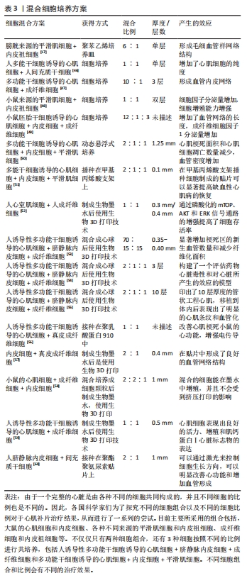

 生物3D打印的方式主要有3种,分别是挤压式生物打印、喷墨式生物打印和激光辅助光固化[9],这3种打印方式各有优劣,并且适合打印的组织也不同。如果打印心血管组织的话,常常采用后两种方式。挤出式打印的结构简单,并且支持高密度的细胞打印,但是在高黏度的情况下容易堵塞喷嘴,并且由于喷嘴的直径较小对细胞的损害较大,适合打印神经、皮肤或者肾脏组织。喷墨式生物打印属于非接触,它的印刷速度快、成本低,但是它打印垂直结构的组织较为困难,并且因为喷嘴温度较高,对细胞的损伤也较大。激光辅助光固化有很高的分辨率,对于打印精细的组织结构有一定优势,但是它的成本较高,且打印系统复杂。